

Metabolic Engineering - T. Scheper and Jens Nielsen
.pdfMetabolic Engineering of Indene Bioconversion in Rhodococcus sp. |
99 |
tion in Rhodococcus sp. KY1 is through the novel monooxygenase enzyme. For all steady states analyzed, at least 94% of the indene was oxidized to indan oxide. This analysis also demonstrated that KY1 lacks a trans-(1R,2R)-indandiol dehydrogenase previously hypothesized to be present in the parent I24 strain. Additionally, the use of tracers showed a previously unidentified chemical step in the bioconversion network, namely the hydrolysis of indan oxide to cis- (1S,2R)-indandiol in addition to trans-(1R,2R)-indandiol.
5
Future Directions for Metabolic Engineering of Indene Bioconversion
A central finding of our analysis is that indene monooxygenase is the key enzyme for indene oxidation, and the most likely candidate for overexpression if further increase of the total oxidation flux of the indene network is desired. The emergence of indene monooxygenase as the main oxidizing enzyme in KY1 is contrary to the initial hypothesis that implicated toluene-induced dioxygenase as the main route for (2R)-indandiol biosynthesis. Estimates of monooxygenase activity in KY1 suggest that it is probably satisfactory for industrial-scale production. Assuming that indan oxide synthesis proceeds approximately at the same rate as indene depletion, a final titer of 8.7 g/l of product should be expected from a fed batch fermentation of three days duration at a cell density of 10 g/l. Other data indicating that trans-(1R,2R)-indandiol and 1-keto-2-hy- droxy-indan may have an inhibitory effect on the monooxygenase, consistent with observations made in P. putida F1 [12], suggest this enzyme could also be considered as a candidate for directed evolution to reduce or eliminate product inhibition. Our revised view of the biocatalysis network emphasizes the need to express enzymes catalyzing the selective hydrolysis of indan oxide to trans- (1R,2R)-indandiol to prevent degradation by dehydrogenase(s). In terms of genetic modification, this task is more palatable than our original focus on multiple enzyme knockouts. Such secondary targets to improve (2R)-indandiol yield that were also identified by our analysis include the knockouts of multiple dehydrogenase activities and the dioxygenase producing cis-(1R,2S)-indandiol.
The presence of the cis-(1S,2R)-indandiol dehydrogenase means that the maximum yield of (2R)-indandiol that one can expect from KY1 is just under 60% due to the nature of the chemical hydrolysis of indan oxide to trans- (1R,2R)-indandiol and cis-(1S,2R)-indandiol. A promising approach to improving the product yield of KY1 is to hydrolyze selectively indan oxide to trans- (1R,2R)-indandiol by introducing an epoxide hydrolase and/or modifying culture conditions. A limonene-1,2-epoxide hydrolase from Rhodococcus erythropolis DCL14 has been characterized and cloned, and showed significant activity against indan oxide [14–16]. The activity of this enzyme encoded by the 0.5 kb limA gene should support the amount of indan oxide generated in KY1 by the indene monooxygenase. This would nullify the need for a dehydrogenase knockout since little or no cis-(1S,2R)-indandiol would be produced. Plasmids that can replicate in Rhodococcus were developed [17] that served as the foundation of a vector for the expression of this epoxide hydrolase in KY1, which has resulted in improved yield of trans-(1R,2R)-indandiol from indene [18]. Ad-
100 |
D.E. Stafford et al. |
ditionally, studies on the nature of indan oxide hydrolysis have shown that the ratio of trans-indandiol to cis-indandiol formed is highly pH-dependent. Further improvement of trans-indandiol yield has been obtained by performing the KY1 indene biotransformation at pH>8.0 [18].
Transaminase-type enzymes that can convert the indan oxide or (2R)-indan- diols directly to (–)-CAI are also promising tools for the improvement of Rhodococcus as a biocatalyst. With the indandiols siphoned away to (–)-CAI, 1- keto-2-hydroxy-indan would not be formed and this product or trans-(1R,2R)- indandiol would not inhibit the monooxygenase enzyme activity. During KY1 fermentations, product inhibition of the monooxygenase by trans-indandiol and 1-keto-2-hydroxy-indan could also be avoided by removing the (2R)-indan- diol product from the culture using resins or an organic phase. This technique has been applied to indene fermentations with P. putida using SP-207 resin to remove indandiols from unfiltered culture [12].Additional factors that may contribute to the inhibition observed in fed-batch culture include the general toxicity of indene (and possibly other indene metabolites) to the cells, as well as the possible growth dependence of the expression of indene oxidation genes. These warrant further consideration as development with a viable production strain proceeds.
The metabolic engineering analysis of indene bioconversion in Rhodococcus species has been instrumental in defining ways to improve the strain and the fermentation process for the production of (2R)-indandiol. A pivotal event was the emergence of the KY1 strain that lacked competing dioxygenase activity and gave a higher product yield. This is believed to be a result of the application of selective pressure on the culture in a chemostat environment. This result supports a generic paradigm in this regard for evolution of strain properties in a properly designed continuous flow system.
Acknowledgements. This work was supported by a grant from Merck Research Laboratories. D. Stafford and K. Yanagimachi were supported in part by NIH Biotechnology Training Grant # 2T32 GM08334–10 and by the Engineering Research Program of BES, DoE Grant no. DE- FG02–94ER-14487.
References
1.Stinson S (2000) Chiral drugs. Chem Eng News 79:55–78
2.Finnerty W (1992) The biology and genetics of the genus Rhodococcus. Annu Rev Microbiol 46:193–218
3.Warhurst M, Fewson C (1994) Biotransformations catalyzed by the genus Rhodococcus. Crit Rev Biotechnol 14:29–73
4.Butler C, Mason J (1997) Structure-function analysis of the bacterial aromatic ring-hy- droxylating dioxygenases. In: Advances in microbial physiology. Academic Press, vol 38, pp 47–84
5.Wackett L, Kwart L, Gibson D (1988) Benzylic monooxygenation catalyzed by toluene dioxygenase from Pseudomonas putida. Biochemistry 27:1360–1367
6.Gibson D, Resnick S, Lee K, Brand J, Torok D, Wackett L, Schocken M, Haigler B (1995) Desaturation, dioxygenation, and monooxygenation reactions catalyzed by naphthalene dioxygenase from Pseudomonas sp. strain 9816–4. J Bacteriol 177:2615–2621
Metabolic Engineering of Indene Bioconversion in Rhodococcus sp. |
101 |
7.Allen C, Boyd D, Larkin M, Reid KA, Sharma N, Wilson K (1997) Metabolism of naphthalene, 1-naphthol, indene, and indole by Rhodococcus sp. strain NCIMB 12038. Appl Environ Microbiol 63:151–155
8.Chartrain M, Jackey B, Taylor C, Sandford V, Gbewonyo K, Lister L, DiMichelle L, Hirsch C, Heimbuch B, Maxwell C, Pascoe D, Buckland B, Greasham R (1998) Bioconversion of indene to cis-(1S,2R)-indandiol and trans-(1R,2R)-indandiol by Rhodococcus species. J Fermentat Bioeng 86:550–558
9.Stafford DE, Yanagimachi KS, Lessard PA, Rijhwani SK, Sinskey AJ, Stephanopoulos G (2001) Optimizing bioconversion pathways through systems analysis and metabolic engineering (submitted)
10.Yanagimachi KS, Stafford DE, Dexter AF, Sinskey AJ, Drew SW, Stephanopoulos G (2001) Application of radiolabeled tracers to biocatalytic flux analysis (submitted)
11.Connors N, Chartrain M, Reddy J, Singhvi R, Patel Z, Olewinshi R, Salmon P, Wilson J, Greasham R (1997) Conversion of indene to cis-(1S),(2R)-indandiol by mutants of Pseudomonas putida F1. J Ind Microbiol Biotechnol 18:353–359
12.Buckland B, Drew S, Connors N, Chartrain M, Lee C, Salmon P, Gbewonyo K, Zhou W, Gailliot P, Singhvi R, Olewinshi R, Sun W-J, Reddy J, Zhang J, Jackey B, Taylor C, Goklen K, Junker B, Greasham R (1999) Microbial conversion of indene to indandiol: a key intermediate in the synthesis of CRIXIVAN. Metab Eng 1:63–74
13.Gibson D, Subramanian V (1984) Microbial degradation of aromatic hydrocarbons. In: Gibson D (ed) Microbial degradation of organic compounds. Marcel Dekker,New York,pp 253–294
14.Barbirato F, Verdoes J, de Bont J, van der Werf M (1998) The Rhodococcus erythropolis DCL14 limonene-1,2-epoxide hydrolase gene encodes an enzyme belonging to a novel class of epoxide hydrolases. FEBS Lett 438:293–296
15.van der Werf MJ, Overkamp KM, de Bont JAM (1998) Limonene-1,2-epoxide hydrolase from Rhodococcus erythropolis DCL14 belongs to a novel class of epoxide hydrolases. J Bacteriol 180:5052–5057
16.van der Werf M, Orru R, Overkamp K, Swarts H, Osprian I, Steinreiber A, de Bont J, Faber K (1999) Substrate specificity and stereospecificity of limonene-1,2-epoxide hydrolase from Rhodococcus erythropolis DCL14; an enzyme showing sequential and enantioconvergent substrate conversion. Appl Microbiol Biotechnol 52:380–385
17.Treadway S, Yanagimachi K, Lankenau E, Lessard P, Stephanopoulos G, Sinskey A (1999) Isolation and characterization of indene bioconversion genes from Rhodococcus strain I24. Appl Microbiol Biotechnol 51:786–793
18.Stafford DE, Yanagimachi KS, Lessard PA, Rijhwani SK, Sinskey AJ, Stephanopoulos G (2001) Optimizing bioconversion pathways through systems analysis and metabolic engineering (submitted)
Received: December 2000

Metabolic Engineering of the Morphology
of Aspergillus
Mhairi McIntyre, Christian Müller, Jens Dynesen, Jens Nielsen
Center for Process Biotechnology, Department of Biotechnology, Building 223, Technical University of Denmark, 2800 Lyngby, Denmark, e-mail: jn@ibt.dtu.dk
The morphology of filamentous organisms in submerged cultivation is a subject of considerable interest, notably due to the influence of morphology on process productivity. The relationship between process parameters and morphology is complex: the interactions between process variables, productivity, rheology, and macroand micro-morphology create difficulties in defining and separating cause and effect.Additionally,organism physiology contributes a further level of complexity which means that the desired morphology (for optimum process performance and productivity) is likely to be process specific. However, a number of studies with increasingly powerful image analysis systems have yielded valuable information on what these desirable morphologies are likely to be. In parallel, studies on a variety of morphological mutants means that information on the genes involved in morphology is beginning to emerge. Indeed, we are now beginning to understand how morphology may be controlled at the molecular level. Coupling this knowledge with the tools of molecular biology means that it is now possible to design and engineer the morphology of organisms for specific bioprocesses. Tailor making strains with defined morphologies represents a clear advantage in optimization of submerged bioprocesses with filamentous organisms.
Keywords. Morphological engineering, Aspergillus, Dimorphism |
|
|
1 |
Introduction . . . . . . . . . . . . . . . . . . . . . . . . . . . . . . . |
104 |
2 |
Analysis Tools . . . . . . . . . . . . . . . . . . . . . . . . . . . . . . |
105 |
3 |
Physiological Aspects of Morphological Development . . . . . . . . |
105 |
3.1 |
Morphological Development of Filamentous Fungi . . . . . . . . . . |
106 |
3.1.1 |
Apical Hyphal Extension . . . . . . . . . . . . . . . . . . . . . . . . . |
106 |
3.1.2 |
Cytoskeleton Organization . . . . . . . . . . . . . . . . . . . . . . . |
108 |
3.2 |
The Relationship Between Morphology and Productivity . . . . . . |
109 |
3.2.1 |
Penicillin Production . . . . . . . . . . . . . . . . . . . . . . . . . . |
111 |
3.2.2 |
Enzyme Production . . . . . . . . . . . . . . . . . . . . . . . . . . . |
112 |
4 |
Molecular Aspects of Morphological Control . . . . . . . . . . . . . |
114 |
4.1 |
Filamentous Fungi . . . . . . . . . . . . . . . . . . . . . . . . . . . . |
114 |
4.1.1 |
Genes Involved in Morphology . . . . . . . . . . . . . . . . . . . . . |
114 |
4.1.2 |
Engineering Hyphal Architecture . . . . . . . . . . . . . . . . . . . . |
116 |
4.2 |
Dimorphic Organisms . . . . . . . . . . . . . . . . . . . . . . . . . . |
119 |
4.2.1 |
Biochemical Changes Associated with Dimorphism . . . . . . . . . |
120 |
4.2.2 |
Structural Changes Associated with Dimorphism . . . . . . . . . . . |
122 |
4.2.3 |
Molecular Level Control of Dimorphism . . . . . . . . . . . . . . . . |
123 |
References . . . . . . . . . . . . . . . . . . . . . . . . . . . . . . . . . . . . |
124 |
|
|
Advances in Biochemical Engineering/ |
|
|
Biotechnology,Vol. 73 |
|
|
Managing Editor: Th. Scheper |
|
|
© Springer-Verlag Berlin Heidelberg 2001 |
|

104 M. McIntyre et al.
1 Introduction
Filamentous fungi are extensively used in the fermentation industry for the production of a long list of products including primary metabolites, antibiotics, industrial enzymes, and heterologous proteins. In the production of industrial enzymes, filamentous fungi are among the most important cell factories. This is due to their highly efficient secretion of proteins, and the establishment of good fermentation technology with these organisms.Protein secretion by filamentous organisms has been correlated with hyphal extension rates and tip growth and, as such, morphological characterization of the commonly used enzyme producing strains (mainly Aspergilli) is of interest. Additionally, fungal morphology is of interest due to the fact that it influences the rheology of the fermentation medium, and thereby has a significant impact on mixing and mass transfer within the bioreactor. In industry there is, therefore, a desire to tailor-make the morphology of filamentous fungi to ensure high protein secretion and at the same time a low viscosity culture.
Despite the importance of fungal morphology, our understanding of how the morphology can be manipulated is still rather limited. However, recent developments in basic biology have allowed progress in our understanding of fungal physiology and morphology by providing a number of morphological mutants and strains with disruption or inactivation of specific genes influencing the morphology. Studies employing such strains have greatly added to our knowledge of the regulation and control of morphology in filamentous fungi. A number of the key genes influencing morphology have been identified and it is, therefore, expected that in the future it will be possible to apply a much more directed approach to the development of better industrial strains.
To facilitate this process, this review will collate and summarize the current knowledge regarding fungal morphogenesis, with respect to both the physiological and molecular levels of control and regulation. The information on fungal physiology (growth and productivity) and morphology of filamentous fungi in submerged bioprocesses is relatively extensive compared to what is known about genetic control. In many cases, morphogenesis can be effected by changes in environmental conditions, while the molecular basis for such effects is not always known. On the other hand, morphological mutants have been identified, many with assumed “desirable” morphologies; however, the performance of these strains has not been assessed in submerged cultivation.Additionally,when considering tailoring morphologies for specific bioprocesses, here referred to as morphological engineering, it is not known which genes, either structural or regulatory, would be of interest.
Indeed, it is often the case that the link between control on the physiological level and the molecular basis for such control has not been made. In the past five to ten years, however, an increasing number of studies have identified genes involved in the control of morphology of filamentous fungi (namely Aspergilli and Neurospora). In addition, recent studies of dimorphic fungi have added further information on the genes involved in morphogenesis. The time is right, therefore, to begin building the picture of all factors known to influence morphology
Metabolic Engineering of the Morphology of Aspergillus |
105 |
and discuss the possibilities for utilizing newly constructed strains for process optimization. This will provide a platform from which to push forward metabolic engineering of the morphology of all industrially relevant filamentous organisms.
2
Analysis Tools
The basis for rational design of fungal morphology is powerful analytical techniques. Computerized image analysis systems have been employed in studies of hyphal morphology for more than ten years [1–3] and have now reached a stage where reproducible analysis can be carried out (semi-) automatically and rapidly [4–6]. The resultant data can be used for studying growth mechanisms and kinetics and process modeling [7, 8] providing valuable information on the growth and differentiation of strains under different environmental conditions [9–11].
The application of fluorescent staining techniques to the study of filamentous organisms has provided valuable information on physiology, positioning of organelles and localization of structures within hyphae [12–15]. Indeed much has been learnt about the growth and organization of fungal hyphae through microscopy. When coupled with computerized image analysis, physiological information can be obtained in addition to the morphological data [7],providing two levels of detail on hyphal development.
Recently, studies employing a flow-through growth cell for analysis of the growth of filamentous fungi have been described [16, 17]. The system allowed the growth kinetics of single hyphae,from spore swelling and germination,to be determined on-line, rather than the average populations that are sampled from submerged bioprocesses. Clearly, such new advances and the application of “traditional”image analysis methods provide a valuable set of tools for studies of filamentous fungi, allowing quantification of changes resulting from metabolic engineering.
In addition,high performance bioreactors [18],particularly chemostats with, for example, Teflon coating to reduce wall growth can provide highly controlled environments for studies of morphologically engineered strains. Submerged cultivation under highly controlled conditions would be necessary to quantify precisely the effect of metabolic engineering of the morphology on productivity and bioreactor performance to allow accurate comparisons between strains.
3
Physiological Aspects of Morphological Development
Apical hyphal extension of filamentous fungi has been the subject of a number of thorough reviews [19–23] dealing with aspects of growth, hyphal architecture, and intracellular organization. For this reason, these subjects will not be discussed in detail here. Rather, the review of the physiology of fungal morphogenesis will focus on those features of hyphal development that may be of interest for designing strategies for the production of “better”industrial strains.With
106 |
M. McIntyre et al. |
this aim in mind, particular focus will be on how the processes involved in apical hyphal extension are controlled and how this may be related to improved productivity.
3.1
Morphological Development of Filamentous Fungi
3.1.1
Apical Hyphal Extension
Fungal cells grow by apical hyphal extension in a highly polarized manner [14] with respect to their growth, morphology, organelle positioning, and cytoskeletal distributions [24, 25]. Hyphal extension is facilitated through deposition and insertion of new membrane and cell wall material at localized sites on the cell surface [21]. The enzymes and precursors required at the advancing tips for the synthesis of the new material are delivered in vesicles transported to these sites along a polarized cytoskeletal network [20, 21].
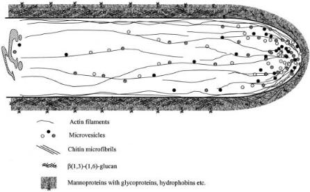
Figure 1 summarizes the processes involved in polar extension of filamentous organisms and the organization of the cell wall and cytoskeleton components. Supply of cell wall precursors is critical for wall expansion at the advancing tip and in many organisms the Spitzenkörper has been identified and visualized as the vesicle supply center [26–29]. This structure is likely also to have a role in controlling growth directionality [26]. The principal components of the cy-
Fig. 1. Model of polar cell wall expansion in filamentous fungi.Vesicles with cell wall components and proteins are transported to the tip. An actin-myosin-based system is important in establishing polar growth through transport of the micro-vesicles to the cell surface. The cell wall at the apex is plastic but it hardens as the matrix of glucans and chitin crystallizes
Metabolic Engineering of the Morphology of Aspergillus |
107 |
toskeleton (actin and tubulin) have a major role in the process of tip growth, being responsible for the migration of organelles to the advancing apex [20,23,25]. While the regulation of polarity is complex and not fully understood, the role of Ca2+ ion gradients [30, 31], calcium mediated secondary messenger systems [32], and turgor pressure [19] have been demonstrated.
Biosynthesis of the cell wall material takes place in three sites, the cytoplasm, plasma membrane, and the wall itself. Deposition of cell wall components starts with several interconnected synthetic processes, which results in the extrusion of cell wall building blocks through the cellular membrane. Maturation of the wall through cross-linking of the components, then follows. The structural polymers chitin and b(1–3) and b(1–4) linked glucans contribute to the rigidity of the wall [33] and it is cross-linking of these that helps shape the hyphal architecture, adding a rigid structure to the mature wall. The enzymes involved in the crosslinking of chitin with wall components have not been identified, but it is most probable that transglycosidation leads to the formation of the cross-linkages. In filamentous fungi, autoradioactive studies following the incorporation of N- acetylglucosamine and glucose into growing hyphal walls have shown that nearly all N-acetylglucosamine is deposited within 1 µm of the hyphal tip region [34,35].
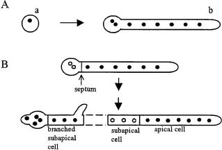
Fungi duplicate their length and nuclei through integration of the processes involved in tip growth, nuclear division, septation, and branching in a process termed the duplication cycle [36]. The duplication cycle in pre-divisional and post-divisional cells of Aspergillus nidulans is illustrated in Fig. 2. The cycle begins as a new apical compartment is created after septum formation has divided an existing apical compartment.
Fig. 2 A, B. Comparison of the duplication cycle and morphology of: A pre-divisional; B postdivisional cells of A. nidulans.A conidium (a) germinates and the first septum is formed at the basal end of the germ tube (b) when the germling has eight or more nuclei. Post-divisional cells are differentiated into subapical and apical tip cells (B). Apical cells contain many nuclei that are evenly spaced along the cell. Subapical cells contain three to four evenly spaced nuclei. Subapical cells can branch,and the branched cell grows like an apical cell.Apical and branched subapical cells have active nuclear cycles (filled circles) while nuclei in unbranched subapical cells are trapped in interphase (empty circles). (Revised from [37])
108 |
M. McIntyre et al. |
It has been argued that different fungal growth forms only differ in the degree of polarization of the processes involved in the formation of the new wall [38], with different types of fungal cells acquiring unique morphologies through distinctive patterns of polarized morphogenesis [39, 40]. For example, the ellipsoidal shape of yeasts occurs as a result of individual cells cycling through transient phases of polarized and isotropic growth. Conversely, filamentous organisms have cells (hyphae) that are long relative to their width. An understanding of how polarity is maintained, therefore, may provide an overview of how morphology, in general, may be manipulated through control of the processes leading to apical wall expansion.
Ultimately, polarized growth requires numerous gene products and coordination of processes involved in cytoskeleton and secretory functions [39].At present we are still building information on how these events are coordinated and regulated. Although no complete picture of polarized apical growth exists, it is possible to study the effects of mutation on tip growth. Several genes have been identified whose products are involved in hyphal extension and mutant strains of filamentous fungi defective in polarity have been characterized. The possibility of morphological engineering via this route will be discussed in Sect. 4.
3.1.2
Cytoskeleton Organization
The fungal cytoskeleton is composed, principally, of two major polymers, microtubules and actin with a growing number of microtubule associated proteins (MAPs) and actin binding proteins (ABPs) being identified. The organized development of the cytoskeleton of filamentous fungi is crucial in shaping morphology, as it is the cytoskeleton that provides the scaffold for hyphal growth while, additionally, playing a role in directing polarity.
Actin has involvement in a variety of the processes that result in tip growth [25],and it is thought to play a multifunctional role in apical growth through the coordination of tip morphogenesis, cell wall synthesis, cytoplasmic migration, and organelle positioning [31, 41]. Filamentous actin (F-actin) is typically concentrated at the apices of filamentous fungi (Fig. 1), implying that it plays a role in tip extension [31]. The actin cap (the concentration of actin plaques located near the hyphal apex) appears to be responsible for tip extension and the actin cables (located subapically) are involved in the transport of vesicles to the extending tips. Studies of Saprolegnia [31],an Oomycete,suggest that the actin cap functions to support the apex in regions where the cell wall is weak, being optimally organized to reinforce the plastic cell wall at the growing tip. While the Oomycetes represent a different evolutionary line to Aspergilli, actin has been shown to have a primary role in the movement of secretory vesicles in fungi,and evidence for an actin-based system controlling polarity and secretion in A. nidulans has been presented [42].
It is likely that Ca2+ plays a role in controlling tip growth via actin in a number of diverse fungi. This is not only due to the fact that actin and Ca2+ are abundant in growing tips; Ca2+ ions are also known to regulate actin function in a number of ways. The subject has been extensively reviewed previously [31].
Metabolic Engineering of the Morphology of Aspergillus |
109 |
Calcium may also play a role as a branching signal, as has been investigated with Neurospora crassa [43], with the addition of the divalent cation ionophore inducing profuse branching. This observation has been linked to the involvement of cyclic AMP in the regulation of branching, as the colonial phenotype was dependent on a low intracellular level of cAMP, and there are known antagonistic regulatory roles of Ca2+ and cAMP [44]. Very little is known about how branching is regulated in filamentous fungi; however, a simple relationship between hyphal elongation rate and branch formation has been shown to exist in A. nidulans [45]. Branch initiation was observed in this organism when a compartment reached a maximum rate of extension, which was achieved at different lengths with different specific growth rates.
Further hyphal structure is provided through an arrangement of microtubules, formed through the polymerization of tubulin heterodimers. In addition to contributing to the internal scaffold of hyphal cells, these filaments have also been shown to be involved in the positioning of organelles in hyphae [43]. Nuclear migration plays an important role in the growth and development of filamentous fungi, as has been exemplified by studies on A. nidulans [46, 47]. Nuclear migration (and perhaps that of other organelles) is mediated by cytoplasmic dynein, a microtubule dependent motor [47, 48]. Actin related proteins, such as dynactin in N. crassa [49],have also been shown to be involved in the stabilization of the internal structure and the positioning of nuclei.
From the evidence presented above it appears that many of the components involved in shaping the hyphal ultrastructure have multifunctional roles, and this presents a complication if engineering of morphology is to proceed via regulation of structural genes. Multiple effects of structural gene inactivation are likely to be observed. Indeed, it would be most desirable if the phenotypes of strains with inactivated regulatory genes were to be investigated, and in any event that strains with single gene inactivations were characterized.
3.2
The Relationship Between Morphology and Productivity
A key aspect in metabolic engineering of Aspergillus morphology is the subsequent effect of morphology on product formation. Generally, morphological forms are described on two levels – macroscopic and microscopic [50, 51] with the macromorphology describing the gross morphology (pellets, clumps or freely dispersed mycelia) and the micromorphology describing the properties of these types (branch frequency, hyphal dimensions, and segregation, i.e., compartmentalization and physiological population distribution) [52,53]. These descriptions are illustrated in Fig. 3. While the macroscopic morphology can influence medium rheology and thus mixing and mass transfer within a culture, the literature mainly describes control of macromorphology by environmental conditions. For example, Aspergillus oryzae produces pellets following spore agglomeration, a process which is pH dependent [53].
Figure 4 provides a schematic representation of the interactions between process conditions,morphology,and productivity. The micro-environment of hyphae is determined by the process conditions and the mixing of the culture,and it