


9350
9Cerebrum
Macroscopically, the cerebrum is made up of the cerebral cortex, the subcortical white matter, and the basal ganglia, which were discussed in Chapter 8. The gross structure of the cerebrum can be understood best with reference to its embryological development. Its most impressive feature is the immense expansion of the cortex, which causes folding (gyration) of the brain surface. The individual cortical areas are connected to each other, and to deeper brain structures, by the numerous fiber pathways that make up the subcortical white matter.
Histologically, most of the cerebral cortex possesses a six-layered cellular architecture. This basic histological pattern undergoes characteristic variations from one location in the cortex to another, giving rise to numerous, cytoarchitecturally distinct cortical areas. The early neuroanatomists proposed that the specific cellular structure of each area corresponded to the specific task that it carried out. It has, indeed, been possible to assign a single, concrete function to a number of areas, the so-called primary cortical fields. Yet the greater part of the cerebral cortex consists of association areas, whose function apparently consists of higher-level processing of information derived from, or traveling to, the primary fields. Higher cortical functions such as language, in particular, cannot be localized to a single cortical area but depend instead on a complex interaction of multiple areas.
Development
The cerebrum or endbrain (telencephalon) develops from the paired telencephalic vesicles at the front end of the neural tube, the so-called prosencephalon. The enormous growth of the telencephalic vesicles makes the endbrain envelop the brainstem like a cloak (pallium) and leads to the development of the lateral ventricles, with their characteristic anatomical subdivisions, from outpouchings of the fluid-filled lumen of the neural tube. The semicircular extension that characterizes the growth of the telencephalon (Fig. 9.1) and the lateral ventricles can also be seen in the developing fiber projections, in the fornices, and in the corpus callosum, the great fibrous connection between the two hemispheres. A few more details of telencephalic development will be presented here as a useful aid to the understanding of cerebral anatomy.
Baehr, Duus' Topical Diagnosis in Neurology © 2005 Thieme
All rights reserved. Usage subject to terms and conditions of license.
Development · 351 9
Parietal
Frontal
Central
portion
Ventricle
Cortex
Occipital
Anterior horn
Basal ganglion
Head of caudate nucleus Posterior horn
Temporal
Tail of caudate nucleus
Inferior horn
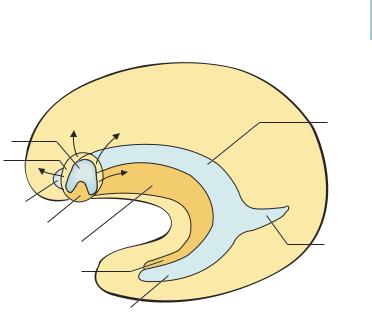
Fig. 9.1 Ontogenetic development of the cerebral cortex. Lateral view of an earlier and a later developmental stage of the telencephalon. The telencephalic vesicle expands massively (arrows) in the form of an arch, resulting in simultaneous, archlike expansion of the cerebral cortex (yellow), ventricle (blue), and basal ganglion (orange).
Evolution of the endbrain. In the telencephalon, as elsewhere in the central nervous system, the neural tube consists of two parts, ventral and dorsal. The ventral part gives rise to the septal region medially and the basal ganglion laterally. The basal ganglion, in turn, gives rise to the caudate (“tailed”) nucleus, putamen, claustrum, and amygdala. The cortex derived from the dorsal part becomes differentiated over the course of phylogenetic development into the more laterally lying paleocortex, the oldest portion of cerebral cortex, and the more medially lying and phylogenetically younger archicortex.
The spatial arrangement of paleocortex and archicortex is preserved unchanged in amphibians. In reptiles, however, the neocortex arises in a lateral position between the paleocortex and the archicortex. The neocortex takes on an enormous size in higher organisms, pushing the paleocortex and archicortex far apart. In humans, the paleocortex is ultimately displaced to the base of the brain, where it makes up various components of the phylogenetically ancient olfactory system (olfactory bulb, tract, and trigone, anterior perforated substance, and lateral olfactory stria; cf. p. 128f.). Meanwhile, the archicortex is displaced medially; the semicircular growth of the telencephalic vesicle
Baehr, Duus' Topical Diagnosis in Neurology © 2005 Thieme
All rights reserved. Usage subject to terms and conditions of license.
9 |
352 · |
9 Cerebrum |
|
|
|
|
|
|
|
|
|
|
Indusium |
Lateral |
Internal |
|
|
|
|
|
griseum |
ventricle |
capsule |
|
|
|
|
|
Neocortex |
|
|
|
|
|
|
|
Fibers of |
|
|
|
|
|
|
|
corpus |
|
|
|
|
|
|
|
callosum |
|
|
|
Ven- |
Archi- |
Paleo- |
Ventricle |
Neocortex |
|
|
|
tricle |
cortex |
cortex |
|
Head of |
|
|
|
|
caudate |
|
|
|||
|
|
|
|
|
|
|
|
|
|
|
|
|
nucleus |
|
|
|
|
|
|
|
Thalamus |
|
|
|
|
|
|
|
Interthalamic |
|
|
|
|
|
|
|
adhesion |
|
|
|
Basal |
Septum |
|
Hippocampus |
|
|
|
|
ganglion |
|
|
|
|
|
|
|
Amphibians |
Reptiles |
Humans |
|
|||
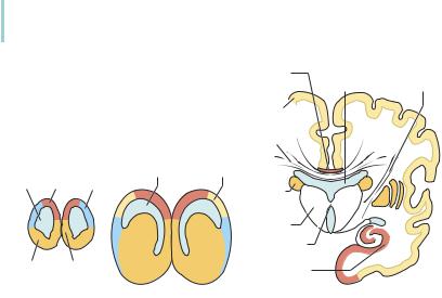
Fig. 9.2 Phylogenetic development of the cerebral cortex (coronal sections). The neocortex (yellow) arises between the archicortex (red) and paleocortex (blue). It expands markedly in higher organisms; in humans it displaces the paleocortex to the base of the brain (olfactory cortex, not shown) and the archicortex to a medial position overlying the corpus callosum (indusium griseum). The hippocampal formation (archicortex) in the floor of the inferior horn of the lateral ventricle reaches its basal position through the archlike expansion of the telencephalon (cf. Fig. 9.1).
pushes it into the inferior horn of the lateral ventricle, where it makes up the massive hippocampal formation. Only a thin layer of archicortex is found mediodorsally on the outer surface of the corpus callosum: this is the indusium griseum, with its medial and lateral longitudinal striae. By far the greatest part of the human cerebral cortex is of neocortical origin (Fig. 9.2).
Inside-out stratification of the cerebral cortex. The neurons of the cerebral cortex, as of all parts of the central nervous system, are initially formed in the ventricular zone, i.e., near the fluid-filled lumen (ventricle) of the neural tube. The cells formed earliest make up the so-called preplate, which is later subdivided into the marginal zone and the subplate. The cortical plate proper, consisting of six cellular layers, develops between these two structures. The cortical neurons that are formed first occupy the deeper layers (layers 5 and 6), and those formed later migrate upward into the more superficial layers (“insideout” arrangement). Thus, the later neurons must travel past their precursor cells to get to the subpial cortical layers (Fig. 9.3), passing from the ventricular zone to the cortical plate along radial glial fibers. The six cortical layers are numbered topographically from outside in (the traditional system, as used in this book), or, alternatively, in the order of their formation, as has lately been
Baehr, Duus' Topical Diagnosis in Neurology © 2005 Thieme
All rights reserved. Usage subject to terms and conditions of license.
Development · 353 9
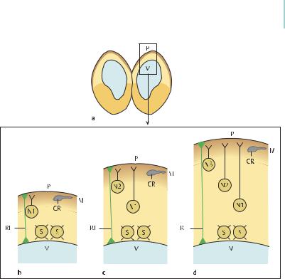
Fig. 9.3 Inside-out lamination of the cerebral cortex. The earliest neurons form the so-called preplate, which soon divides into the marginal zone (M), with its Cajal−Retzius cells (CR), and the “subplate” with its subplate neurons (S). The neurons of the cortical plate are deposited in the intervening space (N1−N3). Early cortical neurons (N1) migrate upward, from the vicinity of the ventricle (V), along radial glial fibers (RF) to the marginal zone of the cerebral cortex, where it is thought that they are stopped by reelin (brown), a protein of the extracellular matrix produced by the Cajal−Retzius cells. As the cortex thickens, later cortical neurons (N2, N3) must travel longer distances to reach the reelin-containing marginal zone. As a result, the neurons that are laid down earliest form the deeper layers of the cerebral cortex, while those that are laid down later form its more superficial layers. P = brain surface with pia mater.
proposed (Marin-Padilla, 1998). According to the findings of recent studies, normal neuronal migration, leading to the characteristic inside-out cortical layering, depends crucially on the participation of the CajalRetzius cells of the marginal zone. These cells secrete a protein called reelin, which apparently directs neuronal migration along the radial glial fibers (Fig. 9.3). Abnormalities of neuron formation, migration, or separation from the radial glial fibers are collectively called neuronal migration disorders.
Baehr, Duus' Topical Diagnosis in Neurology © 2005 Thieme
All rights reserved. Usage subject to terms and conditions of license.