
- •Preface
- •Contents
- •1 Elements of the Nervous System
- •2 Somatosensory System
- •3 Motor System
- •4 Brainstem
- •5 Cerebellum
- •6 Diencephalon and Autonomic Nervous System
- •7 Limbic System
- •8 Basal Ganglia
- •9 Cerebrum
- •10 Coverings of the Brain and Spinal Cord; Cerebrospinal Fluid and Ventricular System
- •Further Reading
- •Index
- •Abbreviations
- •1 Elements of the Nervous System
- •Elements of the Nervous System
- •Information Flow in the Nervous System
- •Synapses
- •Neurotransmitters and Receptors
- •Functional Groups of Neurons
- •Glial Cells
- •Development of the Nervous System
- •2 Somatosensory System
- •Peripheral Nerve, Dorsal Root Ganglion, Posterior Root
- •Peripheral Regulatory Circuits
- •Central Components of the Somatosensory System
- •Posterior and Anterior Spinocerebellar Tracts
- •Posterior Columns
- •Anterior Spinothalamic Tract
- •Lateral Spinothalamic Tract
- •Other Afferent Tracts of the Spinal Cord
- •Central Processing of Somatosensory Information
- •Somatosensory Deficits due to Lesions at Specific Sites along the Somatosensory Pathways
- •3 Motor System
- •Central Components of the Motor System and Clinical Syndromes of Lesions Affecting Them
- •Motor Cortical Areas
- •Corticospinal Tract (Pyramidal Tract)
- •Corticonuclear (Corticobulbar) Tract
- •Other Central Components of the Motor System
- •Lesions of Central Motor Pathways
- •Peripheral Components of the Motor System and Clinical Syndromes of Lesions Affecting Them
- •Clinical Syndromes of Motor Unit Lesions
- •Complex Clinical Syndromes due to Lesions of Specific Components of the Nervous System
- •Spinal Cord Syndromes
- •Vascular Spinal Cord Syndromes
- •Nerve Root Syndromes (Radicular Syndromes)
- •Plexus Syndromes
- •Peripheral Nerve Syndromes
- •Syndromes of the Neuromuscular Junction and Muscle
- •4 Brainstem
- •Surface Anatomy of the Brainstem
- •Medulla
- •Pons
- •Midbrain
- •Olfactory System (CN I)
- •Visual System (CN II)
- •Eye Movements (CN III, IV, and VI)
- •Trigeminal Nerve (CN V)
- •Facial Nerve (CN VII) and Nervus Intermedius
- •Vagal System (CN IX, X, and the Cranial Portion of XI)
- •Hypoglossal Nerve (CN XII)
- •Topographical Anatomy of the Brainstem
- •Internal Structure of the Brainstem
- •5 Cerebellum
- •Surface Anatomy
- •Internal Structure
- •Cerebellar Cortex
- •Cerebellar Nuclei
- •Connections of the Cerebellum with Other Parts of the Nervous System
- •Cerebellar Function and Cerebellar Syndromes
- •Vestibulocerebellum
- •Spinocerebellum
- •Cerebrocerebellum
- •Cerebellar Tumors
- •6 Diencephalon and Autonomic Nervous System
- •Location and Components of the Diencephalon
- •Functions of the Thalamus
- •Syndromes of Thalamic Lesions
- •Thalamic Vascular Syndromes
- •Epithalamus
- •Subthalamus
- •Hypothalamic Nuclei
- •Afferent and Efferent Projections of the Hypothalamus
- •Functions of the Hypothalamus
- •Sympathetic Nervous System
- •Parasympathetic Nervous System
- •Visceral and Referred Pain
- •7 Limbic System
- •Anatomical Overview
- •Internal and External Connections
- •Microanatomy of the Hippocampal Formation
- •Amygdala
- •Functions of the Limbic System
- •Types of Memory
- •8 Basal Ganglia
- •Preliminary Remarks on Terminology
- •The Role of the Basal Ganglia in the Motor System: Phylogenetic Aspects
- •Connections of the Basal Ganglia
- •Function and Dysfunction of the Basal Ganglia
- •Clinical Syndromes of Basal Ganglia Lesions
- •9 Cerebrum
- •Development
- •Gross Anatomy and Subdivision of the Cerebrum
- •Gyri and Sulci
- •Histological Organization of the Cerebral Cortex
- •Laminar Architecture
- •Cerebral White Matter
- •Projection Fibers
- •Association Fibers
- •Commissural Fibers
- •Functional Localization in the Cerebral Cortex
- •Primary Cortical Fields
- •Association Areas
- •Frontal Lobe
- •Coverings of the Brain and Spinal Cord
- •Dura Mater
- •Arachnoid
- •Pia Mater
- •Cerebrospinal Fluid Circulation and Resorption
- •Arteries of the Anterior and Middle Cranial Fossae
- •Arteries of the Posterior Fossa
- •Collateral Circulation in the Brain
- •Dural Sinuses
- •Venous Drainage
- •Cerebral Ischemia
- •Arterial Hypoperfusion
- •Particular Cerebrovascular Syndromes
- •Impaired Venous Drainage from the Brain
- •Intracranial Hemorrhage
- •Intracerebral Hemorrhage (Nontraumatic)
- •Subarachnoid Hemorrhage
- •Subdural and Epidural Hematoma
- •Impaired Venous Drainage
- •Spinal Cord Hemorrhage and Hematoma
- •Further Reading
- •Index

Internal Structure · 243 5
Internal Structure
Although the cerebellum accounts for only about 10% of the brain by weight, it contains more than 50% of all the brain’s neurons. The neurons of the cerebellum are located in the gray matter of the highly convoluted cerebellar cortex and in the four deep cerebellar nuclei on either side (see below).
Cerebellar Cortex
The cerebellar cortex is composed of three layers (Fig. 5.3). Proceeding from the outermost inward, these layers are:
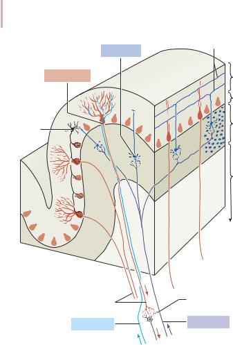
Molecular layer (stratum moleculare). This layer consists mainly of cellular processes,ofwhichthemajorityaregranulecellaxons—parallelfibers,seebelow— and Purkinje cell dendrites. A few neurons are found among the fibers (stellate cells, basket cells, Golgi cells), which function as inhibitory interneurons.
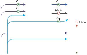
Pukinje cell layer (stratum ganglionare). This thin layer contains nothing but the large cell bodies of the Purkinje cells, arranged side by side in rows. The elaborate, highly branched dendritic trees of these cells are directed outward into the molecular layer, where the dendritic tree of each individual Purkinje cell lies in a plane perpendicular to the long axis of the folium. The Purkinje cell axons are the only efferent fibers leaving the cerebellar cortex. They project mainly to the deep cerebellar nuclei and release the inhibitory neurotransmitter GABA (γ-aminobutyric acid). Efferent fibers from the cortex of the vestibulocerebellum bypass the deep cerebellar nuclei and project directly to sites outside the cerebellum.
Granulecelllayer(stratumgranulosum).Thislayerconsistsalmostentirelyofthe denselypackedcellbodiesofthesmallgranulecells,whichaccountformorethan 95% of all cerebellar neurons. The axons of these cells are mainly found in the molecular layer, where they travel along individual folia as parallel fibers and form synapses with the perpendicularly oriented dendritic trees of the Purkinje cells(approximately200 000parallelfibersformsynapseswithasinglePurkinje cell). The cerebellar granule cells are glutamatergic and are the only neurons of the cerebellar cortex that exert an excitatory influence on their target cells.
Afferent Input to the Cerebellar Cortex
The afferent input to the cerebellar cortex is mainly derived from the ipsilateral vestibular nuclei (a small part, in fact, comes directly from the vestibular organ,
Baehr, Duus' Topical Diagnosis in Neurology © 2005 Thieme
All rights reserved. Usage subject to terms and conditions of license.

5 244 · 5 Cerebellum
Granule cell
Purkinje cell
Basket cell
Purkinje cell axons
Climbing fiber
Parallel fibers
Molecular layer
Purkinje cell layer
Granular layer
White matter
Neurons in the dentate nucleus
Mossy fiber
Fig. 5.3 Structure of the cerebellar cortex with its afferent and efferent connections (schematic drawing)
without any intervening synaptic relay), the ipsilateral spinal cord, the contralateral pontine nuclei (and thus, indirectly, from the contralateral cerebral cortex), and the contralateral olivary nuclear complex in the medulla (olive, for short). The olivary fibers are the so-called climbing fibers, which terminate on the Purkinje cells of the cerebellar cortex, climbing up their dendritic trees like ivy. All other afferent fibers terminate as mossy fibers on the granule cells of the cerebellar cortex, which then relay further impulses along their axons (parallel fibers of the molecular layer) to the Purkinje cell dendrites. Both mossy
Baehr, Duus' Topical Diagnosis in Neurology © 2005 Thieme
All rights reserved. Usage subject to terms and conditions of license.

Internal Structure · 245 5
fibers and climbing fibers give off important collaterals to the deep cerebellar nuclei on their way to the cortex.
In view of the fact that both the mossy fibers and the granule cells (and thus the overwhelming majority of synapses in the cerebellum) are glutamatergic, it is not surprising that the administration of glutamate antagonists causes a marked worsening of cerebellar function in patients with cerebellar lesions.
Cerebellar Nuclei
A horizontal section of the cerebellum reveals four deep nuclei within each cerebellar hemisphere (see Fig. 5.5). The fastigial nucleus (“roof nucleus”) is found most medially, in the roof of the fourth ventricle. It receives most of its afferent fibers from the Purkinje cells of the flocculonodular lobe (vestibulocerebellum). Its efferent fibers travel directly to the vestibular nuclei (fastigiobulbar tract) (Fig. 5.5) or cross to the opposite side of the cerebellum and then continue to the reticular formation and the vestibular nuclei (uncinate fasciculus).
Lateral to the fastigial nucleus, one finds two smaller nuclei, the globose nucleus (usually divided into two or three subnuclei) and the emboliform nucleus. Both of these nuclei receive afferent input from the cortex of the paravermian zone and vermis (spinocerebellum) and send efferent fibers to the contralateral red nucleus (Fig. 5.5).
The largest of the cerebellar nuclei, the dentate nucleus, occupies a lateral position in the deep white matter of each cerebellar hemisphere. Its afferent input comes mainly from the cortex of the cerebellar hemispheres (cerebrocerebellum), and, to a lesser extent, from the cortex of the paravermian zone. Its efferent fibers travel by way of the superior cerebellar peduncle to the contralateral red nucleus and thalamus (ventral lateral nucleus, VL) (Fig. 5.5). The thalamus is the site of a synaptic relay, with further projection to the motor areas of the cerebral cortex (Brodmann areas 4 and 6) (Fig. 6.4, p. 266).
Afferent and Efferent Projections of the Cerebellar Cortex
and Nuclei
Synaptic transmission within the cerebellum follows a uniform scheme (Fig. 5.4): the cerebellar afferent pathways project to the cerebellar cortex and, through collateral fibers, to the deep cerebellar nuclei. In the cortex, afferent information is processed in a complex polysynaptic pathway that eventually converges onto the Purkinje cells. The Purkinje cells, in turn, transmit the results of this processing to the deep cerebellar nuclei, in the form of inhibitory, GABAergic impulses. In the deep nuclei, integrative processing of both primary
Baehr, Duus' Topical Diagnosis in Neurology © 2005 Thieme
All rights reserved. Usage subject to terms and conditions of license.

5 |
246 · 5 Cerebellum |
|
|
|
|
|
|
|
|
|
|
|
|
|||
|
|
|
|
|
|
|
|
|
|
|
|
|
|
|
|
|
|
|
|
|
|
|
|
|
|
|
|
|
|
|
|
|
|
|
|
|
|
|
|
|
|
Granule cells |
|
|
|
|
|
|
|
|
|
|
|
|
|
|
|
|
|
|
|
|
Purkinje |
|
|||
|
|
|
|
|
|
|
|
Inhibitory |
|
|
|
cells |
|
|||
|
|
Afferent |
|
|
|
interneurons |
|
|
|
|
|
|
|
|
||
|
|
|
|
|
|
|
|
|
|
|
|
|
|
|||
|
|
|
|
|
|
|
|
|
|
|
|
|
|
|||
|
|
|
|
|
|
|
|
|
|
|
|
|
|
|||
|
|
supply to |
|
|
|
|
|
|
|
|
|
|
|
|
||
|
cerebellum |
|
|
|
|
|
|
|
|
|
|
|
|
|||
|
|
|
|
|
|
|
|
Neurons |
|
|||||||
|
|
|
|
|
|
|
|
|
|
|
|
|
||||
|
|
|
|
|
|
|
|
|
|
|
|
of the |
|
|||
|
|
|
|
|
|
|
|
|
|
|
|
|
||||
|
|
|
|
|
|
|
|
|
|
|
|
cerebellar |
|
|||
|
|
|
|
|
|
|
|
|
|
|
|
nuclei |
|
|||
|
|
|
|
|
|
|
|
|
|
|
|
|
|
|
|
|
|
|
|
|
|
|
|
|
|
|
|
|
|
|
|
Cerebellar |
|
|
|
|
|
|
|
|
|
|
|
|
|
|
|
|
||
|
|
Mossy fibers |
|
|
Climbing fibers |
|
||||||||||
|
|
|
|
|
|
|
|
|
|
|
efferent |
|||||
|
|
|
|
|
|
|
|
|
|
|
|
|
|
|
||
|
|
|
|
|
|
|
|
|
|
|
|
|
|
|
output |
|
|
|
Pontine nuclei |
|
|
|
|
|
|
|
Thalamus |
||||||
|
|
Spinal cord |
|
|
|
|
|
|
|
Red nucleus |
||||||
|
|
Vestibular nuclei |
|
|
Vestibular nuclei |
|||||||||||
|
|
Olive |
|
|
|
|
|
|
Reticular formation |
Fig. 5.4 The basic scheme of neuronal connections within the cerebellum
information (from the collateral fibers of the cerebellar afferent pathways) and modulated information (from the Purkinje cells/from the cortex) takes place and the result is then transmitted, by way of cerebellar efferent fibers, to the targets of the cerebellar projections.
Connections of the Cerebellum with Other Parts of the Nervous System
All sensory modalities that are important for orientation in space (vestibular sense, touch, proprioception, vision, and hearing) convey information to the cerebellum. The cerebellum receives input from widely diverse sensory areas of the nervous system by way of the three cerebellar peduncles, and sends its output by way of the deep cerebellar nuclei to all motor areas.
This section concerns the many afferent and efferent connections of the cerebellum and their distribution among the three cerebellar peduncles. The more important pathways are shown schematically in Fig. 5.5.
Baehr, Duus' Topical Diagnosis in Neurology © 2005 Thieme
All rights reserved. Usage subject to terms and conditions of license.

license of conditions and terms to subject Usage .reserved rights All |
Thieme 2005 © Neurology in Diagnosis Topical Duus' Baehr, |
. |
|
Globose and |
Fastigial nucleus |
emboliform |
Dentate nucleus |
nuclei |
Planes of section
Emboliform nucleus
Dentate nucleus
Dentatorubral and dentatothalamic tracts
Pontocerebellar tract
Olive
Anterior and posterior spinocerebellar tracts
Spino-olivary tract
Rubrospinal tract
Olivocerebellar tract Pontocerebellar tract
Thalamocortical tract
Thalamus
Corticopontine tract
Red nucleus
Central tegmental tract
Pontine nuclei
Reticular formation
Central lobule
Lingula
Vestibular nuclei
Nodulus
Uvula
Pyramid
Vestibulospinal tract
Reticulospinal tract
Emboliform and globose nuclei
Culmen
Fastigial nucleus
Declive
Folium
Tuber
Fig. 5.5 Afferent and efferent connections of the cerebellum (schematic drawing). (a): The planes of section (left through the dentate nucleus, right through the vermis).
247 · System Nervous the of Parts Other with Cerebellum the of Connections
5

5248 · 5 Cerebellum
Inferior Cerebellar Peduncle
The inferior cerebellar peduncle (restiform body) contains the following afferent pathways:
Fibers from the vestibulocochlear nerve and the vestibular nuclei to the flocculonodular lobe and fastigial nucleus (Fig. 5.5).
Axons from the contralateral olive in the olivocerebellar tract, which continue as climbing fibers to the dendrites of the Purkinje cells of all areas of the cerebellar cortex (the inferior olivary nucleus projects mainly to the cerebrocerebellum, while the accessory olivary nuclei project mainly to the vestibuloand spinocerebellum).
The posterior spinocerebellar tract, whose fibers arise in the neurons of the nucleus dorsalis (thoracic nucleus or Clarke’s column) at the base of the posterior horn of the spinal gray matter (Figs. 2.16 and 2.17, pp. 42f.); this tract mainly conveys impulses from the muscle spindles of the lower limbs and trunk to the paravermian zone of the anterior and posterior lobes.
A pathway arising in neurons of the cervical spinal cord above the level of the thoracic nucleus, which ascends in the lateral portion of the fasciculus cuneatus and undergoes a synaptic relay in the accessory cuneate nucleus of the medulla; this pathway accompanies the posterior spinocerebellar tract on its way into the cerebellum.
Fibers from the reticular formation (not shown in Fig. 5.5).
The inferior cerebellar peduncle contains the following efferent pathways:
The fastigiobulbar tract (largest efferent pathway of the inferior cerebellar peduncle) to the vestibular nuclei; this tract closes a vestibulocerebellar regulatory feedback loop through which the cerebellum influences the motor function of the spinal cord.
Fibers from the fastigial nucleus to the reticular formation (cerebelloreticular tract) and from the dentate nucleus to the olive (cerebello-olivary tract).
Middle Cerebellar Peduncle
The middle cerebellar peduncle (brachium pontis) exclusively contains afferent fibers, of the following types:
The pontocerebellar tract decussates in the pons and then travels in a thick bundle, by way of the middle cerebellar peduncle, to the cerebellar hemispheres. These fibers originate in the basal pontine nuclei and are thus the continuation, after a synaptic relay, of the corticocerebellar projections, which are derived from all of the lobes of the cerebrum, but in greatest number from the frontal lobe. The fibers cross the midline as soon as they emerge from the relay nuclei in the basis pontis.
Baehr, Duus' Topical Diagnosis in Neurology © 2005 Thieme
All rights reserved. Usage subject to terms and conditions of license.
Connections of the Cerebellum with Other Parts of the Nervous System · 249 |
5 |
|
|
Further afferent fibers from the monoaminergic raphe nuclei travel by way of the middle cerebellar peduncle to the cerebellum.
Superior Cerebellar Peduncle
Efferent pathways. The superior cerebellar peduncle (brachium conjunctivum) contains most of the cerebellar efferent fibers. These fibers originate in the deep cerebellar nuclei and project mainly to the following structures:
The contralateral thalamus (ventral lateral and centromedian nuclei, Figs. 6.4 and 6.6, p. 266ff.)
The contralateral red nucleus
The reticular formation
Efferent fibers to the thalamus. Efferent fibers in the superior cerebellar peduncle traveling to the thalamus arise mainly in the dentate nucleus (cerebrocerebellum). After a synaptic relay in the thalamus, further fibers ascend to the motor and premotor cerebral cortex, which, in turn, projects back to the pontine nuclei by way of the corticopontine tract. A long regulatory loop is thus created, traveling from the cerebral cortex to the pontine nuclei, cerebellar cortex, dentate nucleus, thalamus, and finally back to the cortex (Figs 5.5 and 5.6).
Efferent fibers to the red nucleus and reticular formation. A further regulatory circuit comprises the so-called triangle of Guillain and Mollaret, traveling from the red nucleus by way of the central tegmental tract to the olive, then to the cerebellum and back to the red nucleus (Fig. 5.7). The cerebellum influences spinal motor function by way of fibers traveling from the red nucleus and reticular formation down into the spinal cord (cf. Fig. 3.5, p. 62).
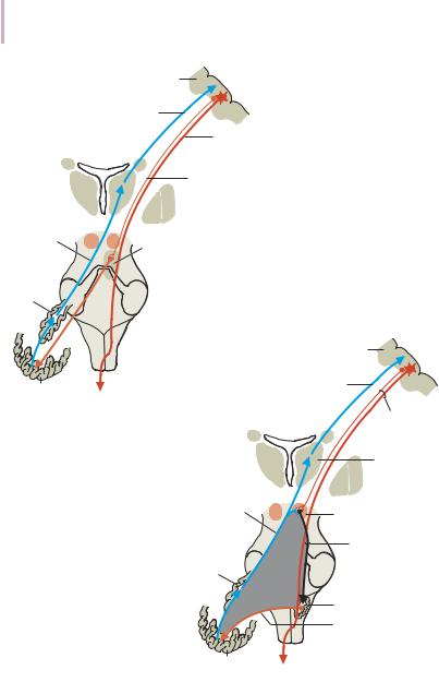
Afferent pathways. One of the few afferent pathways in the superior cerebellar peduncle is the anterior spinocerebellar tract, which terminates in the same area (spinocerebellum) as the posterior spinocerebellar tract. Both convey proprioceptive impulses from the periphery, i.e., from muscle spindles, Golgi tendon organs, and joint receptors.
Fibers from the tectum travel to the cerebellar vermis in the tectocerebellar tract, which occupies a medial position in the superior cerebellar peduncle, at its transition to the superior medullary velum. These fibers convey auditory information from the inferior colliculi, and probably also visual information from the superior colliculi.
Topography of Cerebellar Afferent Pathways
Each half of the cerebellum is responsible for motor function on the ipsilateral half of the body. Some of the efferent fiber systems are doubly crossed: thus,
Baehr, Duus' Topical Diagnosis in Neurology © 2005 Thieme
All rights reserved. Usage subject to terms and conditions of license.

5 250 · 5 Cerebellum
Fig. 5.6 Cerebellar regulatory cir-
Cerebral cortex |
cuits involving the pontine nuclei |
|
Thalamocortical tract
Corticospinal tract
Corticopontine tract
Dentato- |
|
|
rubral and |
|
|
dentato- |
Pontine |
|
thalamic |
||
nuclei |
||
tracts |
||
|
||
Dentate |
|
|
nucleus |
|
Cerebral cortex
Thalamo- Neocerebellar cortical tract cortex
Corticospinal and corticorubral tracts
Thalamus
Dentato- |
|
rubral and |
|
dentato- |
Red nucleus |
thalamic |
|
tracts |
Central |
|
|
Dentate |
tegmental |
nucleus |
tract |
|
Olive |
|
Olivocere- |
|
bellar tract |
Neocerebellar
cortex
Fig. 5.7 Cerebellar regulatory circuits involving the olive. The triangle of Guillain and Mollaret passes from the red nucleus by way of the central tegmental tract, the olive, and the cerebellum back to the red nucleus.
Baehr, Duus' Topical Diagnosis in Neurology © 2005 Thieme
All rights reserved. Usage subject to terms and conditions of license.