

Central Components of the Motor System and Clinical Syndromes of Lesions Affecting Them · 59 |
3 |
|
|
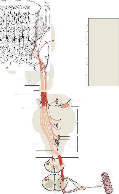
Corticospinal Tract (Pyramidal Tract)
This tract originates in the motor cortex and travels through the cerebral white matter (corona radiata), the posterior limb of the internal capsule (where the fibers lie very close together), the central portion of the cerebral peduncle (crus cerebri), the pons, and the base (i.e., the anterior portion) of the medulla, where the tract is externally evident as a slight protrusion called the pyramid. The medullary pyramids (there is one on either side) give the tract its name. At the lower end of the medulla, 8085% of the pyramidal fibers cross to the other side in the so-called decussation of the pyramids. The fibers that do not cross here descend the spinal cord in the ipsilateral anterior funiculus as the anterior corticospinal tract; they cross farther down (usually at the level of the segment that they supply) through the anterior commissure of the spinal cord (cf. Fig. 3.6). At cervical and thoracic levels, there are probably also a few fibers that remain uncrossed and innervate ipsilateral motor neurons in the anterior horn, so that the nuchal and truncal musculature receives a bilateral cortical innervation.
The majority of pyramidal tract fibers cross in the decussation of the pyramids,thendescendthespinalcordinthecontralaterallateralfuniculusasthe lateral corticospinal tract. This tract shrinks in cross-sectional area as it travels down the cord, because some of its fibers terminate in each segment along the way. About 90% of all pyramidal tract fibers end in synapses onto interneurons, whichthentransmitthemotorimpulsesonwardtothelargeα motorneuronsof the anterior horn, as well as to the smaller γ motor neurons (Fig. 3.4).
Corticonuclear (Corticobulbar) Tract
Some of the fibers of the pyramidal tract branch off from the main mass of the tract as it passes through the midbrain and then take a more dorsal course toward the motor cranial nerve nuclei (Figs. 3.4 and 4.54, p. 212). The fibers supplying these brainstem nuclei are partly crossed and partly uncrossed (for further details, cf. Chapter 4, section 4.4 “Cranial Nerves”). The nuclei receiving pyramidal tract input are the ones that mediate voluntary movements of the cranial musculature through cranial nerves V (the trigeminal nerve), VII (the facial nerve), IX, X, and XI (the glossopharyngeal, vagus, and accessory nerves), and XII (the hypoglossal nerve).
Corticomesencephalic tract. There is also a contingent of fibers traveling together with the corticonuclear tract that arises, not in areas 4 and 6, but rather in area 8, the frontal eye field (Figs. 3.1 and 3.4). The impulses in these fibers mediate conjugate eye movements (p. 46), which are a complex motor process.
Baehr, Duus' Topical Diagnosis in Neurology © 2005 Thieme
All rights reserved. Usage subject to terms and conditions of license.
360 · 3 Motor System
Precentral gyrus
From area 8
Caudate nucleus (tail)
Lentiform nucleus
Internal capsule
Caudate nucleus (head)
Cortico-
mesencephalic tract
Corticonuclear tract
Corticospinal
(pyramidal) tract
Pyramid
Molecular layer 
External granular layer 
External pyramidal layer
Internal  granular layer
Thalamus
Internal  pyramidal layer
|
Multiform layer |
|
Midbrain |
Fig. 3.3 Microarchitecture |
|
|
of the motor cortex |
|
III |
(Golgi stain) |
|
Corticopontine tract |
||
IV |
||
|
Cerebral peduncle |
|
|
( = crus cerebri) |

 V Pons
VI
 VII
VII
IX
X
XII Medulla
XI
Decussation of the pyramids |
C1 |
|
Anterior corticospinal
Lateral corticospinal
tract (uncrossed)
tract (crossed)
T
Motor end plate
Fig. 3.4 Course of the pyramidal tract
Baehr, Duus' Topical Diagnosis in Neurology © 2005 Thieme
All rights reserved. Usage subject to terms and conditions of license.

Central Components of the Motor System and Clinical Syndromes of Lesions Affecting Them · 61 3
Because of its special origin and function, the pathway originating in the frontal eye fields has a separate name (the corticomesencephalic tract), though most authors consider it a part of the corticonuclear tract.
The corticomesencephalic tract runs in tandem with the pyramidal tract (just rostral to it, in the posterior limb of the internal capsule) and then heads dorsally toward the nuclei of the cranial nerves that mediate eye movements, i.e., cranial nerves III, IV, and VI (the oculomotor, trochlear, and abducens nerves). Area 8 innervates the eye muscles exclusively in synergistic fashion, rather than individually. Stimulation of area 8 induces conjugate gaze deviation to the opposite side. The fibers of the corticomesencephalic tract do not terminate directly onto the motor neurons of cranial nerve nuclei III, IV, and VI; the anatomical situation here is complicated and incompletely understood, and is discussed further in Chapter 4 (p. 46ff).
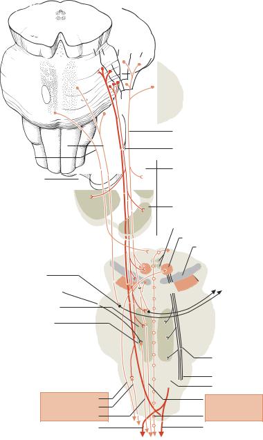
Other Central Components of the Motor System
A number of central pathways beside the pyramidal tract play major roles in the control of motor function (Fig. 3.5). One important group of fibers (the corticopontocerebellar tract) conveys information from the cerebral cortex to the cerebellum, whose output in turn modulates planned movements (cf. Chapter 5 “Cerebellum”). Other fibers travel from the cortex to the basal ganglia (mainly the corpus striatum = caudate nucleus and putamen), the substantia nigra, the brainstem reticular formation, and other nuclei (e. g., in the midbrain tectum). In each of these structures, the impulses are processed and conveyed onward, via interneurons, to efferent tracts that project to the motor neurons of the anterior horn—the tectospinal, rubrospinal, reticulospinal, vestibulospinal, and other tracts (Fig. 3.6). These tracts enable the cerebellum, basal ganglia, and brainstem motor nuclei to influence motor function in the spinal cord. (For further details, see Chapter 4 “Brainstem,” and Chapter 8 “Basal Ganglia.”)
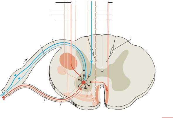
Lateral and medial motor tracts in the spinal cord. The motor tracts in the spinal cord are anatomically and functionally segregated into two groups: a lateral group, comprising the corticospinal and rubrospinal tracts, and a medial group, comprising the reticulospinal, vestibulospinal, and tectospinal tracts (Kuypers, 1985). The lateral tracts mainly project to the distal musculature (especially in the upper limbs) and also make short propriospinal connections. They are primarily responsible for voluntary movements of the forearms and hands, i.e., for precise, highly differentiated, fine motor control. The medial tracts, in contrast, innervate motor neurons lying more medially in the anterior horn and make relatively long propriospinal connections. They are primarily responsible for movements of the trunk and lower limbs (stance and gait).
Baehr, Duus' Topical Diagnosis in Neurology © 2005 Thieme
All rights reserved. Usage subject to terms and conditions of license.
3 62 · 3 Motor System
4 |
6aα 6aβ |
8 |
|
|
2
1
3
Parietotemporopontine tract
Occipitomesencephalic tract
Putamen and globus pallidus
Pontine nuclei
From the cerebellum (fastigial nucleus)
Reticular formation
Lateral vestibular nucleus
Rubrospinal tract
Olivospinal tract
Vestibulospinal tract
Lateral corticospinal tract
Frontopontine tract
Corticospinal tract with extrapyramidal fibers
Thalamus
Head of caudate nucleus
Tegmental nuclei
Red nucleus
Substantia nigra
Py |
To the cerebellum |
|
Central tegmental tract
Inferior olive
Pyramid
Reticulospinal tract Tectospinal tract
Anterior corticospinal tract
Fig. 3.5 Brain structures involved in motor function and the descending tracts that originate in them
Baehr, Duus' Topical Diagnosis in Neurology © 2005 Thieme
All rights reserved. Usage subject to terms and conditions of license.
license of conditions and terms to subject Usage .reserved rights All |
Thieme 2005 © Neurology in Diagnosis Topical Duus' Baehr, |
. |
|
Lateral corticospinal |
Anterior corticospinal |
tract |
tract |
Olivospinal tract |
Reticulospinal tract |
Rubrospinal tract |
Tectospinal tract |
Vestibulospinal tract |
Descending somatosensory |
|
fiber from posterior root |
|
Semilunar fasciculus |
Annulospiral fiber (Ia) |
(comma of Schultz) |
|
|
Golgi fiber (Ib) |
|
α1 fiber
γ fiber
Fig. 3.6 Synapses of the descending motor tracts onto anterior horn neurons
63 · Them Affecting Lesions of Syndromes Clinical and System Motor the of Components Central
3