

Somatosensory Deficits due to Lesions at Specific Sites along the Somatosensory Pathways · 51 |
2 |
|
|
Although the different sensory modalities are already spatially segregated in the thalamus, conscious differentiation among them requires the participation of the cerebral cortex. Higher functions, such as discrimination or the exact determination of the site of a stimulus, are cortex-dependent.
A unilateral lesion of the somatosensory cortex produces a subtotal impairment of the perception of noxious, thermal, and tactile stimuli on the opposite side of the body; contralateral discrimination and position sense, however, are totally lost, as they depend on an intact cortex.
Stereognosis. The recognition by touch of an object laid in the hand (stereognosis) is mediated not just by the primary sensory cortex, but also by association areas in the parietal lobe, in which the individual sensory features of the object, such as its size, shape, consistency, temperature, sharpness/dullness, softness/hardness, etc., can be integrated and compared with memories of earlier tactile experiences.
Astereognosis. Injury to an area in the inferior portion of the parietal lobe impairs the ability to recognize objects by touch with the contralateral hand. This is called astereognosis.
Somatosensory Deficits due to Lesions at Specific Sites along the Somatosensory Pathways
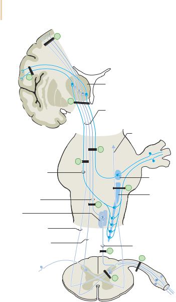
Figure 2.21 shows some typical sites of lesions along the somatosensory pathways; the corresponding sensory deficits are discussed below.
A cortical or subcortical lesion in the sensorimotor area corresponding to the arm or leg (a and b, respectively, in Fig. 2.21) causes paresthesia (tingling, etc.) and numbness in the contralateral limb, which are more pronounced distally than proximally. An irritative lesion at this site can produce a sensory focal seizure; because the motor cortex lies directly adjacent, there are often motor discharges as well (jacksonian seizure).
A lesion of all sensory pathways below the thalamus (c) eliminates all qualities of sensation on the opposite side of the body.
If all somatosensory pathways are affected except the pathway for pain and temperature (d), there is hypesthesia on the opposite side of the body and face, but pain and temperature sensation are unimpaired.
Conversely, a lesion of the trigeminal lemniscus and of the lateral spinothalamic tract (e) in the brainstem impairs pain and temperature sen-
Baehr, Duus' Topical Diagnosis in Neurology © 2005 Thieme
All rights reserved. Usage subject to terms and conditions of license.
252 · 2 Somatosensory System
b
a
|
Thalamus |
|
|
c |
|
|
Spinal lemniscus |
|
|
(anterior and lateral |
|
|
spinothalamic tract) |
|
|
Lateral spinothalamic |
|
|
tract |
|
|
d |
|
|
e |
|
Trigeminal lemniscus |
|
|
|
Principal sensory |
|
|
nucleus of the trigeminal n. |
|
|
g |
|
Medial lemniscus |
Spinal nucleus and |
|
tract of the trigeminal n. |
||
|
f |
|
Nucleus |
|
|
gracilis and nucleus cuneatus |
|
|
Lateral spinothalamic |
|
|
tract |
|
|
Anterior spinothalamic |
Posterior column pathways |
|
tract |
||
h |
||
|
||
|
k |
|
|
i |
Fig. 2.21 Potential sites of lesions along the somatosensory pathways. For the corresponding clinical syndromes, see text.
Baehr, Duus' Topical Diagnosis in Neurology © 2005 Thieme
All rights reserved. Usage subject to terms and conditions of license.
Somatosensory Deficits due to Lesions at Specific Sites along the Somatosensory Pathways · 53 |
2 |
|
|
sation on the opposite side of the body and face, but does not impair other somatosensory modalities.
If the medial lemniscus and anterior spinothalamic tract (f) are affected, all somatosensory modalities of the contralateral half of the body are impaired, except pain and temperature.
Lesions of the spinal nucleus and tract of the trigeminal nerve and of the lateral spinothalamic tract (g) impair pain and temperature sensation on the ipsilateral half of the face and the contralateral half of the body.
Posterior column lesions (h) cause loss of position and vibration sense, discrimination, etc., combined with ipsilateral ataxia.
If the posterior horn of the spinal cord is affected by a lesion (i), ipsilateral pain and temperature sensation are lost, but other modalities remain intact (dissociated sensory deficit).
A lesion affecting multiple adjacent posterior roots (j) causes radicular pain and paresthesiae, as well as impairment or loss of all sensory modalities in the affected area of the body, in addition to hypotonia or atonia, areflexia, and ataxia if the roots supply the upper or lower limb.
Baehr, Duus' Topical Diagnosis in Neurology © 2005 Thieme
All rights reserved. Usage subject to terms and conditions of license.