01-10-2014_10-53-17 / электрофорез-2
.doc|
MoBio Contents |
Section C: Gel Electrophoresis |
|
Chapter 9 |
A | B | C | D | E | F | G | H | I | J | K |
|
|
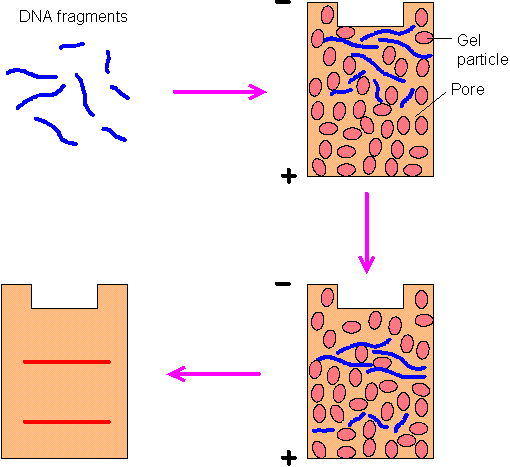
Gel electrophoresis is a technique for separating charged molecules with different sizes. Two kinds of gels are commonly used: agarose and polyacrylamide. Agarose gels can be applied to a wider range of sizes than polyacrylamide gels. By using standard agarose electrophoresis, nuclei acids up to 50 kb may be separated. If Pulsed Field Gel Electrophoresis is used, the upper limit can be extended to 10 Mb. Polyacrylamide gels may separate nucleic acids that differ in length by only 1 nucleotide if their length is less than 500 bp.
Figure 9-C-1. Gel eletrophoresis. In a gel (either agarose or polyacrylamide), the negatively charged DNA fragments move toward the positive electrode at a rate inversely proportional to their length. After the electric field is applied for a certain period, DNA fragments with different lengths will be separated, which can be visualized by autoradiography or by treatment with a fluorescent dye (e.g., ethidium bromide). The relationship between the size of a DNA fragment and the distance it migrates in the gel is logarithmic. Therefore, from the band positions, the lengths of DNA fragments can be determined.
The following two methods are frequently used to separate proteins: Sodium Dodecyl Sulfate - PolyAcrylamide Gel Electrophoresis (SDS-PAGE) - From Davidson College. Two-Dimensional PolyAcrylamide Gel Electrophoresis (2D-PAGE) - By Angelika Gцrg at Technical University of Munich.
Site of Interest: The Electrophoresis Society
|