

конъюгаты с фс
.pdfBioconjugate Chemistry |
|
REVIEW |
Benson group demonstrated that a MT fusion of MBP was able to self-assemble on ligand coated CdSe QDs, CdSe/ZnS QDs,
and Au NPs with dissociation constants on the order of 101 102 nM.279 The interaction was strongest for Au NPs and weakest for
CdSe/ZnS QDs. Furthermore, a direct comparison between MT-MBP and pentahistidine-MBP found that the former was bound more tightly to CdSe QDs.279 In application, a C-terminal MT tag was used to assemble a lead-binding variant of phosphate binding protein onto to CdSe/ZnS QDs.280 The protein was labeled with ruthenium phenanthroline to drive charge transfer quenching of QD PL in a manner dependent on the lead
concentration present. MTs have also been used to assemble peptides and zinc finger proteins with Au11 nanoclusters,281,282
and to drive the bacterial synthesis of a variety of different NP materials.283 An interesting feature of the Benson group’s MT strategy is that it was possible to site-specifically label a cysteine residue associated with a fusion protein of interest.279 In the case of MT-MBP, the introduction of Cd2þ ion acted as a protecting group due to strong binding and folding by the MT domain. An activated maleimide label was thus able to attach specifically at the cysteine residue associated with the MBP. Removal of the Cd2þ using a chelating agent restored the availability of the MT domain for assembly with NPs. Thus, the use of MT tags enables the control over protein orientation in a NP-conjugate while retaining the ability to use mutant cysteine residues as unique labeling sites.
FlAsH/CrAsH System. In 1998, Roger Tsien’s group reported a
novel fluorescein-based dye that contained two As3þ atoms coordinated by 1,2-ethanedithiol at the 40 and 50 dye positions.284
This compound—described as a fluorescein arsenical helix binder, FlAsH—could selectively interact with proteins that contained vicinal thiols in the amino acid sequence Cys-Cys- Xn-Cys-Cys. This motif displaced the ethanedithiol ligands and bound FlAsH, resulting in a 50 000-fold increase in dye fluorescence and provided sufficient affinity and selectivity to label proteins in vivo. A follow-up study demonstrated that the enhancement of dye fluorescence was maintained after protein
denaturation.285 More recently, the FlAsH dye was modified to contain an additional carboxyl group.285,286 This so-called CrAsH
dye exhibits a greater fluorescence enhancement than FlAsH upon binding to the tetracysteine motif under physiological conditions and offers improved signal-to-noise for in vivo experiments.286 Typical dissociation constants for biarsenical ligands with tetracysteine motifs are approximately 10 11 10 12 M.285
The combination of the a nity of the CrAsH dye for the tetracysteine motif and its available carboxyl group render it potentially useful as a cross-linker for NP bioconjugation. For example, Genin et al. used the CrAsH dye to prepare QDintegrase protein conjugates.287 CdSe/ZnS QDs were coated with a mixture of PEG-modified phospholipids that displayed both terminal (unreactive) methoxy and (reactive) amine groups. The CrAsH molecule was ligated to the QD using EDC, and the integrase protein was recombinantly modified with a tetracysteine motif for subsequent assembly of the QDbioconjugate. FlAsH and CrAsH are both organic dyes, and thus highly susceptible to photobleaching. The QD-CrAsH conjugate addresses this limitation via the superior photostability of QDs: the CrAsH PL photobleached within tens of seconds a ording pure QD PL that was stable over an extended period of time. Ultimately, the utility of this approach may not be in the use of the CrAsH dye itself, but in exploiting similar, nonfluorescent arsenyl ligands for NPs that coordinate with tetracysteine motifs to accomplish controlled bioconjugation.
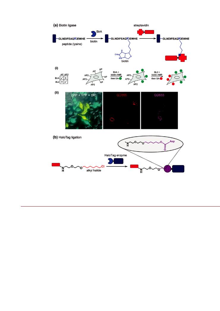
Enzyme Catalyzed Bioconjugation. Biotin Ligase. Biotin binding by avidin proteins is the strongest noncovalent interaction currently known with a dissociation constant of 10 15 M.288 The specificity and stability of the interaction, combined with the ability to biotinylate a wide variety of biomolecules—as well as the ability to label avidin proteins with reporters—has resulted in the diverse use of biotin avidin chemistry in bioconjugate preparation, immobilization, and labeling. The desire for sitespecific biotinylation has driven the development of biotin ligase enzyme systems. For example, E. coli biotin ligase (BirA) transfers endogenous biotin to a specific lysine side chain found in a fifteen-residue acceptor peptide (AP) in an ATP-dependent manner.289 Figure 15a illustrates the generic use of BirA in bioconjugation. Modifying different proteins with this AP sequence enables the enzymatic site-specific biotinylation of re-
combinant proteins that can, for example, be used in biological sensing290 or cellular labeling.291,292
The Ting group adopted the use of BirA ligation to label cellular receptors with QDs.293,294 The extracellular receptors in HeLa cells and neurons were modified with AP sequences and biotinylated by exogenous BirA present in the growth media. This allowed the rapid (2 min) and specific labeling of the live HeLa cells using streptavidin-conjugated QDs.293 Similar labeling was also demonstrated using CHO cells.295 Subsequent work used BirA in combination with yeast biotin ligase for multiplexed labeling (Figure 15a).294 In this case, a yeast acceptor peptide was evolved to provide a second and orthogonal tag for two-color labeling of cell surface proteins, where the two di erent AP sequences defined the labeling specificity. In an alternative strategy, orthogonal two-color QD tracking of single interferon receptor units on live cells was achieved through the combination of BirA and polyhistidine-nickel(II)-NTA interactions.296 The latter required the use of NTA-modified QDs as a chemistry that was orthogonal to streptavidin-coated QDs, and clearly demonstrated the great potential of orthogonal labeling chemistry— even at the single-molecule level. In addition to QDs, the Ting and Bartlett groups also used BirA to selectively label adenoassociated virus particles.297 The virus capsid was engineered to display an available AP sequence that was then labeled by BirA with a chemically synthesized ketone isostere of biotin. The ketone group was chemoselectively labeled with a hydrazido-modified fluorescent dye for optical tracking and a hydrazido-terminated cyclic arginine-gylcine-aspartate (RGD) as a tumor-homing and cell-penetrating peptide. Magnetic NPs biosynthesized by Magnetospirillum magneticum have also been modified to display AP on their surface.298
Carrier Proteins. Peptidyl and acyl carrier proteins (PCP, ACP) can be specifically modified with a variety of fluorophores or affinity handles by phosphopantetheinyl (PPT) transferase. This enzyme catalyzes the transfer of the PPT
unit from coenzyme A (CoA) to a conserved serine in the carrier protein.292,299,300 Since both the carrier protein and
PPT transferase tolerate a wide range of substitutions at the terminal end of the CoA, this system has been used to label ACP-fusion proteins with fluorophores, biotin, and digoxigenin.299 George et al. utilized PPT to specifically biotinylate ACP fusion proteins displayed on yeast cells for labeling with streptavidin-coated QDs.299 In addition, CoAmodified QDs have been used to label a PCP-tagged MBP that retained its binding function in subsequent assays, and also a PCP-tagged transferrin receptor at the membrane of CHOTRVb cells.295
845 |
dx.doi.org/10.1021/bc200065z |Bioconjugate Chem. 2011, 22, 825–858 |
Bioconjugate Chemistry |
|
REVIEW |
Figure 15. Enzymatic labeling systems: (a) biotin ligase and (b) HaloTag ligation. The example for biotin ligase shows (i) a two-step two-color cellular labeling scheme using two orthogonal biotin ligase enzymes (BirA and BL2) with two acceptor peptides (AP and AP2). Cellular labeling with streptavidin (SA)-coated green-emitting QDs (QD565) followed the ligation of biotin to AP. In turn, subsequent cellular labeling with SA-coated redemitting QDs (QD655) followed the ligation of biotin to AP2. Di erential interference contrast and fluorescence images of cells labeled with the two di erent colors of QD are shown in (ii). The two types of cells expressing AP and AP2 individually are distinguished by the expression of either cyan fluorescent protein (CFP) or yellow fluorescent protein (YFP), respectively. Part of (a) is adapted with permission from ref 294. Copyright 2007 American Chemical Society.
Enzyme substrate/Inhibitor Binding. The ability of enzymes to selectively bind their target substrates and/or certain inhibitors is another potential route to the preparation of NP-biocon- jugates. For example, the cutinase enzyme was inserted into the membrane protein integrin LFA-1 and used to bind QDs that were modified with its inhibitor para-nitrophenyl phosphonate (pNPP) in a cellular labeling reaction.301 The pNPP-QDs were prepared from the activation of amine coated QDs with SMCC and a subsequent reaction with an alkyl thiol derivative of pNPP. Similarly, it has been shown that glutathione-S-transferase can bind to Au NPs coated with a mixed surface of thiol-terminated tri(ethylene glycol) and glutathione with high specificity.302 Since glutathione-S-transferase-fusion proteins are routinely prepared,303 this technique may also have potential for controlling the orientation of proteins attached to glutathionemodified NPs.
HaloTag. The HaloTag protein is a recombinantly modified haloalkane dehalogenase (www.Promega.com) that can be fused with other proteins of interest and used to covalently bind
synthetic HaloTag ligands. The ligands are typically fluorescent dyes, affinity handles (e.g., biotin), or solid surfaces modified with a chloroalkane linker.304 In the wild-type dehalogenase enzyme, the His272 residue acts as a base and catalyzes the hydrolysis and release of the substrate intermediate, thereby allowing enzyme regeneration. In contrast, the modified HaloTag enzyme expresses a mutated Phe272 that cannot act as a base and traps the reaction intermediate as a stable covalent adduct (Figure 15b). Both in vitro and in vivo labeling with fluorophores via the HaloTag system have been demonstrated.304 The use of NPs modified with a HaloTag ligand offers the potential for control over the orientation of the HaloTag fusion protein partner in bioconjugation preparation. The Rao group demonstrated that carboxyl-coated QDs could be modified with a bifunctional amino-chloroalkane ligand that enabled subsequent ligation with a HaloTag-Renilla luciferase fusion protein.305 BRET was used to track loading of the HaloTag-luciferase fusion protein, which depended on the number of chloroalkane ligands associated with the QD. The HaloTag system was also used for
846 |
dx.doi.org/10.1021/bc200065z |Bioconjugate Chem. 2011, 22, 825–858 |

Bioconjugate Chemistry |
|
REVIEW |
in vivo labeling of cells with Streptavidin-coated QDs.306 In this case, the HaloTag protein was expressed at a cell membrane anchoring domain and ligated with a biotin HaloTag ligand that was able to bind the Streptavidin-coated QDs.
’PROMISING CHEMISTRIES FOR FUTURE DEVELOPMENT
This review has described several bioconjugate chemistries that are novel in their application to NPs, and o er greater control over NP-conjugate properties than traditional labeling chemistries. In many cases, the studies described represent more proof-of-concept than widespread applicability. Nonetheless, the advantages of the chemistries are evident—particularly the retention of native biomolecule structure and function through bioorthogonality. Despite this success, the toolkit for preparing NP-bioconjugates is far from complete. Fortunately, the continued development of new bioconjugation chemistries for biolabeling and modification will continue to drive new and improved NP-biological applications.
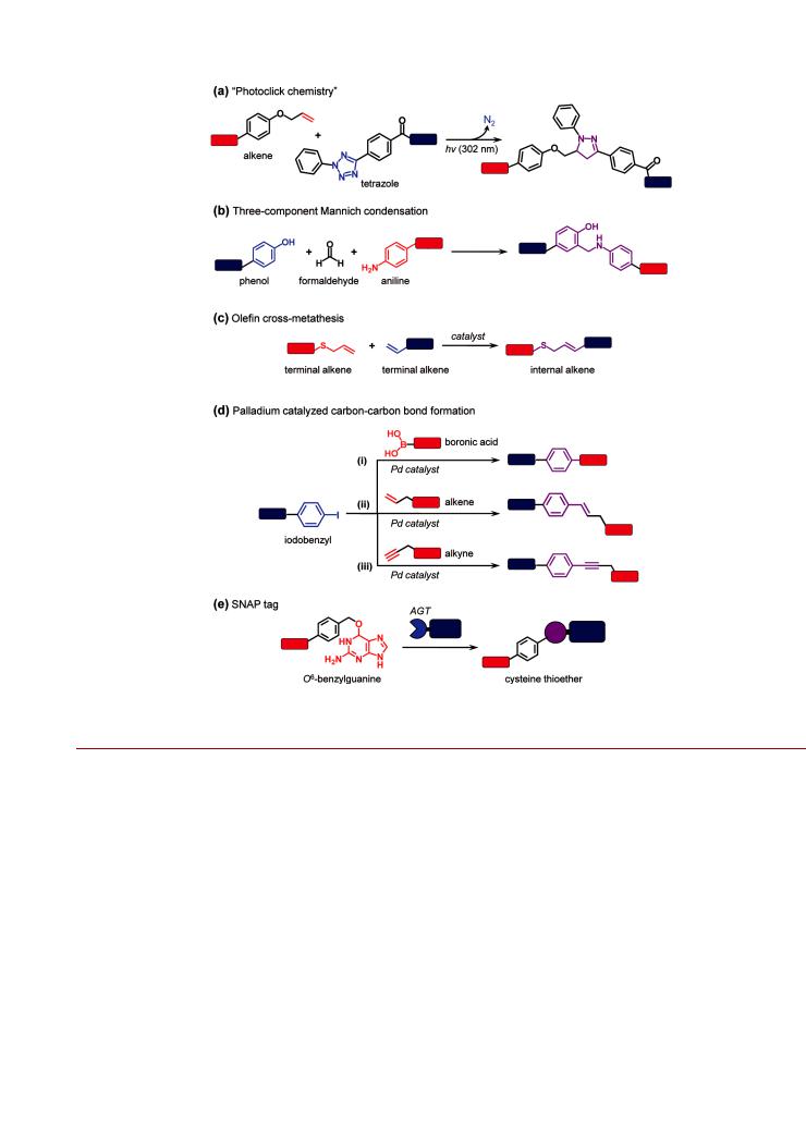
Several promising covalent chemistries are similar to some of the reactions previously described in this review, utilizing similar reactive moieties or analogous concepts in bioorthogonality. One such example relies on the aforementioned scarcity of free thiols in biological macromolecules, which makes the selective introduction of thiols (e.g., cysteine mutation) and their subsequent chemical modification an attractive approach for selective bioconjugation. To this end, several groups have developed alternative electrophiles to the ubiquitous N-alkylmaleimide moiety. For example, Bernardes et al. have used the site-selective introduction of dehydroalanine side chains into proteins and a subsequent Michael addition with thiol-modified labels.307 While the dehydroalanine residues are not as reactive as N-alkyl maleimide groups, they have the advantage of site specific introduction through the elimination of cysteine residues using O-mesitylenesulfonylhydroxylamine,307 or through genetic incorporation of either phenylselenocysteine308,309 or selenalysine310 followed by oxidative elimination with hydrogen peroxide. Another example is conceptually similar to the use of strained alkynes for CuAAC and utilizes the reaction between 1,3-nitrones (in place of the azide group) and alkynes. This strain-promoted alkyne-nitrone cycloaddition strategy has been used to N-term- inally modify peptides311 and proteins312 with a biotin derivative of bicyclo[6.1.0]nonyne and a PEG-modified dibenzocyclooctyne, respectively, through the conversion of an N-terminal serine residue to a 1,3-nitrone. Nitrones will also undergo cycloadditions with maleimides.313 Another novel cycloaddition strategy has addressed the challenge of chemical instability often associated with highly reactive moieties. Song et al. have taken advantage of photoactivation of 2,5-diaryl tetrazoles at 302 nm to generate a highly reactive nitrile imine that chemo-
selectively reacts with alkenes via a 1,3-dipolar cycloaddition to yield a stable pyrazoline cylcoadduct (Figure 16a).314,315 One
attractive aspect of this “photoclick chemistry” is that the resulting pyrazoline ligation product is fluorescent, facilitating direct observation of the reaction in complex systems. The compatibility of the reaction with physiological bu ers and the wide utility of photoactivation in biological systems suggest the potential for broad application of this approach for in vivo labeling.
There are many other reactions that are well-known in organic synthesis and which hold great potential for the controlled
display of biomolecules on NPs. Figure 16 highlights a few of these chemistries that, to our knowledge, have not yet been applied in the preparation of NP-bioconjugates. Several groups have focused on the unique reactivity of the tyrosine phenol ring,
which is found on the surface of many proteins. For example, two-component316 and three-component317,318 Mannich con-
densations (Figure 16b) and ene-forming reactions with cyclic diazocarboxamides319 have found utility. Some other reactions with significant promise for NP bioconjugation include N-terminal transamination with secondary oxime ligation,320 322 olefin cross-metathesis (Figure 16c),323 325 and palladium crosscoupling reactions (vide infra). Olefin cross-metathesis— awarded the 2005 Nobel Prize in Chemistry—is a highly e cient and reliable reaction in which carbon carbon double bonds are broken and reformed catalytically.326 328 In traditional formats, the limitation of olefin cross-metathesis has been poor compatibility with aqueous solvents and thus poor suitability for use in bioconjugate preparation. However, renewed interest in this type of transformation is leading to advances in catalyst design329 and has allowed the expansion of this chemistry into low percentage mixtures of organic and aqueous solvent, thereby enabling limited utility with biomolecules.324 In an early example of the application of olefin cross-metathesis to bioconjugation, Mortell et al. synthesized carbohydrate inhibitors of cell agglutination.330 More recently, Lin et al. demonstrated olefin cross-metathesis in 30% tert-butanol/phosphate bu er between allyl alcohols and allyl sulfides that were chemoselectively conjugated to subtilisin Bacillus lentus protein.323 With further developments, it is anticipated that olefin cross-metathesis will be a valuable addition to the toolkit for preparing NP-bioconjugates. Carbon carbon bond formation via palladium catalysis (Figure 16d) has also been shown to have significant potential in the area of bioconjugation, and was recognized by the 2010 Nobel Prize in Chemistry for its larger contributions to the field. The SuzukiMiyaura reaction consists of a cross-coupling reaction between an aryl halide (ArX, where X = I, Br) and a boronic acid (aryl or alkenyl) that is catalyzed by a palladium complex.331 This chemistry has been used for the site-selective modification of peptides and proteins with small molecule boronic acids in aqueous solution, using p-boronophenylalanine,332 p-iodophenylalanine,333 or p-iodobenzyl modified cysteine side chains.334 Similarly, the palladium catalyzed Mizoroki-Heck reaction335 (unsaturated halides with alkenes) and Sonogashira coupling336 (unsaturated halides with alkynes) have been applied to the covalent modification of peptides and proteins that incorporated p-iodophenyl- alanine337 339 or were acylated with iodobenzoic acid.340 The role of the palladium catalysts begs the question of whether composite NP materials—for example, dumbbells with a palladium NP component—might be able to catalyze their own bioconjugation.
Considering potential advances in self-assembly, the research of the Belcher group should be noted.341 343 They have focused on the development of peptide sequences that coordinate specific metals through genetic engineering of bacteriophage M13. This work has led to many novel peptide sequences for assembly of metal-based materials. While the interest of the Belcher group has been in biomineralization and patterning applications, there may be potential for the development of metal NP-biological hybrid materials and bioconjugates. There may also be new opportunities in the synthesis of artificial amino acids designed to strongly bind NP materials and the subsequent incorporation of oligomeric tracts of these residues into synthetic
847 |
dx.doi.org/10.1021/bc200065z |Bioconjugate Chem. 2011, 22, 825–858 |
Bioconjugate Chemistry |
|
REVIEW |
Figure 16. Promising chemistries for the future development of bioorthogonal NP bioconjugation chemistry: (a) “photoclick chemistry”; (b) threecomponent Manich condensations; (c) olefin cross-metathesis; (d) palladium catalyzed carbon carbon bond formation; and (e) SNAP tag enzyme labeling. These and other promising chemistries are described in the text.
peptides or expressed proteins—analogous to the use of oligohistidine with semiconductor QDs.
The use of enzymatic labeling methods with NPs is still emerging, and many methods remain untapped. The most prominent of these is arguably the SNAP tag. The engineered human DNA repair enzyme alkylguanine-DNA alkyltransferase (AGT or SNAP-tag, www.neb.com) can be used as a tag for selflabeling, where a variety of modified O6-benzylguanine derivatives can function as substrates for the AGT enzyme and are attached via the irreversible transfer of an alkyl group to a cysteine residue (Figure 16e).344 Since its inception, the SNAP tag system has been significantly improved through the development of faster and more e cient enzymes. In addition, a wide range of benzylguanine substrates modified with fluorescent dyes or a nity handles are available, and have been demonstrated to be suitable for a multitude of in vivo cellular labeling applications.292 Recent modifications of the SNAP-tag enzyme can also specifically target O2-benzylcytosine derivatives, thereby enabling an orthogonal labeling approach using two enzymes.345 Given the
relative ease of modifying a variety of substrates with benzylguanine derivatives, it is only a matter of time until this system is used to label NPs in a manner akin to the HaloTag. Additional enzymatic labeling systems with good potential include dihydrofolate reductase, which can covalently bind trimethoprim;346 transglutaminase, which can attach cadaverine-modified probes to small glutamine (Q) expressing peptide substrates, termed Q-tags;347 and lipoic acid ligase, which can functionalize APs with various substituted substrates.348
’SUMMARY AND CONCLUSIONS
This review has highlighted the recent application of nontraditional chemistries to the preparation of NP-bioconjugates, including chemical reactions to form new covalent bonds, selfassembly strategies, and enzymatic methods. These methods are novel in their application to NP materials and advance the degree of control over properties such as NP-bioconjugate valence, biomolecule orientation, reproducibility, and potential for
848 |
dx.doi.org/10.1021/bc200065z |Bioconjugate Chem. 2011, 22, 825–858 |

Bioconjugate Chemistry |
|
REVIEW |
further bioconjugation. In turn, control over these properties is paramount in optimizing the function of NP-bioconjugates in applications such as diagnostic imaging, sensing, and drug delivery.
E cient and bioorthogonal cycloaddition and ligation reactions enable greater levels of control in the display of one or more bio/molecules on NPs—particularly in terms of biomolecule orientation and conjugate valence. To date, many of these new developments in NP bioconjugation have followed the “rediscovery” of many classical synthetic organic chemistry reactions for protein labeling, and this trend is likely to continue. The potential limitation is the continued reliance on traditional labeling chemistries to introduce bioorthogonal groups to biomolecules. However, labeling NPs and biomolecules individually with bifunctional molecules (e.g., NHS-alkyne) using traditional methods, followed by an e cient bioorthogonal reaction (e.g., CuAAC), is more reliable and controllable than using traditional methods to couple the NP and biomolecule directly. This highlights the important point that bioorthogonal chemistries are not meant to completely replace standard bioconjugation approaches, but rather to supplement and augment them. Unnatural amino acid incorporation and other sophisticated methods of introducing bioorthogonal functional groups can provide an even greater degree of control than standard labeling techniques, but are much more time and resource intensive, while also more limited in their applicability.
The self-assembly of biomolecules to QDs using polyhistidine and metallothionein tags has been shown to provide excellent control over bioconjugate valence and orientation. Beyond synthetic peptides and recombinant proteins, the development of modular activated polyhistidine peptides extends the preparation of bioconjugates to include native proteins and synthetic oligonucleotides. Although limited in scope to date, it is anticipated that many of the chemical reactions described herein will be adaptable to modular polyhistidine tags and enable a highly versatile, chemoselective, and bioorthogonal toolkit for the controlled display of biomolecules on QDs and other metalbased NPs. Self-assembly methods are highly advantageous due to the overall simplicity and the determination of conjugate valence on the basis of stoichiometry and equilibrium constants. Self-assembly is also free of the irreproducibility that is often associated with chemical activation and cross-linkers. The disadvantage of self-assembly methods tends to be the scope of their applicability, which can be limited by NP composition and the properties of its coating. In some cases, self-assembly methods may also not be as robust as the formation of new covalent bonds.
Enzymatic labeling methods can be particularly advantageous in that they are highly specific, have little or no opportunity for cross-reactivity, do not require activated intermediates, and can provide a unique point of attachment. However, while enzyme reactions are e cient, they are not necessarily rapid, nor are they as readily scalable as chemical reactions. The scope of the applicability of di erent enzyme labeling methods is also limited, and variable between di erent methods. Enzymatic self-labeling (e.g., HaloTag) generally requires the preparation of a fusion protein. This significantly increases NP-bioconjugate size, can potentially a ect the activity of the protein of interest, and is limited to NP-protein conjugates. Furthermore, both self-label- ing enzymes and enzymes that modify a substrate generally require that molecular or peptidyl tags be introduced to the biomolecules of interest, thus creating the same potential challenges as the introduction of bioorthogonal groups for chemical
labeling. In the case of slow reactions, the activity and long-term stability of the enzyme under di erent conditions of temperature or pH, and in various biological milieus, can be important to labeling e ciency. Moreover, the steric e ect of a NP on enzyme labeling e ciency remains an open question.
The critical message is that traditional labeling chemistries (e.g., carbodiimide) are poorly suited to the controlled display of biomolecules on NPs. The novel application of reactions drawn from organic chemistry and biochemistry to NPs, as well as selfassembly, has provided new levels of control over the properties of NP-bioconjugates. This added control comes at a cost of increased complexity or a limited scope of applicability. Therefore, it is crucial that the toolkit of bioconjugate chemistries for di erent NP materials continue to be developed so that there are methods for the controlled preparation of NP-bioconjugates in every application. In turn, the function and e cacy of these NP materials in biological applications will be greatly advanced.
’AUTHOR INFORMATION
Corresponding Author
*Ph: 202-404-6046. Fax: 202-767-9594. E-mail: Igor.medintz@ nrl.navy.mil.
’ACKNOWLEDGMENT
W.R.A. is grateful to the Natural Sciences and Engineering Research Council of Canada (NSERC) for support through a postdoctoral fellowship. D.E.P. acknowledges an ASEE fellowship through NRL. J.B.B. C. acknowledges a Marie Curie IOF. The authors also acknowledge the CB Directorate/Physical S&T Division (DTRA), DARPA, ONR, NRL and the NRL-NSI for financial support.
’ABBREVIATIONS:
ACP, acyl carrier protein; AP, acceptor peptide; BirA, biotin ligase; BRET, bioluminescence resonance energy transfer; CoA, coenzyme A; CPMV, cowpea mosaic virus; (SW/MW)CNT, (single walled/multiwalled)carbon nanotube; CSS, CMP-sialic acid synthetase; CuAAC, copper-catalyzed azide alkyne cycloaddition; EDC, 1-ethyl-3-(3-dimethylaminopropyl)carbodiimide; EGF, epidermal growth factor; eGFP, enhanced green fluorescent protein; FRET, F€orster resonance energy transfer; MBP, maltose binding protein; HYNIC, 2-hydrazinonicotinoyl; IMAC, immobilized metal a nity chromatography; LSPR, localized surface plasmon resonance; MBP, maltose binding protein; MRI, magnetic resonance imaging; MT, metallothionein; NCL, native chemical ligation; NHS, N-hydroxysuccinimide; NP, nanoparticle; NTA, nitrilotriacetic acid; PCP, peptidyl carrier protein; PEG, poly(ethylene glycol); PL, photoluminescence; PLA, poly(lactic acid); PLGA, poly(lactic-co-glycolic acid); pNPP, para-nitrophenyl phosphonate; PPT, phosphopantetheinyl; QD, quantum dot; SMCC, succinimidyl-4-(N-maleimidomethyl)- cyclohexane-1-carboxylate; TMV, tobacco mosaic virus
’REFERENCES
(1)Haugland, R. P. (2005) The Handbook A Guide to Fluorescent Probes and Labeling Technologies, 10th ed., Invitrogen, San Diego.
(2)Hermanson, G. T. (2008) Bioconjugate Techniques, 2nd ed., Academic Press, San Diego.
(3)Jain, P. K., Huang, X., El-Sayed, I. H., and El-Sayed, M. A. (2007) Review of some interesting surface plasmon resonance-enhanced
849 |
dx.doi.org/10.1021/bc200065z |Bioconjugate Chem. 2011, 22, 825–858 |

Bioconjugate Chemistry |
|
REVIEW |
properties of noble metal nanoparticles and their applications. Plasmonics 2, 107–118.
(4)Jain, P. K., Huang, X., El-Sayed, I. H., and El-Sayed, M. A. (2008) Noble metals on the nanoscale: Optical and photothermal properties and some applications in imaging, sensing, biology and medicine. Acc. Chem. Res. 41, 1578–1586.
(5)Moores, A., and Floch, P. L. (2009) The metal nanoparticle plasmon band as a powerful tool for chemoand biosensing. Biosensing Using Nanomaterials (Merkoc-i, A., Ed.) pp 137 176, Chapter 5, John Wiley & Sons, Inc., Hoboken, NJ.
(6)Elghanian, R., Storho , J. J., Mucic, R. C., Letsinger, R. L., and Mirkin, C. A. (1997) Selective colorimetric detection of polynucleotides based on the distance-dependent optical properties of gold nanoparticles. Science 277, 1078–1081.
(7)Dubertret, B., Calame, M., and Libchaber, A. J. (2001) Singlemismatch detection using gold-quenched fluorescent oligonucleotides.
Nat. Biotechnol. 19, 365–370.
(8)Zhao, W., Brook, M. A., and Li, Y. F. (2008) Design of gold
nanoparticle-based colorimetric biosensing assays. ChemBioChem 9, 2363–2371.
(9)Algar, W. R., Massey, M., and Krull, U. J. (2009) The application of quantum dots, gold nanoparticles, and molecular switches to optical nucleic-acid diagnostics. Trends Anal. Chem. 28, 292–306.
(10)Cao, Y. W. C., Jin, R. C., and Mirkin, C. A. (2002) Nanoparticles with Raman spectroscopic fingerprints for DNA and RNA detection. Science 297, 1536–1540.
(11)Stokes, R. J., Macaskill, A., Lundahl, P. J., Smith, W. E., Faulds, K., and Graham, D. (2007) Quantitative enhanced Raman scattering of labeled DNA from gold and silver nanoparticles. Small 3, 1593–1601.
(12)Murphy, C. J., Gole, A. M., Stone, J. W., Sisco, P. N., Alkilany, A. M., Goldsmith, E. C., and Baxter, S. C. (2008) Gold nanoparticles in
biology: beyond toxicity to cellular imaging. Acc. Chem. Res. 41, 1721–1730.
(13)Hirsch, L. R., Sta ord, R. J., Bankson, J. A., Sershen, S. R., Rivera, B., Price, R. E., Hazle, J. D., Halas, N. J., and West, J. L. (2003)
Nanoshell-mediated near-infrared thermal therapy of tumors under magnetic resonance guidance. Proc. Natl. Acad. Sci. U.S.A 100, 13549– 13554.
(14)Huang, X. H., El-Sayed, I. H., Qian, W., and El-Sayed, M. A. (2006) Cancer cell imaging and photothermal therapy in the nearinfrared region by using gold nanorods. J. Am. Chem. Soc. 128, 2115–2120.
(15)Zheng, J., Nicovich, P. R., and Dickson, R. M. (2007) Highly fluorescent noble-metal quantum dots. Annu. Rev. Phys. Chem. 58, 409–431.
(16)Marambio-Jones, C., and Hoek, E. M. V. (2010) A review of the antibacterial e ects of silver nanomaterials and potential implications for human health and the environment. J. Nanopart. Res. 12, 1531–1551.
(17)Pastoriza-Santos, I., Alvarez-Puebla, R. A., and Liz-Marzan, L. M. (2010) Synthetic routes and plasmonic properties of noble metal nanoplates. Eur. J. Inorg. Chem. 4288–4297.
(18)Xia, Y., Xiong, Y., Lim, B., and Skrabalak, S. E. (2008) Shapecontrolled synthesis of metal nanocrystals: simple chemistry meets complex physics. Angew. Chem., Int. Ed. 48, 60–103.
(19)Biju, V., Itoh, T., Anas, A., Sujith, A., and Ishikawa, M. (2008) Semiconductor quantum dots and metal nanoparticles: syntheses, optical properties, and biological applications. Anal. Bioanal. Chem. 391, 2469–2495.
(20)Tao, A. R., Habas, S., and Yang, P. (2008) Shape control of colloidal metal nanocrystals. Small 4, 310–325.
(21)Wiley, B., Sun, Y., and Xia, Y. (2007) Synthesis of silver
nanostrutures with controlled shapes and properties. Acc. Chem. Res. 40, 1067–1076.
(22)Park, J., Joo, J., Kwon, S. G., Jang, Y., and Hyeon, T. (2007)
Synthesis of monodisperse spherical nanocrystals. Angew. Chem., Int. Ed. 46, 4630–4660.
(23)Love, J. C., Estro , L. A., Kriebel, J. K., Nuzzo, R. G., and Whitesides, G. M. (2005) Self-assembled monolayers of thiolates on metals as a form of nanotechnology. Chem. Rev. 105, 1103–1169.
(24)Laibinis, P. E., and Whitesides, G. M. (1992) Omega-termi- nated alkanethiolate monolayers on surfaces of copper, silver, and gold have similar wettabilities. J. Am. Chem. Soc. 114, 1990–1995.
(25)Lee, J. S., Lytton-Jean, A. K. R., Hurst, S. J., and Mirkin, C. A. (2007) Silver nanoparticle-oligonucleotide conjugates based on DNA with triple cyclic disulfide moieties. Nano Lett. 7, 2112–2115.
(26)Medintz, I. L., Uyeda, H. T., Goldman, E. R., and Mattoussi, H. (2005) Quantum dot bioconjugates for imaging, labelling and sensing.
Nat. Mater. 4, 435–446.
(27)Michalet, X., Pinaud, F. F., Bentolila, L. A., Tsay, J. M., Doose, S., Li, J. J., Sundaresan, G., Wu, A. M., Gambhir, S. S., and Weiss, S. (2005) Quantum dots for live cells, in vivo imaging, and diagnostics.
Science 307, 538–544.
(28)Algar, W. R., and Krull, U. J. (2009) Quantum dots for the development of optical biosensors based on fluorescence. Biosensing Using Nanomaterials (Merkoc-i, A., Ed.) pp 199 245, Chapter 7, John Wiley & Sons, Inc., Hoboken, NJ.
(29)Murray, C. B., Norris, D. J., and Bawendi, M. G. (1993) Synthesis and characterization of nearly monodisperse CdE (E = S, Se, Te) semiconductor nanocrystallites. J. Am. Chem. Soc. 115, 8706–8715.
(30)Guzelian, A. A., Banin, U., Kadavanich, A. V., Peng, X., and Alivisatos, A. P. (1996) Colloidal chemical synthesis and characterization of InAs nanocrystal quantum dots. Appl. Phys. Lett. 69, 1432–1434.
(31)Jiang, W., Singhal, A., Zheng, J. N., Wang, C., and Chan, W. C. W. (2006) Optimizing the synthesis of redto near-IR-emitting CdS-capped CdTexSe1-x alloyed quantum dots for biomedical imaging.
Chem. Mater. 18, 4845–4854.
(32)Pettigrew, K. A., Liu, Q., Power, P. P., and Kauzlarich, S. M.
(2003) Solution snythesis of alkyland alkyl/alkoxy-capped silicon nanoparticles via oxidiation. Chem. Mater. 15, 4005–4011.
(33)Heath, J. R., Shiang, J. J., and Alivisatos, A. P. (1994) Germa-
nium quantum dots - optical properties and synthesis. J. Chem. Phys. 101, 1607–1615.
(34)Dabbousi, B. O., Rodriguez-Viejo, J., Mikulec, F. V., Heine, J. R., Mattoussi, H., Ober, R., Jensen, K. F., and Bawendi, M. G. (1997) (CdSe)ZnS core-shell quantum dots. J. Phys. Chem. B 101, 9463–9475.
(35)Hines, M. A., and Guyot-Sionnest, P. (1996) Synthesis and characterization of strongly luminescing ZnS-capped CdSe nanocrystals.
J. Phys. Chem. 100, 468–471.
(36)Alivisatos, A. P. (1996) Semiconductor clusters, nanocrystals, and quantum dots. Science 271, 933–937.
(37)Alivisatos, A. P. (1996) Perspectives on the physical chemistry of semiconductor nanocrystals. J. Phys. Chem. 100, 13226–13239.
(38)Wang, Y., and Herron, N. (1991) Nanometer-sized semiconductor clusters: materials synthesis, quantum size e ects, and photophysical properties. J. Phys. Chem. 95, 525–532.
(39)Murphy, C. J., and Co er, J. L. (2002) Quantum dots: a primer.
Appl. Spectrosc. 56, 16A–27A.
(40)Smith, A. M., Duan, H. W., Mohs, A. M., and Nie, S. M. (2008) Bioconjugated quantum dots for in vivo molecular and cellular imaging.
Adv. Drug Delivery Rev. 60, 1226–1240.
(41)Algar, W. R., Tavares, A. J., and Krull, U. J. (2010) Beyond labels: A review of the application of quantum dots as integrated
components of assays, bioprobes, and biosensors utilizing optical transduction. Anal. Chim. Acta 673, 1–25.
(42)Medintz, I. L., and Mattoussi, H. (2009) Quantum dot-based
resonance energy transfer and its growing application in biology. Phys. Chem. Chem. Phys. 11, 17–45.
(43)Guzelian, A. A., Katari, J. E. B., Kadavanich, A. V., Banin, U., Hamad, K., Juban, E., Alivisatos, A. P., Wolters, R. H., Arnold, C. C., and
Heath, J. R. (1996) Synthesis of size-selected, surface-passivated InP nanocrystals. J. Phys. Chem. 100, 7212–7219.
(44)Allen, P. M., and Bawendi, M. G. (2008) Ternary I-III-VI quantum dots luminescent in the red to near-infrared. J. Am. Chem. Soc. 130, 9240–9241.
(45)Li, L. S., Hu, J. T., Yang, W. D., and Alivisatos, A. P. (2001) Band gap variation of sizeand shape-controlled colloidal CdSe quantum rods.
Nano Lett. 1, 349–351.
850 |
dx.doi.org/10.1021/bc200065z |Bioconjugate Chem. 2011, 22, 825–858 |

Bioconjugate Chemistry |
|
REVIEW |
(46)Peng, X. G., Manna, L., Yang, W. D., Wickham, J., Scher, E., Kadavanich, A., and Alivisatos, A. P. (2000) Shape control of CdSe nanocrystals. Nature 404, 59–61.
(47)Hezinger, A. F. E., Teβmar, J., and G€opferich, A. (2008) Polymer coating of quantum dots. Eur. J. Pharmaceut. Biopharmaceut. 68, 138–152.
(48)Pons, T., Uyeda, H. T., Medintz, I. L., and Mattoussi, H. (2006) Hydrodynamic dimensions, electrophoretic mobility, and stability of hydrophilic quantum dots. J. Phys. Chem. B 110, 20308– 20316.
(49)Kang, Y. S., Risbud, S., Rabolt, J. F., and Stroeve, P. (1996)
Synthesis and characterization of nanometer-size Fe3O4 and gammaFe2O3 particles. Chem. Mater. 8, 2209–2211.
(50)Hyeon, T. (2003) Chemical synthesis of magnetic nanoparticles. Chem. Commun. 927–934.
(51)Sun, S. H., Zeng, H., Robinson, D. B., Raoux, S., Rice, P. M.,
Wang, S. X., and Li, G. X. (2004) Monodisperse MFe2O4 (M = Fe, Co, Mn) nanoparticles. J. Am. Chem. Soc. 126, 273–279.
(52)Jana, N. R., Chen, Y. F., and Peng, X. G. (2004) Sizeand shapecontrolled magnetic (Cr, Mn, Fe, Co, Ni) oxide nanocrystals via a simple and general approach. Chem. Mater. 16, 3931–3935.
(53)Lu, A. H., Salabas, E. L., and Schuth, F. (2007) Mangetic nanoparticles: synthesis, protection, functionalization, and application.
Angew. Chem., Int. Ed. 46, 1222–1244.
(54)Gupta, A. K., and Gupta, M. (2005) Synthesis and surface engineering of iron oxide nanoparticles for biomedical applications.
Biomaterials 26, 3995–4021.
(55)Sun, S. H. (2006) Recent advances in chemical synthesis, self-
assembly, and applications of FePt nanoparticles. Adv. Mater. 18, 393–403.
(56)Laurent, S., Forge, D., Port, M., Roch, A., Robic, C., Elst, L. V., and Muller, R. N. (2008) Magnetic iron oxide nanoparticles: synthesis, stabilization, vectorization, physicochemial characterizations, and biological applications. Chem. Rev. 108, 2064–2110.
(57)Sosnovik, D. E., Nahrendorf, M., and Weissleder, R. (2008)
Magnetic nanoparticles for MR imaging: agents, techniques and cardiovascular applications. Basic Res. Cardiol. 103, 122–130.
(58)Na, H. B., Song, I. C., and Hyeon, T. (2009) Inorganic nanoparticles for MRI contrast agents. Adv. Mater. 21, 2133–2148.
(59)Hao, R., Xing, R., Xu, Z., Hou, Y., Gao, S., and Sun, S. (2010) Synthesis, functionalization, and biomedical applications of multifunctional magnetic nanoparticles. Adv. Mater. 22, 2729–2742.
(60)Veiseh, O., Gunn, J. W., and Zhang, M. (2010) Design and fabrication of magnetic nanoparticles for targeted drug delivery and imaging. Adv. Drug Delivery Rev. 62, 284–304.
(61)Sandhu, A., Handa, H., and Abe, M. (2010) Synthesis and applications of magnetic nanoparticles for biorecognition and point of care medical diagnostics. Nanotechnology 21, 442001.
(62)Gao, J., Gu, H., and Xu, B. (2009) Multifunctional magnetic nanoparticles: design, synthesis, and biomedical applications. Acc. Chem. Res. 42, 1097–1107.
(63)Yu, H., Chen, M., Rice, P. M., Wang, S. X., White, R. L., and Sun,
S. H. (2005) Dumbbell-like bifunctional Au-Fe3O4 nanoparticles. Nano Lett. 5, 379–382.
(64)Gu, H., Zheng, R., Zhang, X. X., and Xu, B. (2004) Facile onepot synthesis of bifunctional heterodimers of nanoparticles: A conjugate of quantum dot and magnetic nanoparticles. J. Am. Chem. Soc. 126, 5664–5565.
(65)Compton, R. G., Wildgoose, G. G., and Wong, E. L. S. (2009) Carbon nanotube-based sensors and biosensors, Biosensing Using Nanomaterials (Merkoc-i, A., Ed.) pp 3 37, Chapter 1, John Wiley & Sons, Inc., Hoboken, NJ.
(66)Menard-Moyon, C., Kostarelos, K., Prato, M., and Bianco, A.
(2010) Functionalized carbon nanotubes for probing and modulating molecular functions. Chem. Biol. 17, 107–115.
(67)Liu, Z., Tabakman, S., Welsher, K., and Dai, H. J. (2009) Carbon nanotubes in biology and medicine: in vitro and in vivo detection, imaging, and drug delivery. Nano Res. 2, 85–120.
(68)Yang, W. R., Thordarson, P., Gooding, J. J., Ringer, S. P., and Braet, F. (2007) Carbon nanotubes for biological and biomedical applications. Nanotechnology 18, 412001.
(69)Agui, L., Yanez-Sedeno, P., and Pingarron, J. M. (2008) Role of carbon nanotubes in electroanalytical chemistry. Anal. Chim. Acta 622, 11–47.
(70)Rivas, G. A., Rubianes, M. D., Rodriguez, M. C., Ferreyra, N. E., Luque, G. L., Pedano, M. L., Miscoria, S. A., and Parrado, C. (2007) Carbon nanotubes for electrochemical biosensing. Talanta 74, 291–307.
(71)Jacobs, C. B., Peairs, M. J., and Venton, B. J. (2010) Review:
Carbon nanotube based electrochemical sensors for biomolecules. Anal. Chim. Acta 662, 105–127.
(72)Rosen, Y., and Elman, N. M. (2009) Carbon nanotubes in drug delivery. Exp. Opin. Drug Delivery 6, 517–530.
(73)Amiot, C. L., Xu, S. P., Liang, S., Pan, L. Y., and Zhao, J. X. J. (2008) Near-infrared fluorescent materials for sensing of biological targets. Sensors 8, 3082–3105.
(74)Liang, F., and Chen, B. (2010) A review on biomedical applications of single-walled carbon nanotubes. Curr. Med. Chem. 17, 10–24.
(75)Bosi, S., Ros, T. D., Spalluto, G., and Prato, M. (2003) Fullerene
derivatives: an attractive tool for biological applications. Eur. J. Med. Chem. 38, 913–923.
(76)Jensen, A. W., Wilson, S. R., and Schuster, D. I. (1996) Biological applications of fullerenes. Bioorg. Med. Chem. 4, 767–779.
(77)Nakamura, E., and Isobe, H. (2003) Functionalized fulleres in water. The first 10 years of their chemistry, biology, and nanoscience.
Acc. Chem. Res. 36, 807–815.
(78)Chen, R. J., Zhang, Y. G., Wang, D. w., and Dai, H. J. (2001)
Noncovalent sidewall functionalization of single-walled carbon nanotubes for protein immobilization. J. Am. Chem. Soc. 123, 3838–3839.
(79)Zhao, Y. L., and Stoddart, J. F. (2009) Noncovalent functiona-
lization of single-walled carbon nanotubes. Acc. Chem. Res. 42, 1161–1171.
(80)Contarino, M. R., and Withey, G., Chaiken, I. (2009) Isotropic display of biomolecules on CNT-arrayed nanostructures, Biosensing Using Nanomaterials (Merkoc-i, A., Ed.) pp 39 65, Chapter 2, John Wiley & Sons, Inc., Hoboken, NJ.
(81)Tasis, D., Tagmatarchis, N., Bianco, A., and Prato, M. (2006) Chemistry of carbon nanotubes. Chem. Rev. 106, 1105–1136.
(82)Karousis, K., Tagmatarchis, N., and Tasis, D. (2010) Current progress on the chemical modification of carbon nanotubes. Chem. Rev. 110, 5366–5397.
(83)Zhu, Y. W., Murali, S., Cai, W. W., Li, X. S., Suk, W. J., Potts, J. R., and Ruo , R. S. (2010) Graphene and graphene oxide: Synthesis, properties, and applications. Adv. Mater. 22, 3906–3924.
(84)Rao, C. N. R., Sood, A. K., Subrahmanyam, K. S., and
Govindaraj, A. (2009) Graphene: the new two-dimensional nanomaterial. Angew. Chem., Int. Ed. 48, 7752–7777.
(85)Shenderova, O. A., Zhirnov, V. V., and Brenner, D. W. (2002) Carbon nanostructures. Crit. Rev. Solid State Mater. Sci. 27, 227–356.
(86)Barnard, A. S. (2009) Diamond standard in diagnostics: nanodiamond biolabels make their mark. Analyst 134, 1751–1764.
(87)Caravan, P., Ellison, J. J., McMurry, T. J., and Lau er, R. B.
(1999) Gadolinium(III) chelates as MRI contrast agents: structure, dynamics, and applications. Chem. Rev. 99, 2293–2352.
(88)Dickson, E. F. G., Pollak, A., and Diamandis, E. P. (1995) Timeresolved detection of lanthanide luminescence for ultrasensitive bioanalytical assays. J. Photochem. Photobiol. B 27, 3–19.
(89)Eliseeva, S. V., and B€unzli, J. C. G. (2010) Lanthanide luminescence for functional materials and bio-sciences. Chem. Soc. Rev. 39, 189–227.
(90)B€unzli, J. C. G. (2010) Lanthanide luminescence for biomedical analyses and imaging. Chem. Rev. 110, 2729–2755.
(91)Wang, F., Banerjee, D., Liu, Y., Chen, X., and Liu, X. (2010) Upconversion nanoparticles in biological labeling, imaging, and therapy.
Analyst 135, 1839–1854.
(92)Wang, F., and Liu, X. (2009) Recent advances in the chemistry of lanthanide-doped upconversion nanocrystals. Chem. Soc. Rev. 38, 976–989.
851 |
dx.doi.org/10.1021/bc200065z |Bioconjugate Chem. 2011, 22, 825–858 |

Bioconjugate Chemistry |
|
REVIEW |
(93)Ong, L. C., Gnanasammandhan, M. K., Nagarajan, S., and Zhang, Y. (2010) Upconversion: road to El Dorado of the fluorescence world. Luminesc. 25, 290–293.
(94)Cheung, E. N. M., Alvares, R. D. A., Oakden, W., Chaudhary, R., Hill, M. L., Pichaandi, J., Mo, G. C. H., Yip, C., Macdonald, P. M., Stanisz, G. J., Veggel, F. C. J. M. v., and Prosser, R. S. (2010) Polymer-stabilized lanthanide fluoride nanoparticle aggregates as contrast agents for magnetic resonance imaging and computer tomography. Chem. Mater. 22, 4728–4739.
(95)Shen, J., Sun, L. D., and Yan, C. H. (2008) Luminescent rare earth nanomaterials for bioprobe applications. Dalton Trans. 5687– 5697.
(96)Yan, Z. G., and Yan, C. H. (2008) Controlled synthesis of rare earth nanostructures. J. Mater. Chem. 18, 5046–5059.
(97)Feng, W., Sun, L. D., Zhang, Y. W., and Yan, C. H. (2010) Synthesis and assembly of rare earth nanostructures directed by the
principle of coordination chemistry in solution-based process. Coord. Chem. Rev. 254, 1038–1053.
(98)Sivakumar, S., Diamente, P. R., and Veggel, F. C. v. (2006) Silica
coated Ln(3þ)-doped LaF3 nanoparticls as robust downand upconverting biolabels. Chem.—Eur. J. 12, 5878–5884.
(99)Gerion, D., Pinaud, F., Williams, S. C., Parak, W. J., Zanchet, D., Weiss, S., and Alivisatos, A. P. (2001) Synthesis and properties of biocompatible water-soluble silica-coated CdSe/ZnS semiconductor quantum dots. J. Phys. Chem. B 105, 8861–8871.
(100)Wolcott, A., Gerion, D., Visconte, M., Sun, J., Schwartzberg, A., Chen, S. W., and Zhang, J. Z. (2006) Silica-coated CdTe quantum
dots functionalized with thiols for bioconjugation to IgG proteins.
J.Phys. Chem. B 110, 5779–5789.
(101)Santra, S., Tapec, R., Theodoropoulou, N., Dobson, J., Hebard, A., and Tan, W. H. (2001) Synthesis and characterization of silica-coated iron oxide nanoparticles in microemulsion: The e ect of nonionic surfactants. Langmuir 17, 2900–2906.
(102)Kobayashi, Y., Horie, M., Konno, M., Rodriguez-Gonzalez, B., and Liz-Marzan, L. M. (2003) Preparation and properties of silicacoated cobalt nanoparticles. J. Phys. Chem. B 107, 7420–7425.
(103)Lee, D. C., Mikulec, F. V., Pelaez, J. M., Koo, B., and Korgel, B. A. (2006) Synthesis and magnetic properties of silica-coated FePt nanocrystals. J. Phys. Chem. B 110, 11160–11166.
(104)Liu, S., and Han, M. Y. (2010) Silica-Coated Metal Nanoparticles. Chem. Asian J. 5, 36–45.
(105)Burns, A., Ow, H., and Wiesner, U. (2006) Fluorescent coreshell silica nanoparticles: towards “Lab on a Particle” architectures for nanobiotechnology. Chem. Soc. Rev. 35, 1028–1042.
(106)Haensch, C., Hoeppener, S., and Schubert, U. S. (2010) Chemical modification of self-assembled silane based monolayers by surface reactions. Chem. Soc. Rev. 39, 2323–2334.
(107)Knopp, D., Tang, D., and Niessner, R. (2009) Review: Bioanalytical applications of biomolecule-functionalized nanometersized doped silica particles. Anal. Chim. Acta 647, 14–30.
(108)Yan, J., Estevez, M. C., Smith, J. E., Wang, K., He, X., Wang, L., and Tan, W. (2007) Dye-doped nanoparticles for bioanalysis. Nano Today 2, 44–50.
(109)Vivero-Escoto, J. L., Slowing, I. I., Trewyn, B. G., and Lin, V. S. Y. (2010) Mesoporous silica nanoparticles for intracellular controlled drug delivery. Small 6, 1952–1967.
(110)Vallet-Regí, M., Balas, F., and Arcos, D. (2007) Mesoporous materials for drug delivery. Angew. Chem., Int. Ed. 46, 7548–7558.
(111)Guo, X., and Szoka, F. C. (2003) Chemical approaches to
triggerable lipid vesicles for drug and gene delivery. Acc. Chem. Res. 36, 335–341.
(112)Torchilin, V. P. (2006) Multifunctional nanocarriers. Adv. Drug Delivery Rev. 58, 1532–1555.
(113)Discher, D. E., and Ahmed, F. (2006) Polymersomes. Ann. Rev. Biomed. Eng. 8, 323–341.
(114)Peer, D., Karp, J. M., Hong, S., Farokhzad, O. C., Margalit, R., and Langer, R. (2007) Nanocarriers as an emerging platform for cancer therapy. Nat. Nanotechnol. 2, 751–760.
(115)Bala, I., Hariharan, S., and Kumar, M. N. V. R. (2004) PLGA nanoparticles in drug delivery: The state of the art. Crit. Rev. Therapeut. Drug Carrier Sys. 21, 387–422.
(116)Boas, U., and Heegaard, P. M. H. (2004) Dendrimers in drug research. Chem. Soc. Rev. 33, 43–63.
(117)Esfand, R., and Tomalia, D. A. (2001) PAMAM dendrimers: from biomimicry to drug delivery and biomedical applications. Drug Discovery Today 6, 427–436.
(118)Franzen, S., and Lommel, S. A. (2009) Targeting cancer with ’smart bombs’: equiping plant virus nanoparicles for a ’seek and destroy’ mission. Nanomedicine 4, 575–588.
(119)Portney, N. G., and Ozkan, M. (2006) Nano-oncology: drug delivery, imaging, and sensing. Anal. Bioanal. Chem. 384, 620–630.
(120)Steinmetz, N. F. (2010) Viral nanoparticles as platforms for
next-generation therapeutics and imaging devices. Nanomedicine 6, 634–641.
(121)Manchester, M., and Singh, P. (2006) Virus-based nanoparticles: Platform technologies for diagnostic imaging. Adv. Drug Delivery Rev. 58, 1505–1522.
(122)Hooker, J. M., Kovacs, E. W., and Francis, M. B. (2004) Interior surface modification of bacteriophage MS2. J. Am. Chem. Soc. 126, 3718–3719.
(123)Steinmetz, N. F., Lomonosso , G. P., and Evans, D. J. (2006) Decoration of cowpea mosaic virus with multiple, redox-active organomettalic complexes. Small 2, 530–533.
(124)Soto, C. M., and Ratna, B. R. (2010) Virus hybrids as nanomaterials for biotechnology. Curr. Opin. Biotechnol. 21, 426–438.
(125)Cao, Y. C., Jin, R., Nam, J. M., Thaxton, C. S., and Mirkin, C. A.
(2003) Raman dye-labeled nanoparticle probes for proteins. J. Am. Chem. Soc. 125, 14676–14677.
(126)Faulds, K., Jarvis, R., Smith, W. E., and Graham, D. (2008)
Multiplexed detection of six labelled oligonucleotides using surface enhanced resonance Raman scattering (SERRS). Analyst 133, 1505– 1512.
(127)Faulds, K., McKenzie, F., Smith, W. E., and Graham, D. (2007) Quantitative simultaneous multianalyte detection of DNA by dualwavelength surface-enhanced resonance Raman scattering. Angew. Chem., Int. Ed. 46, 1829–1831.
(128)Liu, J., Lau, S. K., Varma, V. A., Mo tt, R. A., Caldwell, M., Liu, T., Young, A. N., Petros, J. A., Osunkoya, A. O., Krogstad, T., JonesLeyland, B., Wang, M. D., and Nie, S. M. (2010) Molecular mapping of tumor heterogeneity on clinical tissue specimens with multiplexed quantum dots. ACS Nano 4, 2755–2765.
(129)Farokhzad, O. C., Cheng, J. J., Teply, B. A., Sherifi, I., Jon, S., Kanto , P. W., Richie, J. P., and Langer, R. (2006) Targeted nanopar- ticle-aptamer bioconjugates for cancer chemotherapy in vivo. Proc. Natl. Acad. Sci. U.S.A 103, 6315–6320.
(130)Cheng, S. H., Lee, C. H., Chen, M. C., Souris, J. S., Tseng, F. G., Yang, C. S., Mou, C. Y., Chen, C. T., and Lo, L. W. (2010) Trifunctionalization of mesoporous silica nanoparticles for comprehensive
cancer theranostics-the trio of imaging, targeting and therapy. J. Mater. Chem. 20, 6149–6157.
(131)Samia, A. C. S., Dayal, S., and Burda, C. (2006) Quantum dot-
based energy transfer: Perspectives and potential for application in photodynamic therapy. Photochem. Photobiol. 82, 617–625.
(132)Huang, X. H., Jain, P. K., El-Sayed, I. H., and El-Sayed, M. A.
(2008) Plasmonic photothermal therapy using gold nanoparticles.
Lasers Med. Sci. 23, 217–228.
(133)Lockett, M. R., Phillips, M. F., Jarecki, J. L., Peelen, D., and Smith, L. M. (2008) A tetrafluorophenyl activated ester self-assembled monolayer for the immobilization of amine-modified oligonucleotides.
Langmuir 24, 69–75.
(134)Medintz, I. (2006) Universal tools for biomolecular attachment to surfaces. Nat. Mater. 5, 842–842.
(135)Mei, B. C., Susumu, K., Medintz, I. L., Delehanty, J. B., Mountziaris, T. J., and Mattoussi, H. (2008) Modular poly(ehtylene glycol) ligands for biocompatible semiconductor and gold nanocrystals with extended pH and ionic stability. J. Mater. Chem. 18, 4949–4958.
852 |
dx.doi.org/10.1021/bc200065z |Bioconjugate Chem. 2011, 22, 825–858 |

Bioconjugate Chemistry |
|
REVIEW |
(136)Pathak, S., Davidson, M. C., and Silva, G. A. (2007) Characterizaton of the functional binding properties of antibody conjugated quantum dots. Nano Lett. 7, 1839–1845.
(137)Algar, W. R., and Krull, U. J. (2006) Adsorption and hybridization of oligonucleotides on mercaptoacetic acid-capped CdSe/ZnS
quantum dots and quantum dot-oligonucleotide conjugates. Langmuir 22, 11346–11352.
(138)Li, H. X., and Rothberg, L. (2004) Colorimetric detection of DNA sequences based on electrostatic interactions with unmodified gold nanoparticles. Proc. Natl. Acad. Sci. U.S.A 101, 14036–14039.
(139)Zheng, M., Jagota, A., Semke, E. D., Diner, B. A., Mclean, R. S., Lustig, S. R., Richardson, R. E., and Tassi, N. G. (2003) DNA-assisted dispersion and separation of carbon nanotubes. Nat. Mater. 2, 338–342.
(140)Medintz, I. L., Clapp, A. R., Brunel, F. M., Tiefenbrunn, T., Uyeda, H. T., Chang, E. L., Deschamps, J. R., Dawson, P. E., and Mattoussi, H. (2006) Proteolytic activity monitored by fluorescence
resonance energy transfer through quantum-dot-peptide conjugates.
Nat. Mater. 5, 581–589.
(141)Banks, P. R., and Paquette, D. M. (1995) Comparison of three common amine reactive fluorescent probes used for conjugation to
biomolecules by capillary zone electrophoresis. Bioconjugate Chem. 6, 447–458.
(142)Best, M. D. (2009) Click chemistry and bioorthogonal reactions: Unprecedented selectivity in the labeling of biological molecules.
Biochemistry 48, 6571–6584.
(143)Prescher, J. A., and Bertozzi, C. R. (2005) Chemistry in living systems. Nat. Chem. Biol. 1, 13–21.
(144)Sletten, E. M., and Bertozzi, C. R. (2009) Bioorthogonal
chemistry: Fishing for selectivity in a sea of functionality. Angew. Chem., Int. Ed. 48, 6974–6998.
(145)Devaraj, N. K., Weissleder, R., and Hilderbrand, S. A. (2008)
Tetrazine-based cylcoadditions: Application to pretargeted live cell imaging. Bioconjugate Chem. 19, 2297–2299.
(146)Bundy, B. C., and Swartz, J. R. (2010) Site-specific incorporation of p-propargyloxyphenylalanine in a cell free environment for direct protein-protein click conjugation. Bioconjugate Chem. 21, 255–263.
(147)Huisgen, R. (1968) Cycloadditions: definition classification and characterization. Angew. Chem., Int. Ed. 7, 321–328.
(148)Moses, J. E., and Moorhouse, A. D. (2007) The growing applications of click chemistry. Chem. Soc. Rev. 36, 1249–1262.
(149)Meldal, M., and Tornoe, C. W. (2008) Cu-catalyzed azidealkyne cycloaddition. Chem. Rev. 108, 2952–3015.
(150)Gupta, S. S., Raja, K. S., Kaltgrad, E., Strable, E., and Finn, M. G. (2005) Virus-glycopolymer conjugates by copper(I) catalysis of atom transfer radical polymerization and azide-alkyne cycloaddition.
Chem. Commun. 4315–4317.
(151)Hong, V., Presolski, S. I., Ma, C., and Finn, M. G. (2009) Analysis and optimization of copper-catalyzed azide-alkyne cycloaddition for bioconjugation. Angew. Chem., Int. Ed. 48, 9879–9883.
(152)Steinmetz, N. F., Mertens, M. E., Taurog, R. E., Johnson, J. E., Commandeur, U., Fischer, R., and Manchester, M. (2009) Potato virus X as a novel platform for potential biomedical applications. Nano Lett. 10, 305–312.
(153)Wang, Q., Chan, T. R., Hilgraf, R., Fokin, V. V., Sharpless, K. B., and Finn, M. G. (2003) Bioconjugation by copper(I)-catalyzed azide-alkyne [3 þ 2] cycloaddition. J. Am. Chem. Soc. 125, 3192–3193.
(154)Strable, E., Prasuhn, D. E., Udit, A. K., Brown, S., Link, A. J., Ngo, J. T., Lander, G., Quispe, J., Potter, C. S., Carragher, B., Tirrell, D. A., and Finn, M. G. (2008) Unnatural amino acid incorporation into virus-like particles. Bioconjugate Chem. 19, 866–875.
(155)Gupta, S. S., Kuzelka, J., Singh, P., Lewis, W. G., Manchester, M., and Finn, M. G. (2005) Accelerated bioorthogonal conjugation: A practical method for the ligation of diverse functional molecules to a polyvalent virus sca old. Bioconj. Chem. 16, 1572–1579.
(156)Prasuhn, D. E., Yeh, R. M., Obenaus, A., Manchester, M., and Finn, M. G. (2007) Viral MRI contrast agents: coordination of Gd by native virions and attachment of Gd complexes by azide-alkyne cycloaddition. Chem. Commun. 1269–1271.
(157)Singh, P., Prasuhn, D., Yeh, R. M., Destito, G., Rae, C. S., Osborn, K., Finn, M. G., and Manchester, M. (2007) Bio-distribution,
toxicity and pathology of cowpea mosaic virus nanoparticles in vivo.
J. Controlled Release 120, 41–50.
(158)Kussrow, A., Kaltgrad, E., Wolfenden, M. L., Cloninger, M. J., Finn, M. G., and Bornhop, D. J. (2009) Measurement of monovalent and polyvalent carbohydrate-lectin binding by back-scattering interferometry. Anal. Chem. 81, 4889–4897.
(159)Kaltgrad, E., O’Reilly, M. K., Liao, L., Han, S., Paulson, J. C.,
and Finn, M. G. (2008) On-virus construction of polyvalent glycan ligands for cell-surface receptors. J. Am. Chem. Soc. 130, 4578–4579.
(160)Kaltgrad, E., Gupta, S. S., Punna, S., Huang, C.-Y., Chang, A., Wong, C.-H., Finn, M. G., and Blixt, O. (2007) Anti-carbohydrate antibodies elicited by polyvalent display on a viral sca old. ChemBioChem 8, 1455–1462.
(161)Udit, A. K., Everett, C., Gale, A. J., Kyle, J. R., Ozkan, M., and
Finn, M. G. (2009) Heparin antagonism by polyvalent display of cationic motifs on virus-like particles. ChemBioChem 10, 503–510.
(162)Destito, G., Yeh, R., Rae, C. S., Finn, M. G., and Manchester, M. (2007) Folic acid-mediated targeting of cowpea mosaic virus particles to tumor cells. Chem. Biol. 14, 1152–1162.
(163)Prasuhn, D. E., Singh, P., Strable, E., Brown, S., Manchester, M., and Finn, M. G. (2008) Plasma clearance of bacteriophage Qβ particles as a function of surface charge. J. Am. Chem. Soc. 130, 1328–1334.
(164)Steinmetz, N. F., Hong, V., Spoerke, E. D., Lu, P., Breitenkamp, K., Finn, M. G., and Manchester, M. (2009) Buckyballs meet viral nanoparticles: Candidates for biomedicine. J. Am. Chem. Soc. 131, 17093–17095.
(165)Gole, A., and Murphy, C. J. (2008) Azide-derivatized gold nanorods: Functional materials for “click” chemistry. Langmuir 24, 266–272.
(166)Brennan, J. L., Hatzakis, N. S., Tshikhudo, T. R., Razumas, V., Patkar, S., Vind, J., Svendsen, A., Nolte, R. J. M., Rowan, A. E., and Brust, M. (2006) Bionanoconjugation via click chemistry: The creation of
functional hybrids of lipases and gold nanoparticles. Bioconjugate Chem. 17, 1373–1375.
(167)Kim, Y.-P., Daniel, W. L., Xia, Z., Xie, H., Mirkin, C. A., and Rao, J. (2010) Bioluminescent nanosensors for protease detection based
upon gold nanoparticle-luciferase conjugates. Chem. Commun. 46, 76–78.
(168)Fischler, M., Sologubenko, A., Mayer, J., Clever, G., Burley, G.,
Gierlich, J., Carell, T., and Simon, U. (2008) Chain-like assembly of gold nanoparticles on artificial DNA templates via ’click chemistry’. Chem. Commun. 169–171.
(169)Lin, P.-C., Ueng, S.-H., Yu, S.-C., Jan, M.-D., Adak, A. K., Yu, C.-C., and Lin, C.-C. (2007) Surface modification of magnetic nanoparticles via Cu(I)-catalyzed alkyne-azide [2 þ 3] cycloaddition. Org. Lett. 9, 2131–2134.
(170)Polito, L., Monti, D., Caneva, E., Delnevo, E., Russo, G., and Prosperi, D. (2008) One-step bioengineering of magnetic nanoparticles via a surface diazo transfer/azide-alkyne click reaction sequence. Chem. Commun. 621–623.
(171)El-Boubbou, K., Gruden, C., and Huang, X. (2007) Magnetic glyco-nanoparticles: A unique tool for rapid pathogen detection, decontamination, and strain di erentiation. J. Am. Chem. Soc. 129, 13392– 13393.
(172)Achatz, D. E., Heiligtag, F. J., Li, X., Link, M., and Wolfbeis, O. S. (2010) Colloidal silica nanoparticles for use in click chemistrybased conjugations and fluorescent a nity assays. Sens. Actuators, B 150, 211–219.
(173)Mader, H. S., Link, M., Achatz, D. E., Uhlmann, K., Li, X., and
Wolfbeis, O. S. (2010) Surface-modified upconverting microparticles and nanoparticles for use in click chemistries. Chem.—Eur. J. 16, 5416–5424.
(174)Pereira, G. R., Santos, L. J., Luduvico, I., Alves, R. B., and de Freitas, R. P. (2010) ’Click’ chemistry as a tool for the facile synthesis of fullerene glycoconjugate derivatives. Tetrahedron Lett. 51, 1022–1025.
853 |
dx.doi.org/10.1021/bc200065z |Bioconjugate Chem. 2011, 22, 825–858 |

Bioconjugate Chemistry |
|
REVIEW |
(175)Palacin, T., Khanh, H. L., Jousselme, B., Jegou, P., Filoramo, A., Ehli, C., Guldi, D. M., and Campidelli, S. (2009) E cient functio-
nalization of carbon nanotubes with porphyrin dendrons via click chemistry. J. Am. Chem. Soc. 131, 15394–15402.
(176)Campidelli, S., Ballesteros, B., Filoramo, A., Diaz, D. D., de la Torre, G., Torres, T., Rahman, G. M. A., Ehli, C., Kiessling, D., Werner, F., Sgobba, V., Guldi, D. M., Cio , C., Prato, M., and Bourgoin, J.-P. (2008) Facile decoration of functionalized single-wall carbon nanotubes with phthalocyanines via “click chemistry. J. Am. Chem. Soc. 130, 11503–11509.
(177)Guo, Z., Liang, L., Liang, J.-J., Ma, Y.-F., Yang, X.-Y., Ren, D.- M., Chen, Y.-S., and Zheng, J.-Y. (2008) Covalently β-cyclodextrin modified single-walled carbon nanotubes: a novel artificial receptor synthesized by ‘click’ chemistry. J. Nanopart. Res. 10, 1077–1083.
(178)van Dijk, M., Rijkers, D. T. S., Liskamp, R. M. J., van Nostrum, C. F., and Hennink, W. E. (2009) Synthesis and applications of
biomedical and pharmaceutical polymers via click chemistry methodologies. Bioconjugate Chem. 20, 2001–2016.
(179)Dondoni, A. (2007) Triazole: The keystone in glycosylated molecular architectures constructed by a click reaction. Chem. Asian J. 2, 700–708.
(180)Iha, R. K., Wooley, K. L., Nystrom, A. M., Burke, D. J., Kade, M. J., and Hawker, C. J. (2009) Applications of orthogonal “click” chemistries in the synthesis of functional soft materials. Chem. Rev. 109, 5620–5686.
(181)Hong, V., Presolski, S. I., Ma, C., and Finn, M. G. (2009) Analysis and optimization of copper-catalyzed azide-alkyne cycloaddition for bioconjugation. Angew. Chem., Int. Ed. 48, 9879–9883.
(182)Agard, N. J., Prescher, J. A., and Bertozzi, C. R. (2004) A strain-promoted [3 þ 2] azide-alkyne cycloaddition for covalent modification of biomolecules in living Systems. J. Am. Chem. Soc. 126, 15046–15047.
(183)Baskin, J. M., Prescher, J. A., Laughlin, S. T., Agard, N. J., Chang, P. V., Miller, I. A., Lo, A., Codelli, J. A., and Bertozzi, C. R. (2007) Copper-free click chemistry for dynamic in vivo imaging. Proc. Natl. Acad. Sci. U.S.A. 104, 16793–16797.
(184)Jewett, J. C., and Bertozzi, C. R. (2010) Cu-free click
cycloaddition reactions in chemical biology. Chem. Soc. Rev. 39, 1272–1279.
(185)Zou, Y., and Yin, J. (2008) Cu-free cycloaddition for identifying catalytic active adenylation domains of nonribosomal peptide synthetases by phage display. Bioorg. Med. Chem. Lett. 18, 5664–5667.
(186)Xinghai, N., Jun, G., Margreet, A. W., and Geert-Jan, B. (2008) Visualizing metabolically labeled glycoconjugates of living cells by copper-free and fast Huisgen cycloadditions. Angew. Chem., Int. Ed. 47, 2253–2255.
(187)Laughlin, S. T., Baskin, J. M., Amacher, S. L., and Bertozzi, C. R. (2008) In vivo imaging of membrane-associated glycans in developing zebrafish. Science 320, 664–667.
(188)Bernardin, A., Cazet, A., Guyon, L., Delannoy, P., Vinet, F., Bonna e, D., and Texier, I. (2010) Copper-free click chemistry for highly luminescent quantum dot conjugates: Application to in vivo metabolic imaging. Bioconjugate Chem. 21, 583–588.
(189)Lallana, E., Fernandez-Megia, E., and Riguera, R. (2009) Surpassing the use of copper in the click functionalization of polymeric nanostructures: A strain-promoted approach. J. Am. Chem. Soc. 131, 5748–5750.
(190)Blackman, M. L., Royzen, M., and Fox, J. M. (2008) Tetrazine
ligation: fast bioconjugation based on inverse-electron-demand dielsalder reactivity. J. Am. Chem. Soc. 130, 13518–13519.
(191)Han, H. S., Devaraj, N. K., Lee, J., Hilderbrand, S. A., Weissleder, R., and Bawendi, M. G. (2010) Development of bioorthogonal and highly e ciency conjugation method for quantum dots using tetrazine-norbornene cycloaddition. J. Am. Chem. Soc. 132, 7838–7839.
(192)Haun, J. B., Devaraj, N. K., Hilderbrand, S. A., Lee, H., and Weissleder, R. (2010) Bioorthogonal chemistry amplifies nanoparticle binding and enhances the sensitivity of cell detection. Nat. Nanotechnol. 5, 660–665.
(193)Shi, M., Wosnick, J. H., Ho, K., Keating, A., and Shoichet, M. S. (2007) Immuno-polymeric nanoparticles by Diels-Alder chemistry.
Angew. Chem., Int. Ed. 46, 6126–6131.
(194)Staudinger, H., and Meyer, J. (1919) On new organic phosphorus binding III Phosphine methylene derivatives and phosphinimine.
Helv. Chim. Acta 2, 635–646.
(195)Saxon, E., and Bertozzi, C. R. (2000) Cell surface engineering by a modified Staudinger reaction. Science 287, 2007–2010.
(196)Maja, K., and Rolf, B. (2004) The Staudinger ligation - A gift to chemical biology. Angew. Chem., Int. Ed. 43, 3106–3116.
(197)Saxon, E., Armstrong, J. I., and Bertozzi, C. R. (2000) A “traceless” staudinger ligation for the chemoselective synthesis of amide bonds. Org. Lett. 2, 2141–2143.
(198)Soellner, M. B., Nilsson, B. L., and Raines, R. T. (2002) Staudinger ligation of R-azido acids retains stereochemistry. J. Org. Chem. 67, 4993–4996.
(199)Zhang, H., Ma, Y., and Sun, X.-L. (2009) Chemically-selective surface glyco-functionalization of liposomes through Staudinger ligation. Chem. Commun. 3032–3034.
(200)Parkhouse, S. M., Garnett, M. C., and Chan, W. C. (2008) Targeting of polyamidoamine-DNA nanoparticles using the Staudinger ligation: Attachment of an RGD motif either before or after complexation. Bioorg. Med. Chem. 16, 6641–6650.
(201)Dirksen, A., and Dawson, P. E. (2008) Rapid oxime and
hydrazone ligations with aromatic aldehydes for biomolecular labeling.
Bioconjugate Chem. 19, 2543–2548.
(202)Dirksen, A., Dirksen, S., Hackeng, T. M., and Dawson, P. E. (2006) Nucleophilic catalysis of hydrazone formation and transimina-
tion: implications for dynamic covalent chemistry. J. Am. Chem. Soc. 128, 15602–15603.
(203)Brunel, F. M., Lewis, J. D., Destito, G., Steinmetz, N. F., Machester, M., Stuhlmann, H., and Dawson, P. E. (2010) Hydrazone ligation strategy to assemble multifunctional viral nanoparticles for cell imaging and tumor targeting. Nano Lett. 10, 1093–1097.
(204)Prasuhn, D. E., Blanco-Canosa, J. B., Vora, G. J., Delehanty, J. B., Susumu, K., Mei, B. C., Dawson, P. E., and Medintz, I. L. (2010) Combining chemoselective ligation with polyhistidine-driven self-as- sembly for the modular display of biomolecules on quantum dots. ACS Nano 4, 267–278.
(205)Blanco-Canosa, J. B., Medintz, I. L., Farrell, D., Mattoussi, H., and Dawson, P. E. (2010) Rapid covalent ligation of fluorescent peptides to water solubilized quantum dots. J. Am. Chem. Soc. 132, 10027–10033.
(206)Banerjee, S. S., and Chen, D. H. (2008) Multifunctional pHsensitive magnetic nanoparticles for simultaneous imaging, sensing and targeted intracellular anticancer drug delivery. Nanotechnology 19, 505104.
(207) Yuan, Q., Venkatasubramanian, R., Hein, S., and Misra,
R.D. K. (2008) A stimulus-responsive magnetic nanoparticle drug carrier: magnetite encapsulated by chitosan-grafted-copolymer. Acta Biomater. 4, 1024–1037.
(208)Aryal, S., Grailer, J. J., Pilla, S., Steeber, D. A., and Gong, S. (2009) Doxorubicin conjugated gold nanoparticles as water-soluble and pH-responsive anticancer drug nanocarriers. J. Mater. Chem. 19, 7879–7884.
(209)Prabaharan, M., Grailer, J. J., Pilla, S., Steeber, D. A., and Gong,
S.(2009) Gold nanoparticles with a monolayer of doxorubicin-con- jugated amphiphilic block copolymer for tumor-targeted drug delivery.
Biomaterials 30, 6065–6075.
(210)Aryal, S., Hu, C. M. J., and Zhang, L. (2010) Polymer-cisplatin conjugate nanoparticles for acid-responsive drug delivery. ACS Nano 4, 251–258.
(211)Zeng, Y., Ramya, T. N. C., Dirksen, A., Dawson, P. E., and Paulson, J. C. (2009) High e ciency labeling of sialylated glycoproteins on living cells. Nat. Methods 6, 207–209.
(212)Beaudette, T. T., Cohen, J. A., Bachelder, E. M., Broaders,
K.E., Cohen, J. L., Engleman, E. G., and Frechet, J. M. J. (2009) Chemoselective ligation in the functionalization of polysaccharide-based particles. J. Am. Chem. Soc. 131, 10360–10361.
854 |
dx.doi.org/10.1021/bc200065z |Bioconjugate Chem. 2011, 22, 825–858 |