


90 Abnormalities of the White Cell Series
Acute Leukemias
Acute leukemias are described in this place for morphological reasons, because they involve a predominance of mononuclear cells (p. 63). Al- though—or perhaps because—the term “leukemia” is relatively imprecise, an overview seems required (Table 13).
The cellular phenomenon common to the different forms of leukemia is the rapidly progressive reduction in numbers of mature granulocytes, thrombocytes, and erythrocytes. Simultaneously, the leukocyte count usually increases due to the occurrence of atypical round cells.
Table 13 Overview of all forms of leukemia
Type of leukemia |
Classification/clinical findings |
Acute leukemias (AL#AML, ALL) |
|
– Myeloid M0-7 (incl. monocytic, ery- |
Always acute disease, often with |
throid, megakaryoblastic leukemias) |
fever and tendency to hemorrhage |
– Lymphocytic |
There may be lymphoma and thy- |
|
mus infiltrates |
|
|
Chronic myeloid leukemia (CML) |
|
– Persistent leukemic diseases of the |
Chronic disease, usually with |
myeloproliferative system (p. 114) |
splenomegaly |
|
|
Chronic lymphocytic leukemia (CLL) |
|
and other leukemic lymphomas |
|
–B-CLL (90%), T-CLL (p.74f.)
–Prolymphocytic leukemia (p.77)
–Hairy cell leukemia (p.80)
–Chronic lymphocytic leukemia, (rarely) prolymphocytic leukemia and hairy cell leukemia are primary leukemic lymphomas with a chronic course. All other non-Hodgkin lymphomas can develop secondary leukemic disease
Chronic myelomonocytic leukemia (CMML)
–Leukemic form of myelodysplasia (p.106) is classified between myelodysplasia and myeloproliferative diseases
Subacute disease with transitions into acute forms of myeloid leukemia (secondary AML following MDS)
Predominance of Mononuclear Round to Oval Cells |
91 |
|
|
|
|
Note that in about one-fourth of all leukemias total leukocyte counts are normal, or even reduced, and the atypical round cells affect only the relative (differential) blood analysis (“aleukemic leukemia”).
In all forms of leukemia, the more fluffy layered areas of a smear show that the nuclear chromatin structure is not dense and coarse, as in a normal lymphocyte nucleus (p. 49), but more delicately structured and irregular, often “sand-like”. A careful blood cell analysis should be carried out—per- haps with the assistance of a specialist laboratory—before bone marrow analysis is performed.
In most cases, the high leukocyte count facilitates the diagnosis of leukemia. Apart from the leukemia-specific blast cells, a variable number of segmented neutrophilic granulocytes may also remain, depending on the disease progression at the time of diagnosis. This gap in the cell series between blasts and mature cells is called “leukemic hiatus.” It is found in ALL but not in reactive responses or chronic myeloid leukemia, which show a continuous left shift. Morphological or differential diagnosis of acute leukemia is followed by the diagnostic work-up that continues with cytochemical tests. Immunological identification of leukemia cells is always indicated (Table 14).
Diagnostic work-up when acute leukemia is suspected:
CBC, cytochemistry, bone marrow. Collection of material to identify cell surface markers, cytogenetics, molecular genetics.
Morphological and Cytochemical Cell Identification
After a first-line diagnosis of acute leukemia has been arrived at on the basis of the cell morphology (see above), the diagnosis must be refined by cytochemical testing of blood cells or bone marrow (on fresh smears). Table 14 shows a leukemia classification based on both morphological and cytochemical criteria. The table shows that a peroxidase test allows leukemias to be classified as peroxidase-positive (myeloid or monocytic) or peroxidase-negative (lymphoblastic). The next step is the immunological classification based on cell markers.
In routine clinical hematology, the FAB classification (Table 14) will be with us for a few more years.

92 Abnormalities of the White Cell Series
Table 14 Classification of the acute nonlymphatic leukemias by morphology, cytochemistry, and immunology
FAB type* |
|
Peroxidase |
PAS |
α-Naph- |
Naph- |
Immuno- |
|
|
|
|
thyl- |
thyl- |
phenotype |
|
|
|
|
acetat- |
ASD- |
|
|
|
|
|
esterase |
esterase |
|
M0 |
AML with minimal |
$ 3% |
% |
% |
% |
CD 13 " or |
|
marker differentia- |
|
|
|
|
CD 33 " or |
|
tion, undifferen- |
|
|
|
|
MPO " |
|
tiated blasts |
|
|
|
|
CD 79a # |
|
without granules; |
|
|
|
|
and cyCD 3 # |
|
distinguished from |
|
|
|
|
and cyCD 22 # |
|
M1 and ALL only by |
|
|
|
|
and CD 61/ |
|
immunopheno- |
|
|
|
|
CD 41 # and |
|
typing |
|
|
|
|
CD 14 # |
|
|
|
|
|
|
|
M1 |
AML with distinct |
& 3% |
Negative |
% |
% |
MPO " and |
|
marker differentiation |
|
to fine |
|
|
CD 13/CD 33/ |
|
(but without morpho- |
|
gran. |
|
|
CD 65s "/# |
|
logical differentia- |
|
|
|
|
and CD 14 # |
|
tion); sporadic dis- |
|
|
|
|
|
|
crete cytoplasmic |
|
|
|
|
|
|
granulation possible |
|
|
|
|
|
|
|
|
|
|
|
|
M2 |
AML with morpho- |
" 3% |
Negative |
% |
% |
MPO " and |
|
logically mature |
|
to fine |
|
|
CD 13/CD 33/ |
|
cells; " 10% of the |
|
gran. |
|
|
CD 65s/ |
|
blasts contain very |
|
|
|
|
CD 15 "/# |
|
small granules |
|
|
|
|
and CD 14 # |
|
|
|
|
|
|
|
M3 |
Acute promyelo- |
! 100% |
% |
% |
% |
MPO " and |
|
cytic leukemia; the |
|
|
|
|
CD 13 " and |
|
predominant pro- |
|
|
|
|
CD 33" and |
|
myelocytes contain |
|
|
|
|
normally CD |
|
copious granules, |
|
|
|
|
34 # and |
|
some contain Auer |
|
|
|
|
HLA-DR # |
|
bodies; variant M3 |
|
|
|
|
|
|
contains few |
|
|
|
|
|
|
granules; peripheral |
|
|
|
|
|
|
bilobal blasts |
|
|
|
|
|
|
|
|
|
|
|
|
M4 |
Acute myelomono- |
& 3% |
% |
+ |
+ |
Mixed M 1/ |
|
cytic leukemia; |
|
|
"20% |
|
M 2 and M 5 |
|
30–80% of bone |
|
|
|
|
|
|
marrow blasts are |
|
|
|
|
|
myeloblasts, promyelocytes, and myelocytes; 20% are monocytes; variant M4 eosinophilia; additional immature eosinophils with dark granules
|
|
Predominance of Mononuclear Round to Oval Cells |
93 |
|||||
|
|
|
|
|
|
|
|
|
Table 14 |
Continued |
|
|
|
|
|
|
|
|
|
|
|
|
|
|
|
|
FAB type* |
|
Peroxidase |
PAS |
α-Naph- |
Naph- |
Immuno- |
|
|
|
|
|
|
|
thyl- |
thyl- |
phenotype |
|
|
|
|
|
|
acetat- |
ASD- |
|
|
|
|
|
|
|
eesterase |
esterase |
|
|
M5 |
a) |
Acute mono- |
! |
% |
+++ |
+++ |
CD 13/CD 33/ |
|
|
|
blastic leuke- |
|
|
" 80% |
|
CD 65/CD 14/ |
|
|
|
mia; monoblasts |
|
|
|
|
CD64 "/# |
|
|
|
predominant in |
|
|
|
|
and HLA-DR |
|
|
|
the blood and |
|
|
|
|
#" |
|
|
|
bone marrow |
|
|
|
|
|
|
|
b) |
Acute mono- |
! |
% |
+++ |
+++ |
|
|
|
|
cytic leukemia; |
|
|
|
|
|
|
|
|
monocytes in |
|
|
|
|
|
|
|
|
the process of |
|
|
|
|
|
|
|
|
maturation pre- |
|
|
|
|
|
|
|
|
dominante |
|
|
|
|
|
|
M6 |
Acute erythroid |
|
% |
% |
% |
Erythroblasts |
||
|
leukemia; 50% of |
|
|
|
|
Gly A " and |
|
|
|
bone marrow blasts |
|
|
|
|
CD 36 " |
|
|
|
are erythropoietic, |
|
|
|
|
Myeloblasts |
|
|
|
30% myeloblasts |
|
|
|
|
MPO/CD |
|
|
|
|
|
|
|
|
|
13/CD 33/CD |
|
|
|
|
|
|
|
|
65s "/# and |
|
|
|
|
|
|
|
|
CD 14 # |
|
M7 |
Acute megakaryo- |
% |
! |
! |
! |
CD 13/CD 33 |
||
|
cytic leukemia; very |
|
|
|
|
#/" und CD |
||
|
polymorphic, some- |
|
|
|
|
41" oder CD |
||
|
times vacuolated |
|
|
|
|
61 " |
|
|
|
blasts, some with |
|
|
|
|
|
|
|
|
cytoplastic blebs, |
|
|
|
|
|
|
|
|
sometimes aggre- |
|
|
|
|
|
|
|
|
gated with throm- |
|
|
|
|
|
|
|
|
bocytes |
|
|
|
|
|
|
|
|
|
|
|
|
|
|
|
|
*French American British Classification (FAB) 1976/85
**A biphenotypic leukemia must be considered a possibility if several additional lymphoblastic markers are present
PAS Periodic acid-Schiff reaction

94 Abnormalities of the White Cell Series
Chromosome analysis provides important information. In practice, however, the diagnosis of acute leukemia is still based on morphological criteria.
However, where the possibilities of modern therapeutic and prognostic methods are fully accessible, new laboratory procedures based on genetic and molecular biological testing procedures form part of the diagnostic work-up of AML. The current WHO classification takes account of these new methods, placing genetic, morphological, and anamnestic findings in a hierarchical order (Table 15).
According to the new WHO classification, blasts account for more than 20% of cells in acute myeloid leukemia (in contradistinction to myelodysplasias).
Table 15 WHO classification of AML
AML with specific |
– |
With t(8;21) (q22; q22), AML 1/ETO |
cytogenetic trans- |
– |
Acute promyelocytic leukemia (AML M3 with t(15;17) (q22; |
locations |
|
q11-12) and variants, PML/RAR-α |
|
– With abnormal bone marrow eosinophils and (inv16) |
|
|
|
(p13;q22) or to t(16;16) (p13; q22); CBF"/MYH 11 |
|
– With 11q23 (MLL) anomalies |
|
|
|
|
AML with dysplasia in |
– |
With preceding myelodysplastic/myeloproliferative syn- |
more than 1 cell line |
|
drome |
(2 or 3 cell lines |
– |
Without preceding myelodysplastic syndrome |
affected)* |
|
|
|
|
|
Therapy-induced AML |
– |
After treatment with alkylating agents |
und MDS |
– |
After treatment with epipodophyllotoxin |
|
– |
Other triggers |
|
|
|
AML that does not fit |
– |
AML, minimal differentiation |
any of the other cat- |
– |
AML without cell maturation |
egories |
– |
AML with cell maturation |
–Acute myelomonocytic leukemia
–Acute monocytic leukemia
–Acute erythroid leukemia
–Acute megakaryoblastic leukemia
–Acute panmyelosis with myelofibrosis
–Myelosarcoma/chloroma
–Acute biphenotypic leukemia**
*The dysplasia must be evident in at least 50% of the bone marrow cells and in 2–3 cell lines.
**Biphenotypic leukemias should be classified according to their immunophenotypes. They are grouped between acute lymphocytic and acute myeloid leukemias.

Predominance of Mononuclear Round to Oval Cells |
95 |
|
|
Acute Myeloid Leukemias (AML)
Morphological analysis makes it possible to group the predominant leukemic cells into myeloblasts and promyeloblasts, monocytes, or atypical (lympho)blasts. A morphological subclassification of these main groups was put forward in the French–American–British (FAB) classification (Table 14).
In practical, treatment-oriented terms, the most relevant factor is whether the acute leukemia is characterized as myeloid or lymphatic.
Including the very rare forms, there are at least 11 forms of myeloid leukemia.

96 Abnormalities of the White Cell Series
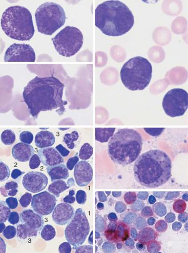
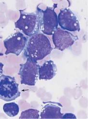
Acute Myeloblastic Leukemia (Type M0 through M2 in the FAB Classification). Morphologically, the cell populations that dominate the CBC and bone marrow analyses (Fig. 31) more or less resemble myeloblasts in the course of normal granulopoiesis. Differences may be found to varying degrees in the form of coarser chromatin structure, more prominently defined nucleoli, and relatively narrow cytoplasm. Compared with lymphocytes (micromyeloblasts), the analyzed cells may be up to threefold larger. In a good smear, the transformed cells can be distinguished from lymphatic cells by their usually reticular chromatin structure and its irregular organization. Occasionally, the cytoplasm contains crystalloid azurophilic needle-shaped primary granules (Auer bodies). Auer bodies (rods) are conglomerates of azurophilic granules. A few cells may begin to display promyelocytic granulation. Cytochemistry shows that from stage M1 onward, more than 3% of the blasts are peroxidase-positive.
Characteristics of Acute Leukemias
Age of onset: Any age.
Clinical findings: Fatigue, fever, and signs of hemorrhage in later stages.
Lymph node and mediastinal tumors are typical only in ALL. Generalized involvement of all organs (sometimes including the meninges) is always present.
CBC and laboratory: Hb ", thrombocytes ", leukocytes usually strongly elevated (~ 80%) but sometimes decreased or normal.
In the differential blood analysis, blasts predominate (morphologies vary).
Beware: Extensive urate accumulation!
Further diagnostics: Bone marrow, cytochemistry, immunocytochemistry, cytogenetics, and molecular genetics.
Differential diagnosis: Transformed myeloproliferative syndrome (e.g., CML) or myelodysplastic syndrome.
Leukemic non-Hodgkin lymphomas (incl. CLL). Aplastic anemias.
Tumors in the bone marrow (carcinomas, but also rhabdomyosarcoma).
Course, therapy: Usually rapid progression with infectious complications and bleeding.
Immediate efficient chemotherapy in a hematology facility; bone marrow transplant may be considered, with curative intent.

Fundamental characteristic of acute leukemia: variable blasts drive out other cell series
a 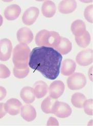
 b
b
c 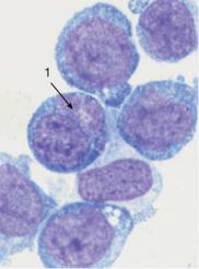
 d
d
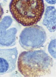
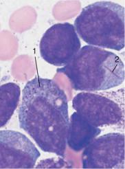
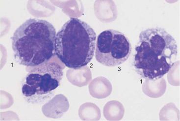
Fig. 31 Acute leukemia, M0–M2. a Undifferentiated blast with dense, fine chromatin, nucleolus (arrow), and narrow basophilic cytoplasm without granules. This cell type is typical of early myeloid leukemia (M0–M1); the final classification is made using cell surface marker analysis (see Table 14). b The peroxidase reaction, characteristic of cells in the myeloid series, shows positive (&3%) only for stage M1 leukemia and higher. The image shows a weakly positive blast (1), strongly positive eosinophil (2), and positive myelocyte (3). c and d Variants of M2 leukemia. Some of the cells already contain granules (1) and crystal-like Auer bodies (2).
97

98 Abnormalities of the White Cell Series
Acute Promyelocytic Leukemia (FAB Classification Type M3 and M3v). The characteristic feature of the cells, which are usually quite large with variably structured nuclei, is extensive promyelocytic granulation. Auer rods are commonly present. Cytochemistry reveals a positive peroxidase reaction for almost all cells. All other reactions are nonspecific. Acute leukemia with predominantly bilobed nuclei is classified as a variant of M3 (M3 V). The cytoplasm may appear either ungranulated (M3) or very strongly granulated (M3 V).
Acute Myelomonocytic Leukemia (FAB Classification Type M4). Given the close relationship between cells in the granulopoietic and the monocytopoietic series (see p. 3), it would not be surprising if the these two systems showed a common alteration in leukemic transformation. Thus, acute myelomonocytic leukemia shows increased granulocytopoiesis (up to more than 20% myeloblasts) with altered cell morphologies, together with increased monocytopoiesis yielding more than 20% monoblasts or promonocytes. Immature myeloid cells (atypical myelocytes to myeloblasts) are found in peripheral blood in addition to monocyte-related cells. Cytochemically, the classification calls for more than 3% peroxidase-positive and more than 20% esterase-positive blasts in the bone marrow. M4 is similar to M2; the difference is that in the M4 type the monocyte series is strongly affected. In addition to the above characteristics, the M4Eo variant shows abnormal eosinophils with dark purple staining granules.
|
|
The diagnosis of acute leukemia is relevant even without further |
|
|
|
subclassification |
|
a |
|
|
|
|
|
|
|
b |
|
|
c |
|
|
|
e |
d |
|
|
f |
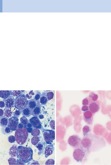
Fig. 32 Acute leukemia M3 and M4. a Blood analysis in promyelocytic leukemia (M3): copious cytoplasmic granules. b In type M3, multiple Auer bodies are often stacked like firewood (so-called faggot cells). c Blood analysis in variant M3v with dumbbell-shaped nuclei. Auer bodies d Bone marrow cytology in acute myelomonocytic leukemia M4: in addition to myeloblasts (1) and promyelocytes (2) there are also monocytoid cells (3). e In variant M4Eo abnormal precursors of eosinophils with dark granules are present. f Esterase as a marker enzyme for the monocyte series in M4 leukemia.
99

100 Abnormalities of the White Cell Series
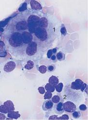
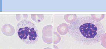
Acute Monocytic Leukemia (FAB Classification Types M5a+b). Two morphologically distinct forms of acute monocytic leukemias exist, monoblastic and monocytic. In the monoblastic variant M5a, blasts predominate in the blood and bone marrow. The blast nuclei show a delicate chromatin structure with several nucleoli. Often, only the faintly grayish-blue stained cytoplasm hints at their derivation.
In monocytic leukemia (type M5b), the bone marrow contains promonocytes, which are similar to the blasts in monocytic leukemia, but their nuclei are polymorphic and show ridges and lobes. Some promonocytes show faintly stained azurophilic granules. The peripheral blood contains monocytoid cells in different stages of maturation which cannot be distinguished with certainty from normal monocytes. Both types are characterized by strong positive esterase reactions in over 80% of the blasts, whereas the peroxidase reactivity is usually negative, or positive in only a few cells.
Acute Erythroleukemia (FAB Classification Type M6)
Erythroleukemia is a malignant disorder of both cell series. It is suspected when mature granulocytes are virtually absent, but blasts (myeloblasts) are present in addition to nucleated erythrocyte precursors, usually erythroblasts (for morphology, see p. 33). The bone marrow is completely overwhelmed by myeloblasts and erythroblasts (more than 50% of cells in the process of erythropoiesis). Bone marrow cytology and cytochemistry confirm the diagnosis. Sporadically, some cases show granulopenia, erythroblasts, and severely dedifferentiated blasts, which correspond to immature red cell precursors (proerythroblasts and macroblasts).
The differential diagnosis in cases of cytopenia with red blood cell precursors found in the CBC must include bone marrow carcinosis, in which the bone marrow–blood barrier is destroyed and immature red cells (and sometimes white cells) appear in the bloodstream. Bone marrow cytology and/or bone marrow histology clarifies the diagnosis. Hemolysis with hypersplenism can also show this constellation of signs.
Fig. 33 Acute leukemia M5 and M6. a In monoblastic leukemia M5a, blasts with a ! fine nuclear structure and wide cytoplasm dominate the CBC. b Seemingly matu-
re monocytes in monocytic leukemia M5b. c Homogeneous infiltration of the bone marrow by monoblasts (M5a). Only residual granulopoiesis (arrow). d Same as c but after esterase staining. The stage M5a blasts show a clear positive reaction (red stain). There is a nonspecific-esterase (NSE)-negative promyelocyte.
Acute leukemias may also derive from monoblasts or erythroblasts
a |
|
b |
c |
d 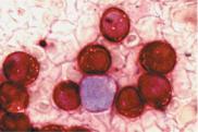
e |
f |
Fig. 33 e Same as c Only the myelocyte in the center stains peroxidase-positive (brown tint); the monoblasts are peroxidase-negative. f In acute erythrocytic leukemia (M6) erythroblasts and myeloblasts are usually found in the blood. This image of bone marrow cytology in M6 shows increased, dysplastic erythropoiesis (e.g., 1) in addition to myeloblasts (2).
101

102 Abnormalities of the White Cell Series
Acute Megakaryoblastic Leukemia (FAB Classification Type M7)
This form of leukemia is very rare in adults and occurs more often in children. It can also occur as “acute myelofibrosis,” with rapid onset of tricytopenia and usually small-scale immigration into the blood of dedifferentiated medium-sized blasts without granules. Bone marrow harvesting is difficult because the bone marrow is very fibrous. Only bone marrow histology and marker analysis (fluorescence-activated cell sorting, FACS) can confirm the suspected diagnosis.
The differential diagnosis, especially if the spleen is very enlarged, should include the megakaryoblastic transformation of CML or osteomyelosclerosis (see pp. 112ff.), in which blast morphology is very similar.
AML with Dysplasia
The WHO classification (p. 94) gives a special place to AML with dysplasia in two to three cell series, either as primary syndrome or following a myelodysplastic syndrome (see pp. 106) or a myeloproliferative disease (see pp. 114ff.).
Criteria for dysgranulopoiesis: &50% of all segmented neutrophils have no granules or very few granules, or show the Pelger anomaly, or are peroxidase-negative.
Criteria for dyserythropoiesis: &50% of the red cell precursor cells display one of the following anomalies: karyorrhexis, megaloblastoid traits, more than one nucleus, nuclear fragmentation.
Criteria for dysmegakaryopoiesis: &50% of at least six megakaryocytes show one of the following anomalies: micromegakaryocytes, more than one separate nucleus, large mononuclear cells.
Hypoplastic AML
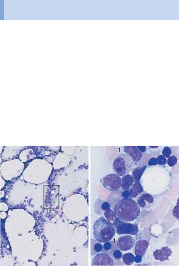
Sometimes (mostly in the mild or “aleukemic” leukemias of the FAB or WHO classifications), the bone marrow is largely empty and shows only a few blasts, which usually occur in clusters. In such a case, a very detailed analysis is essential for a differential diagnosis versus aplastic anemia (see pp. 148f.).
New WHO classification: AML with dysplasia and hypoplastic AML
a 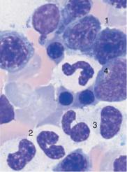
 b
b
c |
d |
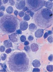
Fig. 34 AML with dysplasia and hypoplastic AML. a AML with dysplasia: megaloblastoid (dysplastic) erythropoiesis (1) and dysplastic granulopoiesis with PelgerHuët forms (2) and absence of granulation in a myelocyte (3). Myeloblast (4). b Multiple separated nuclei in a megakaryocyte (1) in AML with dysplasia. Dyserythropoiesis with karyorrhexis (2). c and d Hypoplastic AML. c Cell numbers below normal for age in the bone marrow. d Magnification of the area indicated in c, showing predominance of undifferentiated blasts (e.g., 1).
103

104 Abnormalities of the White Cell Series
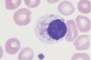
Acute Lymphoblastic Leukemia (ALL)
ALL are the leukemias in which the cells do not morphologically resemble myeloblasts, promyelocytes, or monocytes, nor do they show the corresponding cytochemical pattern. Common attributes are a usually slightly smaller cell nucleus and denser chromatin structure, the grainy consistency of which can be made out only with optimal smear technique (i.e., very light). The classification as ALL is based on the (often remote) similarities of the cells to lymphocytes or lymphoblasts from lymph nodes, and on their immunological cell marker behavior. Insufficiently close morphological analysis can also result in possible confusion with chronic lymphocytic leukemia (CLL), but cell surface marker analysis (see below) will correct this mistake. Advanced diagnostics start with peroxidase and esterase tests on fresh smears, performed in a hematology laboratory, together with (as a minimum) immunological marker studies carried out on fresh heparinized blood samples in a specialist laboratory. The detailed differentiation provided by this cell surface marker analysis has prognostic implications and some therapeutic relevance especially for the distinction to bilineage leukemia and AML (Table 16).
Table 16 Immunological classification of acute bilineage leukemias (adapted from Bene MC et al. (1995) European Group for the Immunological Characterization of Leukemias (EGIL) 9: 1783–1786)
Score |
B-lymphoid |
T-lymphoid |
Myeloid |
2 |
CytCD79a* |
CD3(m/cyt) |
MP0 |
|
Cyt IgM |
anti-TCR |
|
1 |
CD19 |
CD2 |
CD117 |
|
CD20 |
CD5 |
CD13 |
|
CD10 |
CD8 |
CD33 |
|
|
CD10 |
CD65 |
0.5 |
TdT |
TdT |
CD14 |
|
CD24 |
CD7 |
CD15 |
|
|
CD1a |
CD64 |
|
|
|
|
*CD79a may also be expressed in some cases of precursor T-lymphoblastic leukemia/lymphoma.

The cells in acute lymphocytic leukemia vary, and the subtypes can be reliably identified only by immunological methods
a 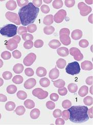
 b
b
c 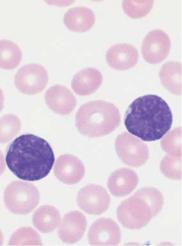
 d
d
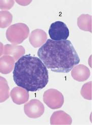
Fig. 35 Acute lymphocytic leukemias. a Screening view: blasts (1) and lymphocytes (2) in ALL. Further classification of the blasts requires immunological methods (common ALL). b Same case as a . The blasts show a dense, irregular nuclear structure and narrow cytoplasm (cf. mononucleosis, p. 69). Lymphocyte (2). c ALL blasts with indentations must be distinguished from small-cell non-Hodgkin lymphoma (e.g., mantle cell lymphoma, p. 77) by cell surface marker analysis. d Bone marrow: large, vacuolated blasts, typical of B-cell ALL. The image shows residual dysplastic erythropoietic cells (arrow).
105

106 Abnormalities of the White Cell Series
Myelodysplasia (MDS)
Clinical practice has long been familiar with the scenario in which, after years of bone marrow insufficiency with a more or less pronounced deficit in all three cell series (tricytopenia), patients pass into a phase of insidiously increasing blast counts and from there into frank leukosis —although the evolution may come to a halt at any of these stages. The transitions between the forms of myelodysplastic syndromes are very fluid, and they have the following features in common:
Anemia, bicytopenia, or tricytopenia without known cause.
Dyserythropoiesis with sometimes pronounced erythrocyte anisocytosis; in the bone marrow often megaloblastoid cells and/or ring sideroblasts.
Dysgranulopoiesis with pseudo-Pelger-Huët nuclear anomaly (hyposegmentation) and hypogranulation (often no peroxidase reactivity) of segmented and band granulocytes in blood and bone marrow.
Dysmegakaryopoiesis with micromegakaryocytes.
The FAB classification is the best-known scheme so far for organizing the different forms of myelodysplasia (Table 17).
Table 17 Forms of myelodysplasia
Form of myelodysplasia |
Blood analysis |
Bone marrow |
RA = refractory anemia |
Anemia (normo- |
Dyserythropoiesis |
|
chromic or hyper- |
(marginal dysgranulo- |
|
chromic); possibly |
poiesis and dysmega- |
|
pseudo-Pelger granulo- |
karyopoiesis " 10%) |
|
cytes; blasts ' 1% |
$ 5% blasts |
RAS = refractory anemia with ring sideroblasts (( aquired idiopathic sideroblastic anemia, p. 137)
Hypochromic and |
More than 15% of the |
hyperchromic erythro- |
red cell precursors are |
cytes side by side, |
ring sideroblasts; |
sometimes discrete |
blasts $ 5% |
thrombopenia and |
|
leukopenia; pseudo- |
|
Pelger cells |
|
RAEB = refractory ane- |
Often thrombocyto- |
Erythropoietic hyper- |
mia with excess of |
penia in addition to |
plasia (with or without |
blasts |
anemia; blasts $ 5%, |
ring sideroblasts); |
|
monocytes $ 1000/µl, |
5–20% blasts |
|
pseudo-Pelger syn- |
|
|
drome |
|
|
|
|
|
|
Cont. p. 108 |
In unexplained anemia and/or leukocytopenia and/or thrombocytopenia, blood cell abnormalities may indicate myelodysplasia
a |
b |
c 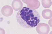
 d
d
 e
e
Fig. 36 Myelodysplasia and CMML. a–d Different degrees of abnormal maturation (pseudo-Pelger type); the nuclear density can reach that of erythroblasts (d). The cytoplasmic hypogranulation is also observed in normal segmented granulocytes. These abnormalities are seen in myelodysplasia or after chemotherapy, among other conditions. e Blood analysis in CMML: monocytes (1), promyelocyte (2), and pseudo-Pelger cell (3). Thrombocytopenia.
107
108 |
Abnormalities of the White Cell Series |
|
|
||
|
|
|
|
|
|
Table 17 |
Continued |
|
|
|
|
|
|
|
|
|
|
Form of myelodysplasia |
|
Blood analysis |
Bone marrow |
|
|
CMML = chronic myelo- |
|
Blasts $ 5%, mono- |
Hypercellular, blasts |
|
|
monocytic leukemia |
|
cytes " 1000/µl, |
$ 20%, elevated pro- |
|
|
|
|
|
pseudo-Pelger syn- |
monocytes |
|
|
|
|
drome |
|
|
RAEB in transformation |
|
Similar to RAEB but |
Blasts 20–30% (some |
|
|
(RAEBt)* |
|
" 5% blasts |
cells contain Auer |
|
|
|
|
|
|
bodies) |
|
|
|
|
|
|
|
*In the WHO classification, the category RAEBt would belong to the category of acute myeloid leukemia.
The new WHO classification of myelodysplastic syndromes defines the differences in cell morphology even more precisely than the FAB classification (Table 18).
For the criteria of dysplasia, see page 106.
The “5qsyndrome” is highlighted as a specific type of myelodysplasia in the WHO classification; in the FAB classification it would be a subtype of RA and RAS. A macrocytic anemia, the 5qsyndrome manifests with normal or increased thrombocyte counts while the bone marrow contains megakaryocytes with hyposegmented round nuclei (Fig. 37b).
Naturally, bone marrow analysis is of particular importance in the myelodysplasias.
Table 18 WHO classification of myelodysplastic syndromes
Disease* |
Dysplasia** |
Blasts in |
Blasts in the |
Ring sidero- |
Cytogenetics |
|
|
peripheral |
bone marrow |
blasts in the |
|
|
|
blood |
|
bone marrow |
|
5qsyndrome |
Usually only E |
$5% |
$5% |
$15% |
5q only |
RA |
Usually only DysE |
$1% |
$5% |
$15% |
Variable |
RARS |
Usually only DysE |
None |
$5% |
&15% |
Variable |
RCMD |
2–3 lines |
Rarely |
$5% |
$15% |
Variable |
RCMD-RS |
2–3 lines |
Rarely |
$5% |
&15% |
Variable |
RAEB-1 |
1–3 lines |
$5% |
5–9% |
$15% |
Variable |
RAEB-2 |
1–3 lines |
5–19% |
10–19% |
$15% |
Variable |
CMML-1 |
1–3 lines |
$5% |
$10% |
$15% |
Variable |
CMML-2 |
1–3 lines |
5–19% |
10–19% |
$15% |
Variable |
MDS-U |
1 cell lineage |
None |
$5% |
$15% |
Variable |
|
|
|
|
|
|
* RA = refractory anemia; RARS = refractory anemia with ring sideroblasts; RCMD = refractory cytopenia with more than one dysplastic cell line; RCMD-RC = refractory cytopenia with more than one dysplastic lineage and ring sideroblasts; RAEB = refractory anemia with elevated blast count; CMML = chronic myelomonocytic leukemia, persistent monocytosis (more than 1 #109/l) in peripheral blood; MDS-U = MDS, unclassifiable. ** Dysplasia in granulopoiesis = Dys G, in erythropoiesis = DysE, in megakaryopoiesis = DysM, multilineage dysplasia = two cell lines affected; trilineage dysplasia (TLD) = all three lineage show dysplasia.
The classification of myelodysplasias requires bone marrow analysis
a 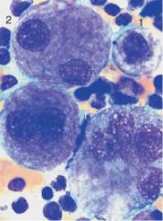
 b
b
c |
d |
Fig. 37 Bone marrow analysis in myelodysplasia. a Dysmegakaryopoiesis in myelodysplastic syndrome (MDS). Relatively small disk-forming megakaryocytes
(1) and multiple singular nuclei (2) are often seen. b Mononuclear megakaryocytes (frequent in 5q-syndrome). c Dyserythropoiesis. Particularly striking is the coarse nuclear structure with very light gaps in the chromatin (arrow 1). Some are megaloblast-like but coarser (arrow 2). d Iron staining of the bone marrow (Prussian blue) in myelodysplasia of the RARS type: dense iron granules forming a partial ring around the nuclei (ring sideroblasts).
109