

4 |
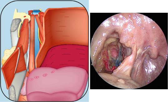
1 Cervical Segment |
|
|
1.2Anatomic Pictures
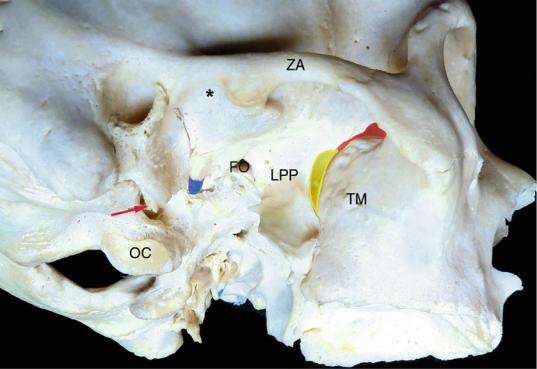
Fig. 1.1 Infratemporal fossa-osteology
FO foramen ovale, LPP lateral pterygoid plate, OC occipital condyle, TM tuberosity of the maxilla, ZA zygomatic arch, black asterisk glenoid fossa, blue spine of the sphenoid, yellow pterygopalatine fissure, red inferior orbital fissure, red arrow styloid process
The pterygomaxillary fissure is a narrow V shaped fissure between the LPP and the superior portion of the tuberosity of the maxilla. The upper part of the pterygomaxillary fissure joins the inferior orbital fissure. The pterygo-palatine fossa communicates with the infratemporal fossa (ITF) by means of the pterygomaxillary fissure laterally. Behind the inferior orbital fissure lies the ITF, which is mainly located below the greater wing of the sphenoid.
1.2 Anatomic Pictures |
5 |
|
|
OM
ZA
Fig. 1.2 Lateral vision of the temporozygomatic region
FA facial artery, fB frontal branch, MM masseter muscle, OM orbicularis oculi muscle, STA superficial temporal artery, tB temporal branch, ZA zygomatic arch, ZM zygomatic muscle
The parotid-masseteric fascia covers the masseteric muscle and surrounds the parotid gland. It attaches superiorly to the zygomatic arch and extends deep to the posterior border of the ramus of the mandible, fusing with the fascia of the posterior belly of the digastric muscle, to give the stylomandibular ligament (Janfaza and Fabian 2001b).
6 |
1 Cervical Segment |
|
|
VIIcn
GAN
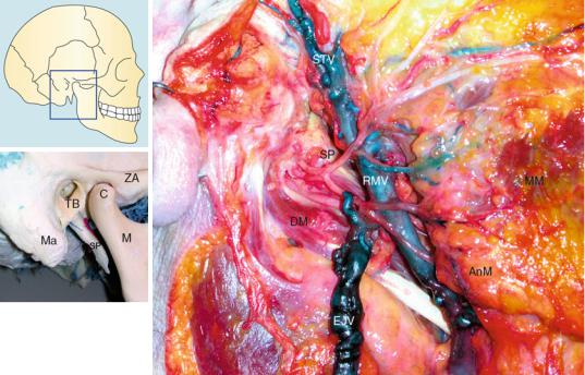
Fig. 1.3 Lateral vision of the parotid-cervical region. The facial nerve has been dissected and the parotid gland removed
AnM angle of the mandible, C condyle, EJV external jugular vein, GAN greater auricular nerve, M mandible, Ma mastoid, MM masseter muscle, RMV retromandibular vein, SP styloid process, STV superficial temporal vein, TB tympanic bone, ZA zygomatic arch, VIIcn facial nerve
The maxillary vein joins the superficial temporal vein in the parotid gland, forming the RMV. The facial nerve lies lateral to the RMV.
1.2 Anatomic Pictures |
7 |
|
|
M VIIcn
FA
Xlcn
FA
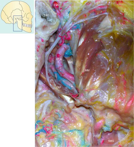
Fig. 1.4 Parotid fossa. The parotid gland has been removed and the deeper areas exposed
DM digastric muscle (posterior belly), EAC external acoustic canal, ECA external carotid artery, FA facial artery, M mastoid, MM masseter muscle, SMG submandibular gland, SML sphenomandibular ligament, STA superficial temporal artery, VIIcn facial nerve, XI accessory nerve
The parotid-masseteric fascia covers the masseteric muscle and splits to involve the parotid gland. Deeply and posteriorly, it fuses with the fascia of the digastric muscle to give the stylo-mandibular ligament.
8 |
1 Cervical Segment |
|
|
VIIcn
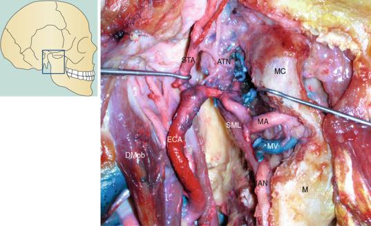
Fig. 1.5 Parotid region. The inferior part of the neck of the mandibular condyle has been partially removed
ATN auriculotemporal nerve, DMpb posterior belly of the digastric muscle, ECA external carotid artery, IAN inferior alveolar nerve, M mandible, MA maxillary artery, MC mandibular condyle, MV maxillary vein, SML sphenomandibular ligament, STA superficial temporal artery, VIIcn facial nerve
The MA enters the infratemporal fossa, passing between the mandibular condyle and the SML. At this level, it usually lies below the ATN and above the MV. Usually, it passes lateral to the nerves, and only in less than 5 % of cases it is deep to all the branches of V3. Only the first part of the MA is accompanied by the MV, the second and third parts are accompanied by a venous plexus. The MV(s) unite with the superficial temporal vein to form the retromandibular vein. The ATN emerges from the upper border of the parotid gland behind the temporomandibular joint and then runs over the root of the zygomatic arch posterior to the STA.
1.2 Anatomic Pictures |
9 |
|
|
VIIcn
VIIcn
VIIcn
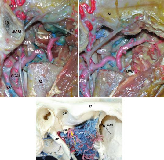
Fig. 1.6 Infratemporal fossa exposure. The condyle and the neck of the mandible have been removed
ATN auriculotemporal nerve, EAM external acoustic meatus, ECA external carotid artery, GF glenoid fossa, LPM lateral pterygoid muscle, M mandible, MA maxillary artery, MM masseter muscle, MPM medial pterygoid muscle, OA occipital artery, pwMS posterior wall of the maxillary sinus, PVP pterygoid venous plexus, SML sphenomandibular ligament, SP styloid process, SpS spine of the sphenoid, STA superficial temporal artery, ZA zygomatic arch, V3 third branch of the trigeminal nerve, VIIcn facial nerve, black arrow pterygopalatine fossa, yellow arrow middle meningeal artery, white asterisk parapharyngeal portion of the internal carotid artery
The infratemporal fossa communicates medially with pterygopalatine fossa and posteromedially with the parapharyngeal space. The MA is 1 of the 2 terminal branches of the ECA; the other is the STA. During its course within the infratemporal fossa, its relationship with the LPM is highly variable. The PVP lies between temporalis, lateral, and medial pterygoid muscles (Janfaza and Montgomery 2001). PVP can comunicate directly with the cavernous sinus through Foramen of Vesalius (when it is present). The pterygoid fascia encases both pterygoid muscles and attaches to the stylomandibular ligament. The sphenomandibular ligament is a fascial condensation between the spine of the sphenoid and the lingula of the mandible (Janfaza and Fabian 2001).
10 |
1 Cervical Segment |
|
|
ATN
IAN
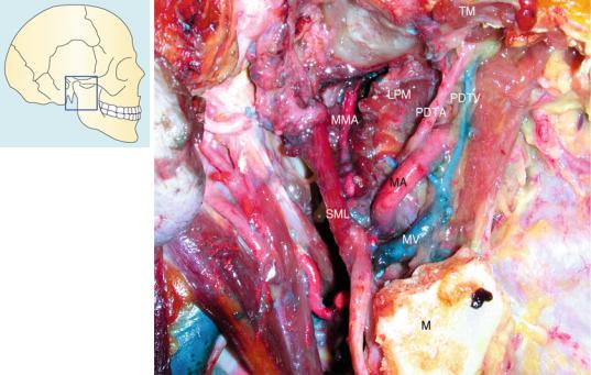
Fig. 1.7 Parotid and retrocondylar region
ATN auriculotemporal nerve, IAN inferior alveolar nerve, LPM lateral pterygoid muscle, M mandible, MA maxillary artery, MMA middle meningeal artery, MV maxillary vein, PDTA posterior deep temporal artery, PDTV posterior deep temporal vein, SML sphenomandibular ligament, TM temporal muscle
Usually, the MMA is posterior to the lateral pterygoid muscle. The ATN passes laterally, around the MMA and between the neck of the mandible and the SML. Similarly, the IAN passes inferiorly between the neck of the mandible and the SML. This last one is a thin fibrous connection between the spine of the sphenoid and the lingula of the mandible (Janfaza et al. 2001). Sometimes, there is an accessory middle meningeal artery that can share a common trunk with the MMA. It accompanies the mandibular nerve and enters the cranial cavity through the foramen ovale.
1.2 Anatomic Pictures |
11 |
|
|
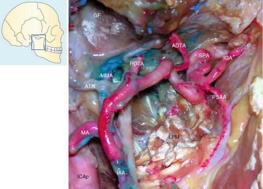
Fig. 1.8 Infratemporal fossa exposure, focus on the maxillary artery
ADTA anterior deep temporal artery, ATN auriculotemporal nerve, GF glenoid fossa, IAA inferior alveolar artery, ICAp parapharyngeal portion of the internal carotid artery, IOA infraorbital artery, LPM lateral pterygoid muscle, MA maxillary artery, MMA middle meningeal artery, PDTA posterior deep temporal artery, PSAA posterosuperior alveolar artery, SPA sphenopalatine artery
The first small branches of the MA are the deep auricular and anterior tympanic arteries. The IAA usually arises from the MA between the sphenomandibular ligament and the neck of the mandible. The DTAs travel superiorly between the temporalis muscle and pericranium. Usually, the PSAA present a common trunk with the IOA. It runs over the tuberosity of the maxilla in close relationship with the posterior superior alveolar nerve. The IOA courses within the infraorbital groove, with the infraorbital nerve. Usually, the PDTA and the MMA are separated by the lateral pterygoid muscle.
12 |
1 Cervical Segment |
|
|
Ma
SML
DM
ECA
XIcn
OV
XIIcn
IJV
Xcn
SCMM
CCA
M
MM
FA
SMG
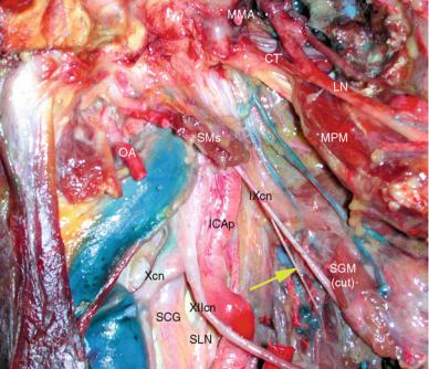
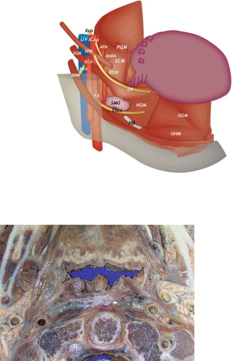
Fig. 1.9 Upper laterocervical and parotid regions
CCA common carotid artery, DM digastric muscle, ECA external carotid artery, FA facial artery, IJV internal jugular vein, M mandible, Ma mastoid (tip), MM masseter muscle, OV occipital vein, SCMM sternocleidomastoid muscle, SMG submandibular gland, SML sphenomandibular ligament, Xcn vagus nerve, XIcn accessory nerve, XIIcn hypoglossal nerve
The internal carotid artery origins at the level of the carotid bifurcation. Two segments can be described. The proximal segment extends from the bifurcation to the point where the artery is crossed by the posterior belly of the digastric muscle (Beretta et al. 2006). The distal segment is from this point to the carotid canal at the skull base.
The hypoglossal nerve courses along the inferior aspect of the posterior belly of the digastric muscle and crosses both internal and external carotid artery.
1.2 Anatomic Pictures |
13 |
|
|
Ma
SCMM
DM
XIcn
IJV
EAC
MAVN
SP STA
MA
IAA
ECA
MPM M
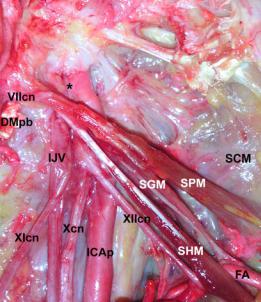
ICAp |
LN |
OA
OV |
IAN |
|
SHM
Fig. 1.10 Infratemporal, parapharyngeal, and upper laterocervical regions. The mandible has been partially cut, pushed forward and inferiorly, and the inferior alveolar nerve dissected in its own canal
DM digastric muscle, EAC external acoustic canal, ECA external carotid artery, IAA inferior alveolar artery, IAN inferior alveolar nerve, ICAp parapharyngeal portion of the internal carotid artery, IJV internal jugular vein, LN lingual nerve, M mandible, Ma mastoid, MA maxillary artery, MAVN maxillary artery vascular network, MPM medial pterygoid muscle, OA occipital artery, OV occipital vein, SCMM sternocleidomastoid muscle, SHM stylohyoid muscle, SP styloid process, STA superficial temporal artery, XIcn accessory nerve
The styloid apparatus separates the internal carotid artery from the external carotid artery. The stylohyoid muscle is the most lateral element of the styloid apparatus, while the stylopharyngeus the most medial.
14 |
1 Cervical Segment |
|
|
STA
|
VIIcn |
|
|
|
JB |
* |
SPA |
|
IOA |
||
|
|
||
|
|
MMA V3 |
|
|
IJV |
TVPM |
|
|
|
|
|
SCMM |
|
XIcn ICAp |
LPM |
|
|
||
|
|
|
|
|
DM |
|
|
Fig. 1.11 Infratemporal fossa and jugular foramen exposure
DM digastric muscle, ICAp parapharyngeal portion of the internal carotid artery, IJV internal jugular vein, IOA infraorbital artery, LPM lateral pterygoid muscle, MMA middle meningeal artery, SCMM sternocleidomastoid muscle, SPA sphenopalatine artery, STA superficial temporal artery, TVPM tensor veli palatini muscle, V3 third branch of the trigeminal nerve, VIIcn facial nerve, XIcn accessory nerve, white asterisk branch for the middle cranial fossa dura
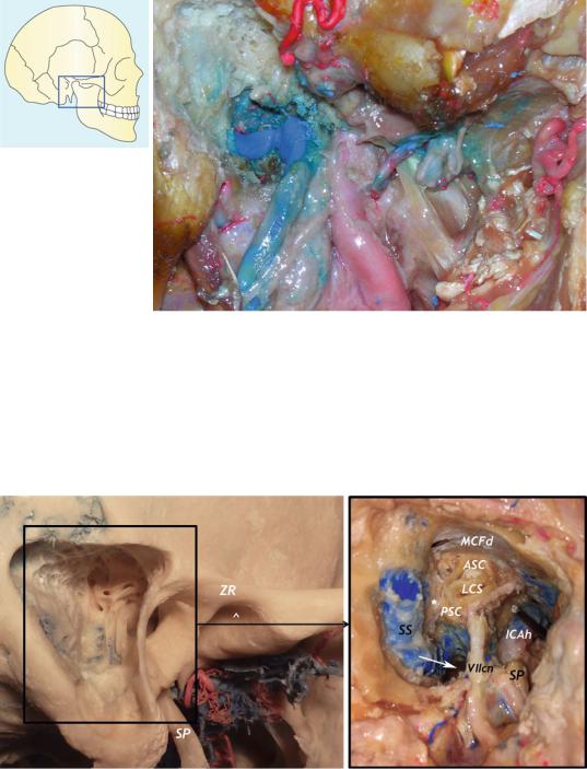
Fig. 1.12 Jugular foramen region: transpetrous exposure
ASC anterior semicircular canal, ICAh horizontal portion of the internal carotid artery, LSC lateral semicircular canal, MCFd dura of the middle cranial fossa, PSC posterior semicircular canal, SP styloid process, SS sigmoid sinus, ZR zygomatic root, VIIcn facial nerve, white arrow jugular tubercle, white asterisk endolymphatic sac, white arrowhead glenoid fossa
1.2 Anatomic Pictures |
15 |
|
|
VIIcn
JB
CR
MMA
IJV
ICAp
IXcn
DM |
XIIcn |
XIcn Xcn
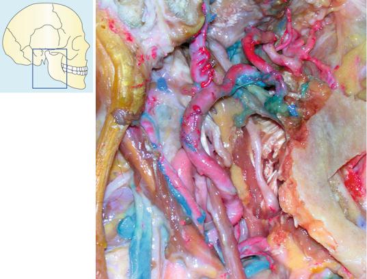
Fig. 1.13 Jugular foramen region
CR carotid ridge, DM digastric muscle (posterior belly), ICAp parapharyngeal portion of the internal carotid artery, IJV internal jugular vein, JB jugular bulb, MMA middle meningeal artery, VIIcn facial nerve, IX glossopharyngeal nerve, X vagus nerve, XI accessory nerve, XII hypoglossal nerve, black arrow inferior ganglion of vagus nerve
The jugular bulb lies beneath the floor of the middle ear cavity (Roche et al. 2008). It can be of variable shape and size. All the lower cranial nerves (LCNs) exit the foramen anteromedially to the jugular bulb, separated from it by connective tissue. The superior ganglion of the vagus nerve is within the jugular foramen (JF). At the level of the intraforaminal course, there is a strict connection between the LCNs. The vagus nerve exits the JF vertically, behind IXcn and ICAp (Roche et al. 2008) and gives its inferior ganglion on the outer skull base surface. The accessory nerve lies immediately lateral to the vagus nerve.
16 |
1 Cervical Segment |
|
|
SpS
VIIcn
DMpb
IJV
XIcn
Fig. 1.14 Upper parapharyngeal region below the jugular foramen. Note that the styloid muscles covering the parapharyngeal vessels are partially removed
CT chorda tympani, DMpb posterior belly of the digastric muscle, ICAp parapharyngeal portion of the internal carotid artery, IJV internal jugular vein, LN lingual nerve, MMA middle meningeal artery, MPM medial pterygoid muscle, OA occipital artery, SCG superior cervical ganglion (sympathetic), SGM styloglossus muscle, SLN superior laryngeal nerve, SMs styloid muscles, SpS spine of the sphenoid, VIIcn facial nerve, IXcn glossopharyngeal nerve, Xcn vagus nerve, XIcn accessory nerve, XIIcn hypoglossal nerve, yellow arrow pharyngeal branch of IX cranial nerve
The lower cranial nerves and the IJV present an intimate relationship with the transverse process of C1. In its upper segment, the IJV lies behind the ICAp on the levator scapulae (Janfaza and Fabian 2001b). The stylopharyngeus muscle and the styloid process separate the IJV from the parotid gland. Usually, the pharyngeal branch of cranial nerve IX runs anteriorly between the internal and external carotid arteries inferior to the main trunk of cranial nerve IX (Janfaza and Fabian 2001b).
1.2 Anatomic Pictures |
17 |
|
|
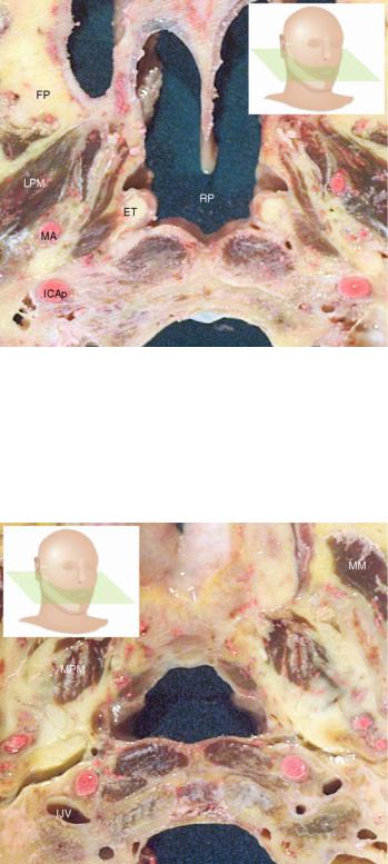
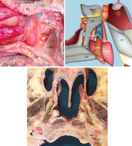
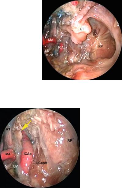
Fig. 1.15 Axial view of the parapharyngeal region
ET eustachian tube, FP fat pad, ICAp parapharyngeal portion of the internal carotid artery, LCapM longus capitis muscle, LPM lateral pterygoid muscle, LVPM levator veli palatini muscle, MA maxillary artery, MPM medial pterygoid muscle, RP rhinopharynx, TVPM tensor veli palatini muscle, black asterisk Rosenmuller’s fossa
MPM
TVPM
LVPM *
LCapM
The upper parapharyngeal region is demonstrated. In particular, the position of the muscles is evident. The prestyloid compartment is now identified as the true parapharyngeal space, while the poststyloid compartment is now named the carotid space (Stambuk and Patel 2008). The prestyloid space is separated from the poststyloid regions by means of a fascial layer connecting the styloid process to the pharyngeal wall. The true parapharyngeal space contains the tensor veli palatine muscle and the tensor-styloid fascia, which reflects from the TVPM and reaches the pharyngeal wall (Janfaza et al. 2001).
HP
Fig. 1.16 Axial view
of the parapharyngeal region (more caudally in respect to the previous image)
ET eustachian tube, HP hard |
|
LVPM |
|
|
|
|
|
||
palate, ICAp parapharyngeal |
|
|
|
|
portion of the internal carotid |
|
|
|
|
artery, IJV internal jugular |
ET |
|
|
|
vein, LCapM longus capitis |
LCapM |
ICAp |
||
|
||||
muscle, LColM longus colli |
|
LColM |
|
|
muscle, LVPM levator veli |
|
|
|
|
palatini muscle, MM masseter |
|
|
|
|
muscle, MPM medial |
|
|
|
|
pterygoid muscle |
|
|
|
18 |
1 Cervical Segment |
|
|
|
|
TM |
TM |
|
|
SPA |
|
|
|
MA |
LPM |
|
|
|
|
|
LPM |
|
|
M |
MA |
|
MM |
|
|
MPM |
|
|
|
MPM |
|
|
MPM |
|
|
|
|
|
|
TM |
|
|
|
|
|
|
LPM |
|
|
LPM |
LPM |
|
MALPM |
MM FP |
|
MM |
|
||
|
FP |
|
M MPM |
M |
|
MPM |
* |
|
MPM |
||
|
|
|
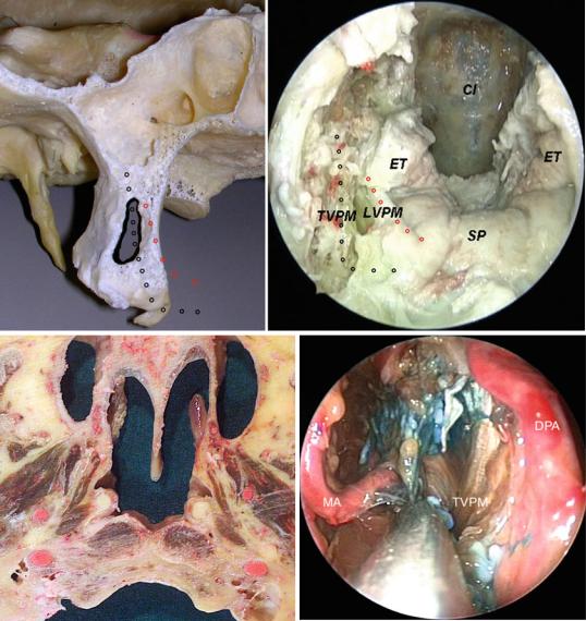
Fig. 1.17 Coronal views of the infratemporal fossa
FP fat pad, LPM lateral pterygoid muscle, M mandible, MA maxillary artery, MM masseter muscle, MPM medial pterygoid muscle, SPA sphenopalatine artery, TM temporal muscle
*
**°°° LPM
LPM °° * °
°
* °
SP * * |
MPM |
MPM |
|
MM |
|
SGM |
|
Fig. 1.18 Coronal view of the infratemporal fossa
LPM lateral pterygoid muscle, MM masseter muscle, MPM medial pterygoid muscle, SGM styloglossus muscle, SP soft palate, white asterisks levator veli palatini muscle, white circles tensor veli palatini muscle
1.2 Anatomic Pictures |
19 |
|
|
LPM
MC
Mc
V3
LPM
PG
SMG
ACP
ON
CS
ICAc
SS
LPM
ET
V3
TVPM
LVPM
SS SS ° Mc
V3 |
ICAh |
|
MC |
LCapM LCapM
PG
Fig. 1.19 Coronal views of the infratemporal fossa and parapharyngeal region
ACP anterior clinoid process, CS cavernous sinus, ET eustachian tube, ICAc cavernous portion of the internal carotid artery, ICAh horizontal portion of the internal carotid artery, LCapM longus capitis muscle, LPM lateral pterygoid muscle, LVPM levator veli palatini muscle, MC mandibular condyle, Mc Meckel’s cave, ON optic nerve, PG parotid gland, SMG submandibular gland, SS sphenoid sinus, TVPM tensor veli palatini muscle, V3 third branch of the trigeminal nerve, yellow arrow ophthalmic artery, red arrow second branch of the trigeminal nerve, white arrows maxillary artery, white circle cavernous (vertical) portion of the internal carotid artery
20 |
1 Cervical Segment |
|
|
AEA
Orbit
SB
ICAc
NS
MTt
Ch
Fig. 1.20 Panoramic endoscopic vision of the sphenoethmoidal spaces and 3D reconstruction of the pterygomaxillo-orbital region
AEA anterior ethmoidal artery, Ch choana, ICAc cavernous portion of the internal carotid artery, MTt tail of the middle turbinate, NS nasal septum, SB skull base
OAp |
ICAc |
|
|
|
|
|
|
PG |
|
|
|
|
SPAnb |
CR |
|
PLL |
|
|
|
|
ICAh |
|
|
|
|
|
|
|
|
|
SPA |
|
|
|
|
pwMS |
PVA |
|
|
MS |
SS |
|
|
|
|
||
|
|
|
|
|
|
|
LRSS |
|
RS |
FR |
|
|
|
|
|
||
|
RPm |
|
|
|
|
PB |
|
|
|
|
|
|
|
|
|
PPs |
|
Fig. 1.21 Close endoscopic vision of the sphenopalatine region and related osteology
CR clival recess, FR foramen rodundum, ICAc cavernous portion of the internal carotid artery, ICAh horizontal portion of the internal carotid artery, MS maxillary strut, OAp orbital apex, PB palatine bone, PG pituitary gland, PLL petrolingual ligament (yellow), PPs pterygoid plates, PVA palatovaginal artery, pwMS posterior wall of the maxillary sinus, RPm rhinopharyngeal mucosa, RS rostrum sphenoidale, SPA sphenopalatine artery, SPAnb nasal branch of the sphenopalatine artery, SS sphenoid sinus, black arrowhead lingula of the sphenoid, blue-sky asterisk and line vidian canal, violet asterisk and line palatovaginal canal
1.2 Anatomic Pictures |
21 |
|
|
|
SS |
|
|
PVA |
VN |
BS |
|
PVAs |
|||
|
|
||
OPPB |
RS |
RS |
|
|
|
||
SPPB |
LRSS |
ET |
|
|
|||
SPAib |
|
|
|
RPm |
|
|
Fig. 1.22 Close vision of the pterygoid and basisphenoid region. The position of the palatovaginal artery is evident
BS basisphenoid, ET eustachian tube, LRSS lateral recess of the sphenoid sinus, OPPB orbital process of the palatine bone, PVA(s) palatovaginal artery(ies), RPm rhinopharyngeal mucosa, SPAib inferior branch of the sphenopalatine artery, SPPB sphenoidal process of the palatine bone, SS sphenoid sinus, RS rostrum sphenoidale, VN vidian nerve
The PVA (also called the pharyngeal artery) lies within the palatovaginal canal accompanied by the pharyngeal nerve (from the pterygopalatine ganglion). The palatovaginal (or pharyngeal) canal is given superiorly by a groove in the basisphenoid and inferiorly by the sphenoidal process of the palatine bone.
22 |
1 Cervical Segment |
|
|
|
SS |
|
PVA |
|
PPF |
FR |
SPPB PVA |
pwMS |
RS |
|
|
|
PVC |
|
ET |
|
SPPB |
|
RS |
DS
V2I
ICAc-p
LRSS Cl
OC
LPP MPP
H
Fig. 1.23 Pterygoid and basisphenoid region
Cl clivus, DS dorsum sellae, ET eustachian tube, FR foramen rotundum, H hamulus, ICAc-p protuberance of the cavernous portion of the internal carotid artery, LPP lateral pterygoid plate, LRSS lateral recess of the sphenoid sinus, MPP medial pterygoid plate, OC occipital condyle, PPF pterygopalatine fossa, PVA palatovaginal artery, PVC palatovaginal canal, pwMS posterior wall of the maxillary sinus, RS rostrum sphenoidale, SPPB sphenoidal process of the palatine bone, SS sphenoidal sinus, V2I incisure of V2, yellow arrow vidian canal
1.2 Anatomic Pictures |
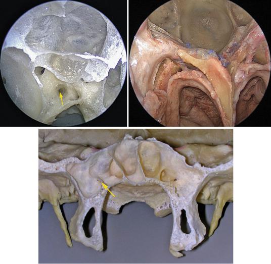
23 |
|
|
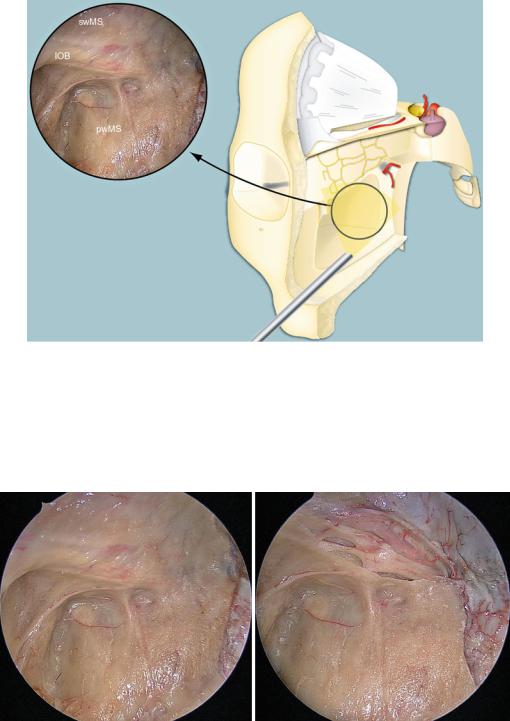
Fig. 1.24 Maxillary window
IOB infraorbital bundle, pwMS posterior wall of the maxillary sinus, swMS superior wall of the maxillary sinus
Through the maxillary window, usually associated with a transpterygoid approach, it is possible to gain access to the infratemporal fossa and upper parapharyngeal space.
Orbit
swMS
|
IOA |
IOB |
ION |
|
PPF |
PPF |
pwMS |
pwMS |
Fig. 1.25 Endoscopic view of the maxillary window
IOA infraorbital artery, IOB infraorbital bundle, ION infraorbital nerve, pwMS posterior wall of the maxillary sinus, swMS superior wall of the maxillary sinus, PPF pterygopalatine fossa
24 |
1 Cervical Segment |
|
|
Vnw |
Vnw |
P
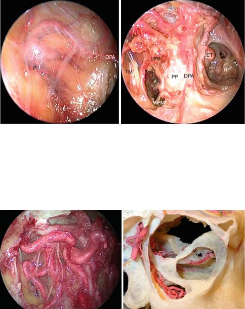
Fig. 1.26 Endoscopic view of the maxillary window
DPA descending palatine artery, ET eustachian tube, LPM lateral pterygoid muscle, MPM medial pterygoid muscle, P periostium, PP pterygoid process, RP rhinopharynx, SP soft palate, TM temporal muscle, Vnw vascular network
The fat pad filling the space between the medial pterygoid, lateral pterygoid, and temporal muscle is in direct continuation with the Bichat’s fat pad.
|
ION |
|
|
|
SPA |
|
|
|
|
SPAbs |
|
TM |
|
|
|
PSAA |
DPA |
|
|
GPN |
LPA |
||
|
|||
|
PP |
|
|
|
|
DPA |
Fig. 1.27 Endoscopic view of the vascular plane below the periostium and focus on the “palatine vessels” (external view)
DPA descending palatine artery, GPN greater palatine nerve, ION infraorbital nerve, LPA lesser palatine artery, PP pterygoid plate, PSAA posterosuperior alveolar artery, SPA sphenopalatine artery, SPAbs sphenopalatine branches, TM temporal muscle
The PSAA often presents a common trunk with the infraorbital artery (IOA). The IOA runs forward in the infraorbital groove and canal accompanying the ION. The DPA descends with the GPN and becomes the greater palatine artery after exiting from the greater palatine foramen. The LPA is a branch of the DPA and exits the nasal cavity through the lesser palatine foramen and feeds mainly the soft palate.
1.2 Anatomic Pictures |
25 |
|
|
|
ET |
LPM |
|
|
LC |
MA |
|
FP |
DPA LVPM |
IAN |
|
IAN LN |
MPM |
|
TVPM |
ION
PP
Fig. 1.28 Anterior views of the infratemporal fossa. The muscular wall given by the lateral pterygoid muscle, with the overlying vascular network, is evident. Between the lateral and the medial pterygoid muscle, the lingual and the inferior alveolar nerves are visible
BN buccal nerve, Cl clivus, DPA descending palatine artery, ET eustachian tube, FP fat pad, HP hard palate, IAN inferior alveolar artery, ION infraorbital nerve, LC longus capitis, LN lingual nerve, LPM lateral pterygoid muscle, LPP lateral pterygoid plate, LVPM levator veli palatini muscle, MA maxillary artery, MPM medial pterygoid muscle, PP pterygoid plate, SP soft palate, TM temporal muscle, TVPM tensor veli palatini muscle, VC vidian canal, Vnw vascular network, white arrow vidian nerve, black arrowheads tensor veli palatini muscle
LPM connects the lateral surface of the pterygoid plate and the surrounding skull base (infratemporal crest of the greater wing of the sphenoid) to the anterior surface of the neck of the mandible and the articular capsule. The muscular “wall” given by the LPM covers the deeper structures and should be cut and removed to gain wide access to the infratemporal fossa and upper parapharygeal space (Dallan et al. 2010). The buccal nerve passes between the two heads of the LPM and runs toward the medial aspect of the temporal muscle. Then, it passes close to the ramus of the mandible to lie on the lateral surface of the buccinator muscle in the cheek and innervate the mucosa of the cheek. The buccal nerve can be accompanied by the buccal artery. The FP between the LMP, MPM, and TM is a prolongation of the buccal fat pad, which lies on the lateral surface of the buccinator muscle. The position of the MA in respect to the LPM is highly variable.
26 |
1 Cervical Segment |
|
|
|
LPM |
VN ET |
|
LPM |
|
ICAp V3 |
|
|
|
TM |
IJV |
LC |
ATN |
LPP |
|
||||
|
MA |
|
|
TVPM |
|
|
|
|
MMA
LVPM
IAN
IAN
LN MPM
TVPM |
LN |
Fig. 1.29 Anterior view of the infratemporal fossa. The lateral pterygoid muscle has been cut, and V3 and the Eustachian Tube are exposed. Through this muscular window access to the upper parapharyngeal space can be gained
ATN auricolotemporal nerve, ET eustachian tube, IAN inferior alveolar nerve, ICAp parapharyngeal portion of the internal carotid artery, IJV internal jugular vein, LC longus capitis, LN lingual nerve, LPM lateral pterygoid muscle, LPP lateral pterygoid plate, LVPM levator veli palatini muscle, MA maxillary artery, MMA middle meningeal artery, MPM medial pterygoid muscle, TM temporal muscle, TVPM tensor veli palatini muscle, VN vidian nerve, V3 third branch of the trigeminal nerve, white arrow chorda tympani
MPM connects the medial surface of the lateral pterygoid plate and the tuberosity of the maxilla with the medial surface of the ramus and angle of the mandible. On its lateral surface, LN and IAN lie. A relationship is also present with the sphenomandibular ligament, maxillary vessels, and sometimes with a parotid process. The medial surface of the MPM presents a relationship with the TVPM, styloglossus, stylopharyngeus, and superior constrictor muscles.
1.2 Anatomic Pictures |
27 |
|
|
V3
HP
|
MPM |
|
*** |
|
|
|
* |
LPM |
|
** |
|
|
TVPM ET |
RP |
** |
|
MA LVPM |
|
*** |
MC |
LcapM |
|
*** |
|
ICAp |
|
* |
|
|
* |
|
Fig. 1.30 Rhinopharyngeal region, medial to lateral and axial views
BS basisphenoid, Cl clivus, ET eustachian tube, HP hard palate, ICAc cavernous portion of the internal carotid artery, ICAp parapharyngeal portion of the internal carotid artery, IT inferior turbinate, LCapM longus capitis muscle, LVPM levator veli palatini muscle, MA maxillary artery, MC mandibular condyle, MMA middle meningeal artery, MPM medial pterygoid muscle, MPP medial pterygoid plate, MT middle turbinate, RP rhinopharynx, SCM superior constrictor muscle, SP soft palate, TVPM tensor veli palatini muscle, VN vidian nerve, V2 second branch of the trigeminal nerve, V3 third branch of the trigeminal nerve, black asterisks medial corridor to ICAp
Cranially, the TVPM attaches to the lateral side of the ET, spine of the sphenoid bone, and scaphoid fossa. It runs inferiorly between the medial pterygoid muscle and plate, and then it turns medially at the level of the hamulus of the medial pterygoid plate to join, in the soft palate, the muscle from the opposite side (it also attaches to the posterior margin of the hard palate). The cranial origin of LVPM is at the level of the petrous bone anterior to the carotid canal, but mainly from the inferior surface of the cartilaginous portion of the ET. The muscle runs inferiorly parallel to the ET, medial to the SCM to join, in the soft palate, the muscle from the opposite side.
28 |
1 Cervical Segment |
|
|
|
SPA |
|
MA |
|
|
LPM |
TVPM |
DPA |
|
PSAA
IAN ICAp
LN
GPN
MPM
Fig. 1.31 Panoramic endoscopic vision of the infratemporal fossa. The lateral pterygoid muscle and the fat pad have been removed, and a panoramic vision on the infratemporal fossa is achieved. The inferior alveolar and the lingual nerves are visible on the lateral surface of the medial pterygoid muscle
DPA descending palatine artery, GPN greater palatine nerve, IAN inferior alveolar nerve, ICAp parapharyngeal portion of internal carotid artery, LN lingual nerve, LPM lateral pterygoid muscle, MA maxillary artery, MPM medial pterygoid muscle, PSAA posterosuperior alveolar artery, SPA sphenopalatine artery, TVPM tensor veli palatini muscle
DPA descends with the GPN and becomes the greater palatine artery after exiting from the greater palatine foramen. The PSAA frequently origins as a common trunk with the infraorbital artery; it runs over the tuberosity of the maxilla and accompains the posterior superior alveolar nerve.
1.2 Anatomic Pictures |
29 |
|
|
HP
MPM
LPM
TVPM
ET RP
MA LVPM
MC LcapM
ICAp
********
*******
*
Fig. 1.32 Vision of the “medial wall” given by the muscular plane
Cl clivus, DPA descending palatine artery, ET eustachian tube, HP hard palate, ICAp parapharyngeal portion of the internal carotid artery, LCapM longus capitis muscle, LPM lateral pterygoid muscle, LVPM levator veli palatini muscle, MA maxillary artery, MC mandibular condyle, MPM medial pterygoid muscle, RP rhinopharynx, SP soft palate, TVPM tensor veli palatini muscle, black asterisks medial corridor to ICAp, black circles tensor veli palatini muscle, red circles levator veli palatini muscle
The TVPM belongs to the prestyloid compartment (Janfaza et al. 2001). Actually, the prestyloid compartment is considered the true parapharyngeal space (Stambuk and Patel 2008). Posterior to the TVPM lies the carotid space. Fibers from the LVPM follow eustachian tube orientation to insert at the soft palate. By passing laterally, in relation to the TVPM, it is possible to reach the ICAp and, immediatly lateral to it, the internal jugular vein and lower cranial nerves (Dallan et al. 2010)
30 |
1 Cervical Segment |
|
|
V3
AMMA
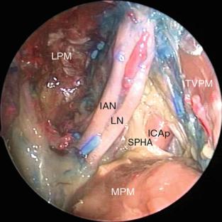
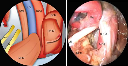
Fig. 1.33 Endoscopic view of the infratemporal fossa. The lingual and inferior alveolar nerves, two of the three branches of the posterior trunk of the third branch of the trigeminal nerve, are clearly visible, lying on the lateral surface of medial pterygoid muscle
AMMA accessory middle meningeal artery, IAN inferior alveolar nerve, ICAp parapharyngeal portion of the internal carotid artery, LN lingual nerve, LPM lateral pterygoid muscle, MPM medial pterygoid muscle, SPHA stylopharyngeal aponeurosis, TVPM tensor veli palatini muscle, V3 third branch of the trigeminal nerve
The LN emerges from the inferior border of the LPM and curves downward and forward in the pterygomandibular space. Passing inferiorly, it lies close to the lingual alveolar plate of the mandibular third molar. Sometimes, a small lingual comitant artery can be present that follows the LN. More consistent is the inferior alveolar artery, which follows the IAN, giving off a branch (mylohyoid branch) at the level of the mandibular foramen.

1.2 Anatomic Pictures |
|
31 |
|
|
|
|
BS |
V3 |
|
|
|
VP |
|
ET |
ET |
|
|
V3 |
|
MMA |
TVPM |
|
|
|
|
|
LVPM |
|
|
MA |
CT |
LVPM
MPM
LN
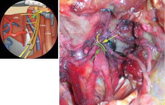
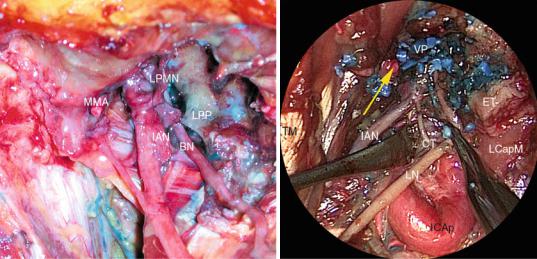
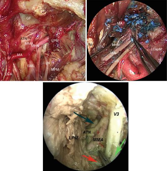
Fig. 1.34 Endoscopic view of the infratemporal fossa. Focus on the close relationship between the eustachian tube and the mandibular nerve (V3). V3 belongs to the pre-styloid compartment
BS basisphenoid, CT chorda tympani, ET eustachian tube, LN lingual nerve, LPM lateral pterygoid muscle, LVPM levator veli palatini muscle, MA maxillary artery, MMA middle meningeal artery, MPM medial pterygoid muscle, TVPM tensor veli palatini muscle, VP venous plexus around V3, V3 third branch of trigeminal nerve, black arrow the canal of V3 within the skull base
V3 is an important landmark to locate the poststyloid compartment, being just anterior to this space (Falcon et al. 2011). No foramina of the skull base is included within the borders of the parapharyngeal space (Stambuk and Patel 2008). On the anterolateral surface of the ET, it is possible to identify the tubal incisure. This is a groove in the medial lamella of the ET that accommodates the LVPM. Around V3 a wide venous plexus is well evident.
32 |
1 Cervical Segment |
|
|
DTN DTN
LPMN
MN
LPP
MMA
BN
ATN
LPM
IAN CT LN
SML
MPM
Fig. 1.35 View from below of the main branches of V3. The infratemporal fossa has been subtotally cleaned
ATN auricolotemporal nerve, BN buccal nerve, CT chorda tympani, DTN deep temporal nerve, ET eustachian tube, IAN inferior alveolar nerve, LN lingual nerve, LPM lateral pterygoid muscle, LPMN lateral pterygoid muscle nerve, LPP lateral pterygoid plate, LVPM levator veli palatini muscle, MA maxillary artery, MMA middle meningeal artery, MN masseteric nerve, MPM medial pterygoid muscle, SML sphenomandibular ligament, SMs styloid muscles, TVPM tensor veli palatini muscle, VN vidian nerve, V2 second branch of the trigeminal nerve, green arrow posterior trunk, yellow arrow anterior trunk
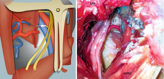
V3 divides at a short distance from the skull base. Before splitting into an anterior and posterior trunk, it offers some branches/nerves: meningeal, medial pterygoid, tensor tympani, and tensor veli palatine nerves. In close proximity to V3, on its dorso-medial surface, lies the otic ganglion. It is in close relationship with the lateral surface of the ET and just below the skull base. The CT comes out from the petrotympanic fissure and passes over the spine of the sphenoid bone to join the posterior aspect of the LN. The CT is usually accompanied by the anterior tympanic artery (branch of the maxillary artery). Within the infratemporal fossa, anastomotic nerve(s) between the IAN and the LN can be present.
1.2 Anatomic Pictures |
33 |
|
|
V3
SpS
CT
LN
MPM
Fig. 1.36 External and endoscopic anterior view of the close relationship between chorda tympanic and the lingual nerve
BN buccal nerve, CT chorda tympani, ET eustachian tube, IAN inferior alveolar nerve, ICAp parapharyngeal portion of the internal carotid artery, LCapM longus capitis muscle, LN lingual nerve, LPMN lateral pterygoid muscle nerve, LPP lateral pterygoid plate, MMA middle meningeal artery, MPM medial pterygoid muscle, SpS spine of the sphenoid, TM temporal muscle, VP venous plexus around V3, V3 third branch of the trigeminal nerve, yellow arrow maxillary artery (cut)
The meningeal nerve enters the cranial cavity through the foramen spinosum, with the MMA. LN runs inferiorly on the lateral surface of the medial pterygoid muscle, passing between this muscle and the body of the mandible.
34 |
1 Cervical Segment |
|
|
IAN
SP
Fig. 1.37 External (lateral) and endoscopic anterior view of the main branches of the posterior trunk of V3
ATN auricolotemporal nerve, ECA external carotid artery, ET eustachian tube, GF glenoid fossa, IAN inferior alveolar nerve, ICAp parapharyngeal portion of the internal carotid artery, LCapM longus capitis muscle, LN lingual nerve, LPM lateral pterygoid muscle, MA maxillary artery, MHN mylohyoid nerve, MMA middle meningeal artery, MPM medial pterygoid muscle, SML sphenomandibular ligament, SP styloid process, TM temporal muscle, VP venous plexus around V3, V3 third branch of the trigeminal nerve, blue arrow sphenomandibular ligament, green arrow lingual nerve, red arrow inferior alveolar nerve
The MHN can be branched off at different heights, just below the foramen ovale or before the mandibular foramen (Janfaza and Fabian 2001a). The ATN splits around the MMA in about 75 % of the cases. It passes between the neck of the mandible and the SML and courses posteriorly to the temporomandibular joint. The spine of the sphenoid is a little bit posterior and lateral to the foramen spinosum. The SML connects the spine of the sphenoid to the lingula of the mandibular foramen (on the medial aspect of the mandible).
1.2 Anatomic Pictures |
35 |
|
|
|
|
MA |
ET |
|
|
|
|
VIIcn |
|
|
|
DMpb |
|
|
|
|
|
TM |
|
IJV |
|
SCM |
|
|
|
|
|
SGM |
SPM |
|
|
|
|
LN |
|
XIIcn |
|
APA |
ICAp |
Xcn |
|
|
|
XIcn |
SHM |
SMs |
|
ICAp |
|
||
|
|
FA |
|
Fig. 1.38 Endoscopic and external view of the upper parapharyngeal space. Note the extreme loop of the parapharyngeal portion of the internal carotid artery
APA ascending pharyngeal artery, DMpb posterior belly of the digastric muscle, ET eustachian tube, FA facial artery, ICAp parapharyngeal portion of the internal carotid artery, IJV internal jugular vein, LCapM longus capitis muscle, LN lingual nerve, MA maxillary artery (cut), SCM superior constrictor muscle, SGM styloglossus muscle, SHM stylohyoid muscle, SMs styloid muscles, SPM stylopharyngeus muscle, TM temporal muscle, VIIcn facial nerve, Xcn vagus nerve, XIcn accessory nerve, XIIcn hypoglossal nerve, yellow arrows glossopharyngeal nerve
Note the role of the styloid muscles that “cover” the ICAp. Unfortunately, the presence of looping and kinking of the ICAp makes segment(s) of this vessel anterior to the styloid “apparatus.” The styloid diaphragm is given by the posterior belly of the digastric muscle, the stylohyoid and stylopharyngeus muscles, and the stylopharyngeal aponeurosis (Falcon et al. 2011; Rhoton 2000).
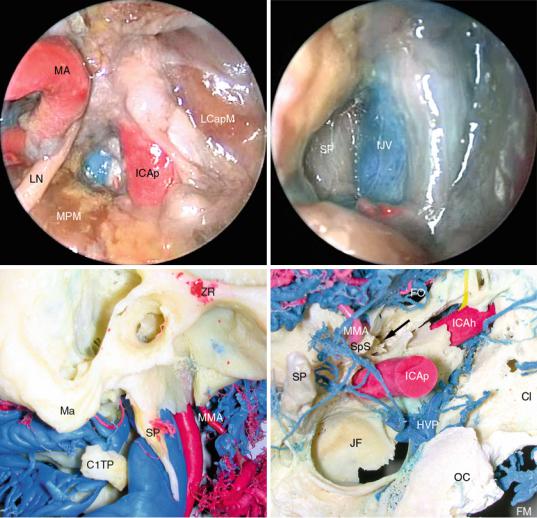
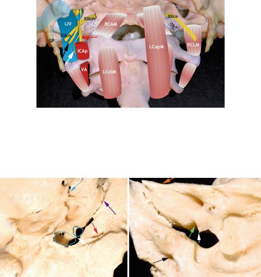
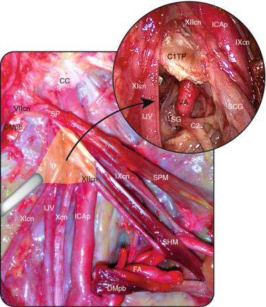
Fig. 1.39 Three-dimensional reconstruction of the endoscopic approach to craniovertebral junction
and jugular foramen area
C1 atlas, ICAp parapharyngeal portion of the internal carotid artery, IJV internal jugular vein, LColM longus colli muscle, RCAM rectus capitis anterior muscle, VA vertebral artery, white arrow hypoglossal nerve
36 |
1 Cervical Segment |
|
|
*
*
Fig. 1.40 Endoscopic view and reconstruction of the medial corridor to the parapharyngeal vessels
IAN inferior alveolar nerve, ICAp parapharyngeal portion of the internal carotid artery, LCapM longus capitis muscle, LN lingual nerve, LVPM levator veli palatini muscle, MA maxillary artery, MPM medial pterygoid muscle, SPHA stylopharyngeal aponeurosis, SCM superior constrictor muscle, SMs styloid muscles, black asterisk ascending pharyngeal artery, white asterisk lateral pterygoid muscle
Theoretically, the role of the SCM in indicating the ICAp could be critical. Unfortunately, in real conditions it is hard to identify this structure in endoscopic approach. Anatomically speaking, the SCM does not reach the skull base laterally, but it is attached to the cranial base by means of the pharyngobasilar fascia. This fascia originates from the pharyngeal tubercle, courses laterally toward the petrous bone, turns anteromedially just in front of the carotid foramen, and attaches along the cartilaginous portion of the eustachian tube and the medial pterygoid plate (Janfaza et al. 2001).
1.2 Anatomic Pictures |
37 |
|
|
PG-AT
IJV
SPF
ICAp
IJV
Fig. 1.41 Jugular foramen and styloid process
C1TP transverse process of C1, Cl clivus, FM foramen magnum, FO foramen ovale, HPV hypoglossal venous plexus, ICAh horizontal segment of the internal carotid artery, ICAp parapharyngeal segment of the internal carotid artery, IJV internal jugular vein, JF jugular foramen, LCapM longus capitis muscle, LN lingual nerve, Ma mastoid (tip), MMA middle meningeal artery, MPM medial pterygoid muscle, OC occipital condyle, PG-AT parotid gland- adipose tissue, SP styloid process, SPF sphenopetrosal fissure, SpS spine of the sphenoid, ZR zygomatic root, black arrow opening of the musculo-tubal canal, yellow line vidian nerve
The styloid process is laterally located with respect to the IJV. The stylopharyngeal aponeurosis (SPHA) extends from the SP and the stylopharyngeus muscle to the lateral pharyngeal recess (Falcon et al. 2011). The SPHA separates the prestyloid from the poststyloid compartment in its most medial aspect and encases major neurovascular structures (Rhoton 2000).
38 |
1 Cervical Segment |
|
|
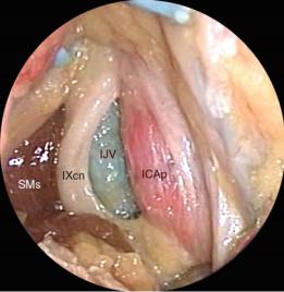
Fig. 1.42 Endoscopic view of the jugular foramen area
ICAp parapharyngeal portion of the internal carotid artery, IJV internal jugular vein, SMs styloid muscles, IXcn glossopharyngeal nerve
The glossopharyngeal nerve is located between the ICAp and IJV, enclosed within the stylopharyngeal aponeurosis. It runs downward on the lateral aspect of the stylopharyngeus muscle. The vagus nerve emerges from the superior ganglion and passes posteriorly to the carotid sheath, between the ICAp and the IJV. It is posterior to the ICAp and can be visualized only by displacing the vessel. The accessory and hypoglossal nerves are posteriorly located with respect to the vagus nerve. The accessory nerve is located in between the ICAp and IJV, enclosed by the stylopharyngeal fascia. It runs immediately posterior to the vagus nerve, while the relationship with the IJV is variable. In fact, the nerve runs posteriorly to the IJV in 33 % of cases and anteriorly in 66 % of cases (Cavalcanti et al. 2010).
1.2 Anatomic Pictures |
39 |
|
|
C1
C2
AIM
Fig. 1.43 Jugular foramen region
AIM anterior intertransversarius muscle, C1 atlas, C2 axis, D dens, ICAp parapharyngeal portion of the internal carotid artery, IJV internal jugular vein, LCapM longus capitis muscle, LColM longus colli muscle, RCAM rectus capitis anterior muscle, RCLM rectus capitis lateralis muscle, SP styloid process, VA vertebral artery, XIIcn hypoglossal nerve, black arrow accessory nerve, white arrow glossopharyngeal nerve, red arrow vagus nerve
|
|
FS |
|
|
|
|
|
|
|
|
SAF |
|
|
|
|
PCF |
IAM |
|
|
VPTB CC |
|
||
|
|
|
|
||
TB |
|
CR |
|
ESF |
|
|
SP |
*** |
* |
JT |
|
|
|
|
|
||
|
|
|
|
|
|
°
OC
SSG
Fig. 1.44 Osteology of the jugular foramen area, exocranial and endocranial views
CC carotid canal, CR carotid ridge, ESF endolymphatic sac fossa, FS foramen spinosum, IAM internal acoustic meatus, JT jugular tubercle, OC occipital condyle, PCF petroclival fissure, SAF subarcuate fossa, SP styloid process, SSG sigmoid sinus groove, TB tympanic bone, VPTB vaginal process of the tympanic bone, white arrow intrajugular process of the temporal bone, red arrow external orifice of the hypoglossal canal, violet arrow petroclival fissure, blue-sky arrow tubal isthmus, black arrow endocranial orifice of the hypoglossal canal, orange arrow trigeminal impression, green arrow pyramidal fossa, black asterisks intrajugular ridge, black circle intrajugular process of the occipital bone
Within the JF area 2 venous compartement can be identified: a large postero-lateral_SIGMOID_venous channel and a small antero-medial_PETROSAL_venous channel which can receive the drainage of the inferior petrosal sinus (IPS). Anyway, the relationship with the IPS is highly variable. An intermediary neural compartment is located between the venous ones and houses lower cranial nerves (IX, X, XI).
40 |
1 Cervical Segment |
|
|
|
TTM |
* |
MMA |
Co |
V3
VIIcn
ET
ICAp
IJV |
TVPM |
ET
CI
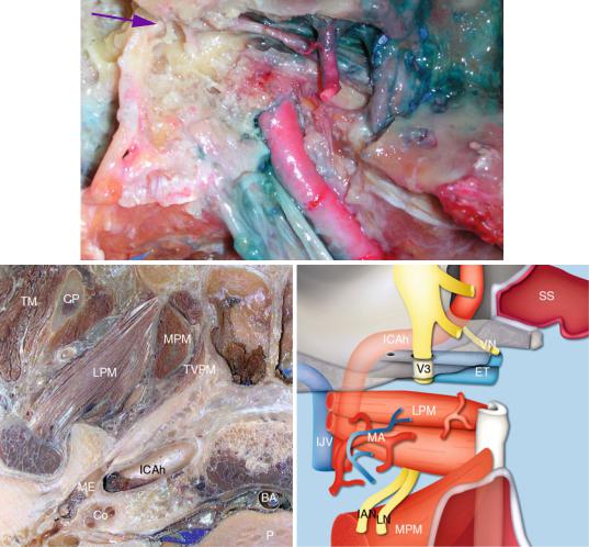
Fig. 1.45 Relationship between the carotid canal and the eustachian tube
BA basilar artery, Cl clivus, Co cochlea, CP coronoid process, ET eustachian tube, IAN inferior alveolar nerve, ICAp parapharyngeal portion of the internal carotid artery, ICAh horizontal portion of the internal carotid artery, IJV internal jugular vein, LN lingual nerve, LPM lateral pterygoid muscle, MA maxillary artery, ME middle ear, MMA middle meningeal artery, MPM medial pterygoid muscle, P pons, SS sphenoid sinus, TM temporal muscle, TTM tensor tympani muscle, TVPM tensor veli palatini muscle, VN vidian nerve, V3 third branch of the trigeminal nerve, VIIcn facial nerve, violet arrow tympanic segment of the facial nerve, black asterisk malleus

1.2 Anatomic Pictures |
41 |
|
|
MTC
*
GG
P |
* |
* |
* |
* |
|
||||
|
|
|
|
ICAh
Fig. 1.46 Bony eustachian tube and carotid canal relationship
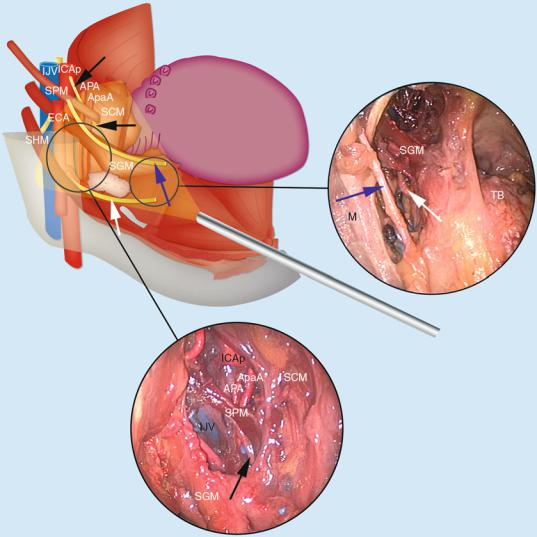
CC carotid canal, FL foramen lacerum, FO foramen ovale, FS foramen spinosum, GG geniculate ganglion area, HC hypoglossal canal, JF jugular foramen, ICAh horizontal portion of the internal carotid artery, M mastoid (tip), MTC musculo-tubal canal, OC occipital condyle, P promontory, SP styloid process, SpS spine of the sphenoid, TMF tympanomastoid fissure, red arrow posterior bend of the ICAh, green arrow bony portion of the ET (musculo-tubal canal), blue arrow canal of the tensor tympani muscle, violet arrow opening of the musculo-tubal canal, black arrow cochleariform process, black asterisk stylomastoid foramen, red asterisks groove of the greater petrosal nerve
The cartilaginous portion of the tube passes posteromedially along the border between the petrous bone and the greater wing of the sphenoid bone to attach the bony eustachian tube immediately anterior to the carotid canal (Janfaza et al. 2001). The isthmus lies immediately behind the SpS.
42 |
1 Cervical Segment |
|
|
Fig. 1.47 Endoscopic view of the infratemporal fossa and upper parapharyngeal space. The role of the eustachian tube in pointing the carotid canal is underlined
ET eustachian tube, ICAp parapharyngeal portion of the internal carotid artery, LN lingual nerve, MA maxillary artery, MPM medial pterygoid muscle, RP rhinopharynx, SP soft palate, V3 third branch of the trigeminal nerve
The cartilaginous portion of the ET is 2 cm long. It inserts at the back of the medial pterygoid plate immediately above the pharyngobasilar fascia (Janfaza et al. 2001).
Fig. 1.48 Endoscopic view of the upper parapharyngeal space. The cartilaginous portion of the eustachian tube has been removed. The upper part of the parapharyngeal portion of the internal carotid artery entering the carotid canal is visible
ICAp parapharyngeal portion of the internal carotid artery, IJV internal jugular vein, LCapM longus capitis muscle, MA maxillary artery, RP rhinopharynx, V3 third branch of the trigeminal nerve, yellow arrow indicates bony tube (immediately anterior to the carotid canal)

1.2 Anatomic Pictures |
43 |
|
|
Fig. 1.49 Relationship between the eustachian tube and V3. The connection between the chorda tympani and the lingual nerve is visible
APA ascending pharyngeal artery, CT chorda tympani, ET Eustachian tube, ICAp parapharyngeal portion of the internal carotid artery, LN lingual nerve, LPM lateral pterygoid muscle, LVPM levator veli palatini muscle, MPM medial pterygoid muscle, TVPM tensor veli palatini muscle, V3 third branch of the trigeminal nerve, black arrow canal of V3 within the skull base
The cartilaginous part of the ET inserted in the posterior border of the medial pterygoid plate. It is attached posteriorly to foramen lacerum and clivus, and superiorly to the sphenopetrosal fissure region. V3 lies anteriorly to cartilaginous portion of ET, and presents an intimate relationship with the ET. The nerve, at the skull base, is surrounded by a wide venous plexus. V3 is an important landmark to locate the post-styloid compartment, as it is anterior to this space (Falcon et al. 2011).

44 |
1 Cervical Segment |
|
|
Fig. 1.50 Endoscopic view of parapharyngeal and laterovertebral regions
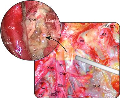
CT chorda tympani, ET eustachian tube, IAN inferior alveolar nerve, ICAc cavernous portion of the internal carotid artery, ICAp parapharyngeal portion of the internal carotid artery, IJV internal jugular vein, LCapM longus capitis muscle, LN lingual nerve, LPM lateral pterygoid muscle, LVPM levator veli palatini muscle, MA maxillary artery, MPM medial pterygoid muscle, SCM superior constrictor muscle, SMs styloid muscles, TVPM tensor veli palatini muscle, VN vidian nerve, XIIcn hypoglossal nerve, V2 second branch of the trigeminal nerve, V3 third branch of the trigeminal nerve, black asterisk ascending pharyngeal artery
1.2 Anatomic Pictures |
|
|
|
45 |
|
|
|
|
|
SpS |
PT |
|
LPT |
LPT |
|
|
|||
|
|
SCG |
JF |
|
|
|
|
|
|
OC |
|
|
|
PT |
|
|
|
|
|
|
D |
|
|
|
|
C1 |
|
SCG |
SCG |
C1TP |
|
|
JF |
|
|
|
|
||
|
|
|
|
|
|
|
|
OC |
FM |
|
|
|
OC |
|
|
|
|
|
TB |
VPTB CC |
CR |
|
|
IR |
|
SP |
OC
Fig. 1.51 Anterior view of the lower clivus and foramen magnum region and inferior view of jugular foramen area
CC carotid canal, CR carotid ridge, C1 atlas, C1TP transverse process of the atlas, D dens, FM foramen magnum, IR intrajugular ridge, JF jugular foramen, LPT lateral pharyngeal tubercle, OC occipital condyle, PT pharyngeal tubercle, SCG supracondylar groove, SP styloid process, SpS spine of the sphenoid, TB tympanic bone, VPTB vaginal process of the tympanic bone, black arrow stylomastoid foramen, red arrow hypoglossal canal, dark green arrow intrajugular process of the temporal bone, violet arrow opening of the musculo-tubal canal
46 |
1 Cervical Segment |
|
|
Fig. 1.52 External view of the extracranial segment of the hypoglossal nerve
C1TP transverse process of C1, C2 second cervical nerve root, ICAp parapharyngeal portion of the internal carotid artery, IOA infraorbital artery, LCapM longus capitis muscle, MA maxillary artery, SCG superior cervical ganglion, SCM superior constrictor muscle, VA vertebral artery, V2 second branch of the trigeminal nerve, V3 third branch of the trigeminal nerve, IXcn glossopharyngeal nerve, Xcn vagus nerve, XIcn accessory nerve, XIIcn hypoglossal nerve
The hypoglossal nerve exits from the hypoglossal canal medial to the carotid sheath. It lies posterior to the vagus nerve and runs laterally between the IJV and the ICAp (enclosed within the carotid sheath). Coursing inferiorly, it is lateral to the internal and external carotid artery and covered by the styloid process.
1.2 Anatomic Pictures |
47 |
|
|
Fig. 1.53 External view of the relationship between the parapharyngeal portion of the internal carotid artery and the vertebral artery. A vertebral window between the transverse processes is needed to visualize the vertebral bed. Note that the posterior belly of the digastric muscle has been cut
C1TP transverse process of C1, C2 second cervical root, CC carotid canal, DMpb posterior belly of the digastric muscle, FA facial artery, ICAp parapharyngeal portion of the internal carotid artery, IJV internal jugular vein, SCG superior cervical ganglion, SG spinal ganglion, SHM stylohyoid muscle, SP styloid process, SPM stylopharyngeus muscle, VA vertebral artery, VIIcn facial nerve, IXcn glossopharyngeal nerve, Xcn vagus nerve, XIcn accessory nerve, XIIcn hypoglossal nerve
The medial segment of the styloid diaphgram consists of the stylopharyngeal fascia and is medial to the styloid process and attached muscles (Bejjani et al. 1998). It is a thick fascia inserting superiorly at the cranial base and medially at the pharyngeal wall.
48 |
1 Cervical Segment |
|
|
Fig. 1.54 External view of the jugular foramen region
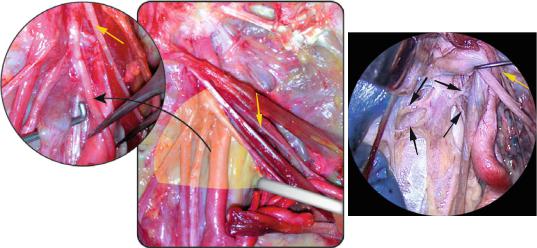
DM digastric muscle, ECA external carotid artery, HN Hering’s nerve, ICAp parapharyngeal portion of the internal carotid artery, IJV internal jugular vein, SCG superior cervical ganglion, SLN superior laryngeal nerve, SMs styloid muscles, VIIcn facial nerve, IXcn glossopharyngeal nerve, Xcn vagus nerve, XIcn accessory nerve, XIIcn hypoglossal nerve
On the outer skull base, the hypoglossal nerve runs on the medial surface of the IJV and on the lateral surface of the vagus nerve in more than 90 % of cases (Lang 1995). Then, it curves around the occipital artery and runs first to the lateral and then to the anterior surface of the ECA. In most cases, there are anastomoses between the inferior ganglion of the vagus nerve and the sympathetic ganglion. The SLN branched off from the vagus nerve a little bit inferior to the inferior ganglion. It lies posterior to the vagus nerve and passes laterally between the IJV and ICAp. Inferiorly, it lies lateral to ICA and ECA and deep to styloid process. The nerve of Hering, carotid branch of IXcn, descends between the ICAp and ECA to reach the carotid body and sinus (Janfaza and Fabian 2001b).
1.2 |
Anatomic Pictures |
|
|
|
|
49 |
|
|
|
|
|
|
|
|
|
|
|
|
CC |
|
|
|
|
|
|
|
VIIcn |
|
|
|
|
|
|
|
SP |
|
|
|
|
XIcn |
Xcn XIIcn |
|
|
SCM |
|
|
|
IJV |
|
|
|
|
XIIcn |
||
|
|
|
|
XIcn |
|||
|
SCG |
|
|
|
|
|
|
|
|
|
|
|
IJV |
|
|
|
|
|
|
|
SPM |
|
|
|
|
|
|
XIIcn |
|
|
|
|
|
|
|
|
|
|
|
|
CST |
ICAp |
IJV |
|
|
|
SCG |
|
|
|
|
|
|||
|
|
|
XIcn |
|
SHM |
|
Xcn ICAp |
|
|
|
|
|
|
||
Xcn |
|
|
ICAp |
|
FA |
|
DMpb |
|
|
|
Fig. 1.55 External views of the upper parapharyngeal region and focus on superior cervical ganglion
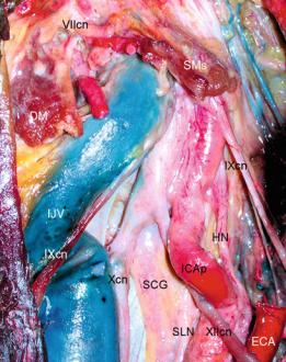
CC carotid canal, CST cervical sympathetic trunk, DMpb posterior belly of the digastric muscle, FA facial artery, ICAp parapharyngeal portion of the internal carotid artery, IJV internal jugular vein, LC longus capitis, SCG superior cervical ganglion, SCM superior constrictor muscle, SHM stylohyoid muscle, SP styloid process, SPM stylopharyngeus muscle, VIIcn facial nerve, X vagus nerve, XIcn accessory nerve, XIIcn hypoglossal nerve, yellow arrow glossopharyngeal nerve, black arrows anastomosis between the superior cervical ganglion and lower cranial nerves
The cervical sympathetic trunk lies posterior to the carotid sheath, within a duplication of the prevertebral fascia and medial to the vagus nerve (Janfaza and Fabian 2001a). It is located in a fascial reflection from the posterior wall of the carotid sheath.
50 |
1 Cervical Segment |
|
|
Fig. 1.56 Lateral view of the parapharyngeal region. 3D reconstruction
APA ascending pharyngeal artery, ApaA ascending palatine artery, ECA external carotid artery, FA facial artery, GGM genioglossus muscle, GHM geniohyoid muscle, HB hyoid bone, HGM hyoglossus muscle, ICAp parapharyngeal portion of the internal carotid artery, IJV internal jugular vein, LN lingual nerve, MHM mylohyoid muscle, PGM palatoglossus muscle, SCM superior constrictor muscle, SGM styloglossus muscle, SHM stylohyoid muscle, SMG submandibular gland, SPM stylopharyngeus muscle, IXcn glossopharyngeal nerve, Xcn vagus nerve, XIcn accessory nerve, XIIcn hypoglossal nerve
|
|
|
SGM |
|
|
|
|
PG |
|
|
SGM |
|
ECA |
|
|
|
|
||
|
|
|
SPM |
|
|
SPM LcapM |
|
ICAp |
|
ECA |
LcolM |
|
LCns |
|
ICAp |
|
|||
D |
IJV |
|||
|
|
|||
|
|
|
||
|
IJV |
|
VA |
|
|
|
|
Fig. 1.57 Axial view of the parapharyngeal region
D dens, ECA external carotid artery, ICAp parapharyngeal portion of the internal carotid artery, IJV internal jugular vein, LCapM longus capitis muscle, LCns lower cranial nerves, LcolM longus colli muscle, PG parotid gland, SGM styloglossus muscle, SPM stylopharyngeus muscle, VA vertebral artery
1.2 Anatomic Pictures |
51 |
|
|
DM OA SHM
XIcn
XIIcn
IJV SMG
LFVT
IJV ICAp |
|
|
SPM |
|
|
SGM |
SCM |
|
XIcn |
IXcn |
|
|
||
SHM |
|
|
FA |
LN |
|
SMG |
||
|
||
|
XIIcn |
SHM |
SGM |
|
ECA |
MPM LN |
|
|
FA |
SMG |
|
|
Fig. 1.58 Lateral vision of the upper cervical and lower parapharyngeal regions. The vertical branch of the mandible has been removed
DM digastric muscle, ECA external carotid artery, FA facial artery, ICAp parapharyngeal portion of the internal carotid artery, IJV internal jugular vein, LFVT linguofacial venous trunk, LN lingual nerve, MPM medial pterygoid muscle, OA occipital artery, SCM superior constrictor muscle, SGM styloglossus muscle, SHM stylohyoid muscle, SMG submandibular gland, SPM stylopharyngeus muscle, IXcn glossopharyngeal nerve, XIcn accessory nerve, XIIcn hypoglossal nerve, yellow arrow ansa cervicalis profunda
52 |
1 Cervical Segment |
|
|
Fig. 1.59 External view of the styloid muscles and their relationship to great cervical vessels. The vertical portion of the mandible has been removed and the ITF content subtotally cleaned
DMpb posterior belly of the digastric muscle, FA facial artery, ICAp parapharyngeal portion of the internal carotid artery, IJV internal jugular vein, SCM superior constrictor muscle, SGM styloglossus muscle, SHM stylohyoid muscle, SPM stylopharyngeus muscle, VIIcn facial nerve, Xcn vagus nerve, XIcn accessory nerve, XIIcn hypoglossal nerve, black asterisk glossopharyngeal nerve at the skull base
The SPM runs inferiorly on the lateral aspect of the ICAp. The SHM is lateral to the ECA. The SGM passes lateral to the ICAp, medially to the ECA. The stylomandibular ligament is a condensation of the deep layer of the parotid fascia. It connects the styloid process with the angle of the mandible.
1.2 Anatomic Pictures |
|
|
53 |
|
|
|
|
SHM |
Xcn |
|
|
ICAp |
|
|
|
PG |
|
|
|
|
|
|
|
|
SPM |
|
|
LN |
|
|
|
MPM |
IXcn |
|
|
|
|
|
|
|
IJV |
|
|
BFP |
|
ICAp |
SCM |
|
ApaA |
||
|
SCM |
PP |
*SGM
SCM
BM
Fig. 1.60 Transoral endoscopic view of the parapharyngeal region
ApaA ascending palatine artery, BFP buccal fat pad, BM buccinator muscle, ICAp parapharyngeal portion of the internal carotid artery, IJV internal jugular vein, LN lingual nerve, MPM medial pterygoid muscle, PG parotid gland, PP pharyngeal plexus, SCM superior constrictor muscle, SGM styloglossus muscle, SHM stylohyoid muscle, SPM stylopharyngeus muscle, IXcn glossopharyngeal nerve, Xcn vagus nerve, black asterisk stylomandibular ligament
The pterygomandibular raphe is tied between the hamulus of the medial pterygoid plate and the posterior part of the mylohyoid line of the mandible. The buccinator muscle attaches to it.
54 |
1 Cervical Segment |
|
|
|
|
ICAp |
|
|
|
|
LCapM |
ICAp |
SCM |
SPM |
SCM |
|
|
|
|
PP ApaA |
|
|
ApaA |
|
SGM |
|
|
|
|
|
|
SGM |
|
|
|
Fig. 1.61 Transoral endoscopic view of the parapharyngeal region
ApaA ascending palatine artery, ICAp parapharyngeal portion of the internal carotid artery, LCapM longus capitis muscle, PP pharyngeal plexus, SCM superior constrictor muscle, SGM styloglossus muscle, SPM stylopharyngeus muscle, white arrows glossopharyngeal nerve
The external carotid artery passes deeply to the digastric and stylohyoid muscles, but superficially to the stylopharyngeus and styloglossal muscle when running toward the parotid gland (Janfaza et al. 2001). With a transoral window it is possible to control the space between the medial pterygoid muscle laterally and the superior constrictor muscle medially. The stylopharyngeus and styloglossus muscles are critical landmarks, being usually placed anterior to the great vessels (Dallan et al. 2011). Note that the presence of kinking or looping of the ICAp could make this statement untrue.
1.2 Anatomic Pictures |
55 |
|
|
M
SPM
SGM
MPM
IAN
LA
LN
DM
HGM
IJV |
XIIcn |
ICAp
IJV |
|
SCM |
|
|
SPM |
|
|
|
|
|
|
|
|
|
APA |
SGM |
|
SPM |
ApaA |
|
|
||
|
|
IJV |
|
XIIcn |
|
SGM |
XIIcn |
|
|
||
|
|
|
|
|
|
TB |
FA |
|
|
|
Fig. 1.62 Lateral external and transoral endoscopic views of the tongue base and parapharyngeal regions
APA ascending pharyngeal artery, ApaA ascending palatine artery, DM digastric muscle, FA facial artery, HGM hyoglossus muscle, IAN inferior alveolar nerve, ICAp parapharyngeal portion of the internal carotid artery, IJV internal jugular vein, LA lingual artery, LN lingual nerve, M mandible, MPM medial pterygoid muscle, SCM superior constrictor muscle, SGM styloglossus muscle, SPM stylopharyngeus muscle, TB tongue base, XIIcn hypoglossal nerve, black arrows glossopharyngeal nerve
56 |
1 Cervical Segment |
|
|
Fig. 1.63 Transoral endoscopic view of the parapharyngeal region
APA ascending pharyngeal artery, ApaA ascending palatine artery, ICAp parapharyngeal portion of the internal carotid artery, IJV internal jugular vein, LN lingual nerve, M mandible, SCM superior constrictor muscle, SGM styloglossus muscle, SHM stylohyoid muscle, SPM stylopharyngeus muscle, TB tongue base, white arrow hypoglossal nerve, black arrows glossopharyngeal nerve, blue arrows lingual nerve