


3.2 Anatomic Pictures |
85 |
|
|
3.2Anatomic Pictures
ASB
|
|
ON |
ON |
|
|
|
|
|
|
|
MA |
|
PG |
|
|
|
|
|
|
TM |
|
ICAc |
ICAc |
MA |
|
|
|||
|
|
|
||
|
|
|
BP |
|
|
|
|
PVMs |
|
Fig. 3.1 Anterior view of the midline structures
ASB anterior skull base, BP basilar plexus, ICAc cavernous portion of the internal carotid artery (vertical segment), MA maxillary artery, ON optic nerve, PG pituitary gland, PVMs prevertebral muscles, TM temporal muscle
Fig. 3.2 Anterior view of the sphenoid sinus and the rhinopharynx
ACP anterior clinoid process, BS basisphenoid, CP carotid protuberance, CS cavernous sinus, ICAc cavernous portion of the internal carotid artery, ON optic nerve, PG pituitary gland, RP rhinopharynx, VN vidian nerve, V2 second branch of the trigeminal nerve, black arrow first branch of the trigeminal nerve, yellow arrows ophthalmic artery
ON
ACP |
ON |
PS |
|
ICAc
CP PG
CS
V2
VN
BS
RP
86 |
|
3 Intracranial Segment |
|
|
|
V1 |
IIIcn |
PG |
|
||
IVcn |
|
|
|
VIcn |
ICAc |
V2 |
|
|
|
LRSS |
BS |
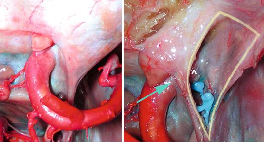
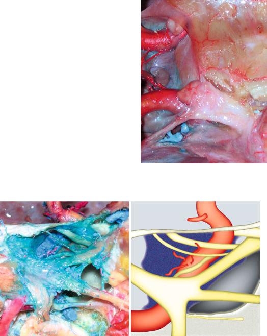
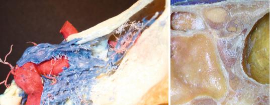
Fig. 3.3 Anterior vision of the right cavernous sinus
BS basisphenoid, CP carotid protuberance, ICAc cavernous portion of the internal carotid artery (vertical segment), LRSS lateral recess of the sphenoid sinus, OA ophthalmic artery, ON optic nerve, PG pituitary gland, SS sphenoid sinus, V1 first branch of the trigeminal nerve, V2 second branch of the trigeminal nerve, IIIcn oculomotor nerve, IVcn trochlear nerve, VIcn abducens nerve, yellow arrow inferior part of the medial wall of the cavernous sinus (yellow line), blue-sky arrow superior part of the medial wall of the cavernous sinus (blue-sky line)
The inferior part of the medial wall of the CS faces the lateral part of the body of the sphenoid, while the upper part of the medial wall is directly related to the PG (Martins et al. 2011). In the upper part, the medial wall is given by the meningeal layer, that is a continuation of the diaphragma sellae, which surrounds the pituitary capsule inferiorly (Yasuda et al. 2005; Martins et al. 2011). In the inferior part, the medial wall is given by the endosteal layer that covers the body of the sphenoid bone. So, a sellar (superior) and a sphenoidal (inferior) part can be described. The limits of the medial wall of the cavernous sinus are the superior orbital fissure anteriorly, the dorsum sellae posteriorly, the superior margin of the maxillary nerve inferiorly and the diaphragma sellae superiorly (Yasuda 2005). The presellar segment of the ICAc corresponds to the anterior bend of the ICAc and to the clinoidal segment of the internal carotid artery.
3.2 Anatomic Pictures |
|
|
|
87 |
|
|
|
|
|
|
PS |
|
|
|
OC |
ChS |
OS |
PS |
|
|
OSACP |
|
|
|
|
|
|
|
|
|
TS |
|
|
|
FR |
* |
ACP |
ChS |
|
PF |
|
|
* |
|
|
|
TS |
||
|
|
|
||
|
DS |
SB |
PF |
|
FO |
|
PCP |
|
|
FO |
|
|
||
|
|
|
DS |
|
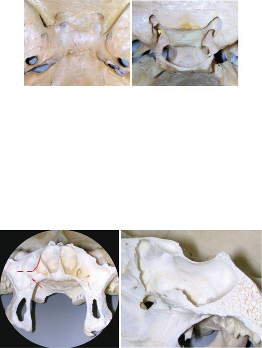
Fig. 3.4 Sphenoid sinus and bone
ACP anterior clinoid process, ChS chiasmatic sulcus, DS dorsum sellae, FO foramen ovale, FR foramen rotundum, OC optic canal, OS optic strut, PCP posterior clinoid process, PF pituitary fossa, PS planum sphenoidale, SB sellar bridge, TS tuberculum sellae, yellow arrow carotidclinoid foramen, black asterisk middle clinoid process
ACPs project posteriorly and laterally to the optic canal and carotid sulcus. The TS separates the chiasmatic sulcus from the pituitary fossa. This is closed posteriorly by the DS. On each side of DS are the PCPs. The OS links the ACP with the sphenoid bone and separates the optic canal from the superior orbital fissure. When anterior and middle clinoid processes fuse, a “carotid-clinoid” foramen is formed. This circumstance is present in about 10 % of adults. The bony connection between anterior and posterior clinoid processes is called the “sellar bridge.” This is found in about 6 % of cases (Lang 1995). The base of the middle clinoid process is just medial to the anterior genu of the (parasellar) caverous ICA, while the apex extends in a dorsolateral direction. The middle clinoid process is, when present, a small eminence of the carotid sulcus at the level of the anterior part of the lateral wall of the sella turcica (Labib et al. 2013). The lateral tubercular crest is a bony eminence placed on the lateral part of the tuberculum sellae.
|
PS |
|
|
TS |
|
V2I |
|
DS |
ICAc/pr |
|
|
** |
|
|
* |
SS |
|
Cl |
|
|
OC |
|
|
OPPB |
BS |
Cl |
LPP |
|
|
MPP |
SPPB |
|
|
|
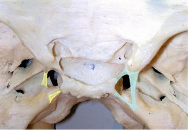
Fig. 3.5 Sphenoid sinus and bone
BS basisphenoid, Cl clivus, DS dorsum sellae, ICAc/pr protuberance of the cavernous portion of the internal carotid artery, LPP lateral pterygoid plate, MPP medial pterygoid plate, OC occipital condyle, OPPB orbital process of the palatine bone, PS planum sphenoidale, SPPB sphenoidal process of the palatine bone, SS sphenoid sinus, TS tuberculum sellae, V2I incisure of V2, red arrow opening of the vidian canal, red asterisks vidian canal, red line internal carotid artery
88 |
3 Intracranial Segment |
|
|
SS
PLL |
|
|
|
ICAh |
FR |
|
|
|
|
||
MS |
SS |
|
|
FR |
pwMS |
|
|
VC |
PVC |
||
|
|||
|
|
RS |
|
|
|
Ch |
|
PPs |
|
|
Fig. 3.6 Sphenoid bone and sinus with a focus on the base of the pterygoid region and the relationship with the cavernous portion of the internal carotid artery
Ch choana, FR foramen rotundum, ICAh horizontal portion of the internal carotid artery, MS maxillary strut, PLL petrolingual ligament, PPs pterygoid plates, PVC palatovaginal canal, pwMS posterior wall of the maxillary sinus, RS rostrum sphenoidale, SS sphenoid sinus, VC vidian canal, black arrowhead lingula of the sphenoid, blue-sky asterisk and line vidian (pterygoid) canal, red asterisk and line palatovaginal (pharyngeal) canal
|
|
|
|
|
|
ACP |
ON |
ON |
|
|
|
|
|
|
* |
ACP |
|
|
ACP |
ON |
ON |
|
|
|
||
|
|
|
|
|
|
|||
|
|
* |
* ACP |
|
|
|
|
PG |
|
|
|
|
|
|
|
|
|
|
ICAc |
CS |
|
|
|
|
SS |
|
|
|
|
|
|
|
|
||
|
|
|
SS |
|
|
|
|
|
|
|
|
BS |
|
|
|
|
BS |
|
|
|
|
|
|
|
V3 |
|
|
|
|
|
|
LPM |
|
|
|
|
ET |
|
|
|
ET |
|
|
|
LPM |
|
ET |
|
|
|
|
||
|
|
|
TVPM |
|
|
|||
|
TVPM |
|
|
V3 |
|
|
|
|
|
|
|
|
|
|
|
||
LVPM |
LVPM |
|
RPW |
||
|
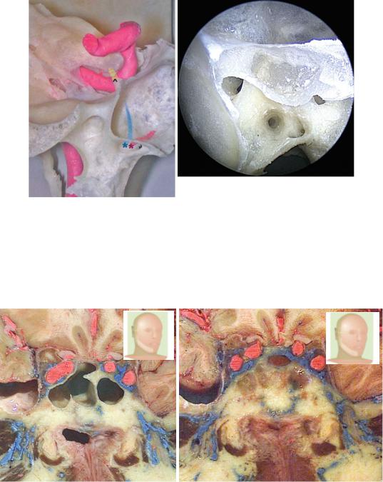
Fig. 3.7 Coronal views at the level of the sphenoidal region. The picture on the left shows a more anterior view than the picture on the right. Note that the picture on the left is seen from posterior, while the picture on the right is seen from anterior
ACP anterior clinoid process, BS basisphenoid, CS cavernous sinus, ET eustachian tube, ICAc cavernous portion of the internal carotid artery, LPM lateral pterygoid muscle, LVPM levator veli palatini muscle, ON optic nerve, PG pituitary gland, RPW rhinopharyngeal posterior wall, SS sphenoid sinus, TVPM tensor veli palatini muscle, V3 third branch of the trigeminal nerve, white asterisk ophthalmic artery
3.2 Anatomic Pictures |
89 |
|
|
MS
|
|
PPF |
ITF |
|
ITF PPF |
|
|
|
|
|
|
|
|
|
|
|
|
SPA |
|
|
|
|
|
ITF |
ICAc |
Cl |
ICAc |
ITF |
|
|
|
|
||
|
BA |
|
|
|
|
P |
Vcn |
ICAc |
Cl |
|
|
|
|
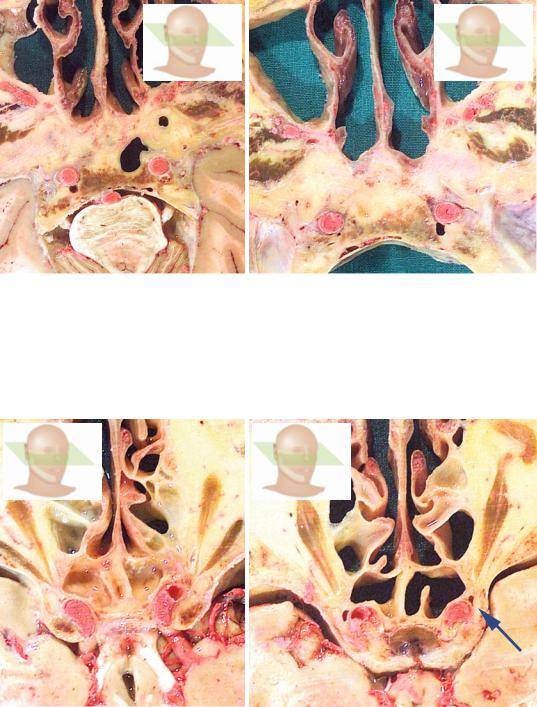
Fig. 3.8 Axial views at the level of the clivus. The picture on the left is seen from below. The picture on the right is seen from above
BA basilar artery, Cl clivus, ICAc cavernous portion of the internal carotid artery (vertical segment), ITF infratemporal fossa, MS maxillary sinus, P pons, PPF pterygopalatine fossa, SPA sphenopalatine artery, Vcn trigeminal nerve
SS |
SS |
SS |
|
SS |
|||
|
|||
|
ICAc |
|
|
ICAc |
ACP |
|
ICAc
ACP
PG
PS ICAi
OT OT
*
Fig. 3.9 Axial view at the level of the sphenoid sinus. The picture on the left is seen from below. The picture on the right is seen from above. The picture on the left shows a more cranial view
ACP anterior clinoid process, ICAc cavernous portion of the internal carotid artery, ICAi intracranial portion of the internal carotid artery, OT optic tract, PG pituitary gland, PS pituitary stalk, SS sphenoid sinus, blue arrow abducens nerve, white asterisk third ventricle

90 |
3 Intracranial Segment |
|
|
MTt
MTt
ITt
RP
OC
AL
PL
SS
SS
CI
RP
PG
SS |
BP |
|
Cl
Fig. 3.10 Medial sagittal vision of the sphenoid sinus and its surrounding structures
AL anterior lobe, BP basilar plexus, BS basisphenoid, Cl clivus, ICAc cavernous portion of the internal carotid artery, ICAi intracranial portion of the internal carotid artery, ITt tail of the inferior turbinate, Lcap longus capitis, MTt tail of the middle turbinate, OC optic chiasm, OP optic protuberance, PG pituitary gland, PL posterior lobe, PS planum sphenoidale, RP rhinopharynx, ST superior turbinate, yellow arrow superior hypophyseal artery(ies), blue arrow optic nerve
The anterior lobe of the pituitary gland is mainly fed by the superior hypophyseal arteries while the posterior lobe is fed mainly by the inferior hypophyseal artery.

3.2 Anatomic Pictures |
91 |
|
|
Fig. 3.11 |
|
|
|
Reconstruction showing |
|
|
|
the pterygoidal window |
|
|
PG |
and the region around |
|
|
|
V2 |
|
|
|
the foramen rotundum |
CS |
CR |
|
|
|
|
|
CR clival recess, CS |
|
ICAc |
|
cavernous sinus, ET |
|
VN |
|
eustachian tube, ICAc |
|
|
|
|
ET |
|
cavernous portion of the internal carotid artery, PG pituitary gland, VN vidian nerve, V2 second branch of the trigeminal nerve
ss |
|
ICAc |
|
|
|
|
|
|
|
|
|
||
|
OAp |
|
|
PG |
||
|
|
|
|
|||
|
|
|
|
|
ICAc |
|
|
|
|
|
V2 |
CS |
CR |
|
|
|
|
|||
FR |
|
|
|
|
||
|
|
|
|
|
||
|
|
|
|
|
|
|
pwMS |
|
|
ICAc |
|
ICAc |
|
VC PVC |
MS |
|
|
|
||
RS
V2 |
VN |
|
|
Ch |
|
Fig. 3.12 Foramen rotundum region
Ch choana, CR clival recess, CS cavernous sinus, FR foramen rotundum, ICAc cavernous portion of the internal carotid artery, MS maxillary strut, OAp orbital apex, PG pituitary gland, PVC palatovaginal canal, pwMS posterior wall of the maxillary sinus, RS rostrum sphenoidale, SS sphenoid sinus, VC vidian canal, VN vidian nerve, V2 second branch of the trigeminal nerve, blue-sky arrows superior orbital fissure

92 |
3 Intracranial Segment |
|
|
PG
|
|
PG |
|
|
° |
ICAc |
° |
|
|
|
|
|
||
|
|
CR |
FRA |
|
V2 |
CS |
V2 |
||
|
||||
|
|
|
FRA ICAc
CNs and PPG
CNs & PPG
SPA
SPA
Fig. 3.13 Endoscopic views of the foramen rotundum region
CNs and PPG connecting nerves (V2 and pterygopalatine ganglion) and pterygopalatine ganglion, CR clival recess, CS cavernous sinus, FRA artery for the foramen rotondum, ICAc cavernous portion of the internal carotid artery, PG pituitary gland, SPA sphenopalatine artery, V2 second branch of the trigeminal nerve, black circle zygomatic nerve
Foramen rotundum transmits the second branch of the trigeminal nerve (maxillary nerve), arteries (foramen rodundum artery), and veins.
ZN
V2
ION
ICAc
PPG
PPG
SPA
SPA
Fig. 3.14 Endoscopic vision of the pterygopalatine ganglion
ICAc cavernous portion of the internal carotid artery, ION infraorbital nerve, PPG pterygopalatine ganglion, SPA sphenopalatine artery, ZN zygomatic nerve, V2 second branch of the trigeminal nerve, blue arrows artery for the foramen rotundum
The zygomatic nerve exits the pterygopalatine fossa via the inferior orbital fissure and diverges from the ION to enter the orbit (Janfaza et al. 2001). The PPG is one of the four parasympathetic ganglia of the head (the other are the otic, the submandibular, and the ciliary). It is reached by the vidian nerve (parasympathetic and sympathetic), but only parasympathetic fibres synapse in the ganglion (Janfaza and Montgomery 2001).

3.2 Anatomic Pictures |
|
|
|
93 |
|
|
|
|
|
|
|
|
IIIcn |
|
|
ICAc |
PG |
|
|
VIcn* |
|
MHT |
|
|
IVcn |
|
* |
|
|
|
|
|
csd |
|
|
|
V1 VIcn |
|
ILT |
|
|
|
ICAc |
|
|
|
PCFd |
|
V2 |
V2 |
|
|
|
|
|
|
|
|
|
|
ICAh |
|
|
ICAc |
|
|
VN |
|
PAp |
|
|
|
V3 |
|
|
|
|
|
ET |
|
|
|
|
ICAp |
TVPM |
|
VN |
|
|
|
|
|
|
||
LVPM
XIIcn |
|
|
IIIcn |
|
IVcn |
|
V1 |
|
V2 |
ICAh |
VN |
|
|
|
V3 |
|
ET |
Fig. 3.15 Relationship between the vidian nerve and the anterior genu of the internal carotid artery
BS basisphenoid, CS cavernous sinus, CSd dura of the cavernous sinus, ET eustachian tube, ICAc cavernous portion of the internal carotid artery, ICAh horizontal portion of the internal carotid artery, ICAp parapharyngeal portion of the internal carotid artery, ILT inferolateral trunk, LVPM levator veli palatini muscle, MHT meningohypophyseal trunk, PAp petrous apex, PCFd posterior cranial fossa dura and periosteum, PG pituitary gland, TVPM tensor veli palatini muscle, VN vidian nerve, IIIcn oculomotor nerve, IVcn trochlear nerve, V1 first branch of the trigeminal nerve, V2 second branch of the trigeminal nerve, V3 third branch of the trigeminal nerve, VIcn abducens nerve, XIIcn hypoglossal nerve, white asterisks sympathetic fibres connecting the VIcn

94 |
|
|
3 Intracranial Segment |
|
|
|
|
|
|
|
CS |
|
|
PG |
|
|
ICAc |
|
VN |
|
|
ICAc |
|
V2 |
CS |
CR |
CI |
|
|||
|
|
||
|
ICAc |
|
FCB |
|
|
|
ICAp |
|
VN |
|
|
|
PCFd |
|
ICAc |
BP |
ICAc |
|
|
|
|
CI |
|
|
|
RF |
LCapM |
|
LCapM |
ET |
|
ET |
|
|
Fig. 3.16 Relationship between the vidian nerve and the anterior genu of the internal carotid artery
BP basilar plexus, Cl clivus, CR clival recess, CS cavernous sinus, ET eustachian tube, FCB fibrocartilago basalis, ICAc cavernous portion of the internal carotid artery, ICAp parapharyngeal portion of the internal carotid artery, LCapM longus capitis muscle, PCFd dura and periosteum of the posterior cranial fossa, RF Rosenmuller’s fossa, VN vidian nerve, V2 second branch of the trigeminal nerve, yellow arrow petrous apex and petroclival region (bone removed), black arrow vidian nerve (bordered in yellow)

3.2 Anatomic Pictures |
95 |
|
|
|
|
PEA |
|
|
|
|
ON |
OAp |
|
ON |
|
|
|
|
|
SP |
CP |
OAp |
|
|
|
PS |
||
|
|
|
|
|
|
|
|
ON |
|
CP |
CR |
CP |
IOCR |
|
|
|
|
ICAc |
SP |
Fig. 3.17 Sphenoidal endoscopic views
CP carotid prominence, CR clival recess, ICAc cavernous portion of the internal carotid artery, lOCR lateral opticocarotid recess, OAp orbital apex, ON optic nerve, PEA posterior ethmoidal artery, PS planum sphenoidale, SP sellar prominence
The lOCR corresponds to the pneumatisation of the optic strut. The clinoid segment of the ICA sits against the posterior surface of the optic strut (Yasuda et al. 2005). The endoscopic removal of the bony wall a little bit inferior and lateral to the lOCR exposes the medial aspect of the superior orbital fissure. The middle clinoid process does not corrispond to the medial optico-carotid recess. The MCP is located at the roof of the cavernous sinus, and it indicates the transition between the ICAc and the paraclinoidal ICA. It has no relationship with the optic nerve.
96 |
3 Intracranial Segment |
|
|
Fig. 3.18 Endoscopic and external views showing the position and relationship of the middle clinoid process and medial optico-carotid recess
Cl clivus, ICAc cavernous portion of the internal carotid artery, ON optic nerve, PG pituitary gland, PS planum sphenoidale, TS tuberculum sellae, yellow asterisks upper dural ring, blue arrowheads lower dural ring, white asterisk lateral optico-carotid recess, white circle medial optico-carotid recess, white arrow ophthalmic artery, black arrows middle clinoid process, red arrows lateral tubercular crest, yellow arrows endocranial region corresponding to MCP
The base of the MCP is just medial to the anterior genu of the parasellar ICA. The MCP is present in about half of the cases. And it is located on the medial side of the carotid sulcus at the anterior third of the lateral wall of the sella turcica. At the level of the most lateral part of the tuberculum sellae a small crest can be visualized. The lOCR corresponds to the ventral surface of the optic strut. The superior surface of lOCR is given by the floor of the optic canal. The mOCR is located just medial to the paraclinoidal-supraclinoidal ICA transition and inferior to the distal cisternal segment of the ON (Labib et al. 2013). The dura lining the inferior aspect of the anterior clinoid process forms the lower dural ring. This ring is often incomplete on the medial side and often a venous channel can follow the paraclinoidal ICA to the upper dural ring.

3.2 Anatomic Pictures |
97 |
|
|
Fig. 3.19
Sphenoidal window
ACFd anterior cranial fossa dura, CR clival recess, ICAc cavernous portion of the internal carotid artery, OA ophthalmic artery, ON optic nerve, PG pituitary gland, red arrow vidian nerve, blue arrow second branch of the trigeminal nerve, white arrow maxillary prominence, yellow arrow vidian prominence, white asterisk lateral opticocarotid recess, white circle medial opticocarotid recess
ACFd
ON
ON
OA
*
ICAc
ICAc
PG
CR ICAc
ICAc
The mOCR is placed at the confluence of the sella, tuberculum sellae, carotid protuberance, optic canal and planum sphenoidale. The mOCR corresponds to the lateral extent of the tuberculum sellae.
|
IHA |
PG |
|
IHA |
|
PG |
|
|
|
DMA |
|
MHT |
MHT |
|
DMA |
|
DMA |
|
CS ICAc |
|
ICAc |
ICAc |
PCFd |
PCFd |
|
|
Fig. 3.20 Clival region. The vascularisation of the pituitary gland related to the inferior hypophyseal arteries is clearly visible
CS cavernous sinus, DMA dorsal meningeal artery, ICAc cavernous portion of the internal carotid artery, IHA inferior hypophyseal artery, MHT meningohypophyseal trunk, PCFd dura and periosteum of the posterior cranial fossa, PG pituitary gland

98 |
3 Intracranial Segment |
|
|
PS
ChS
TS
PF
DS
OS |
|
|
ACP |
|
|
PCP |
|
|
ICAc |
ILT |
|
|
MHT |
ILT |
MHT |
FO |
FR |
CI |
||
|
|
ICAc |
|
ICAh |
|
|
|
FO |
PG
MHT
ICAc
ILT VIcn
PCFd
Fig. 3.21 Branches of the cavernous ICA
ACP anterior clinoid process, ChS chiasmatic sulcus, Cl clivus, DS dorsum sellae, FO foramen ovale, FR foramen rotundum, ICAc cavernous portion of the internal carotid artery, ICAh horizontal portion of the internal carotid artery, ILT inferolateral trunk, MHT meningohypophyseal trunk, OS optic strut, PCFd dura and periosteum of the posterior cranial fossa, PF pituitary fossa, PG pituitary gland, PS planum sphenoidale, TS tuberculum sellae, VIcn abducens nerve
The most important branches of the cavernous carotid artery are the meningohypophyseal trunk and the inferolateral trunk. When present, capsular arteries arise from the medial wall of the ICAc (horizontal segment), run anteromedially to the hypophyseal capsule, and pass through the medial wall of the CS to join the inferior hypophyseal arteries on the floor of the PF. McConnell’s artery has been described in about one-third to one-half of the CS (Rhoton 2002).

3.2 Anatomic Pictures |
99 |
|
|
Fig. 3.22
Meningohypophyseal trunk
BCA Bernasconi-Cassinari artery, CF cavernous fat, CR clival recess, DMA dorsal meningeal artery, GR gyrus rectus, ICAc cavernous portion of the internal carotid artery, IHA inferior hypophyseal artery, MHT meningohypophyseal trunk, PG pituitary gland
|
GR |
CF |
|
ICAc |
|
IHA |
PG |
MHT CF |
|
DMA |
|
BCA |
|
|
CR |
ICAc |
|
By opening the dura of the medial wall of the cavernous sinus, the space between the internal carotid artery and the PG is exposed. In this space, the MHT is usually evident.
DMA
ICAc
VIcn
VIcn
PCFd
PG ICAc
MHT
IHA
VIcn DMA
PCFd
Fig. 3.23 Meningohypophyseal trunk and detail on dorsal meningeal artery
DMA dorsal meningeal artery, ICAc cavernous portion of internal carotid artery, IHA inferior hypophyseal artery, MHT meningohypophyseal trunk, PCFd dura and periosteum of the posterior cranial fossa, PG pituitary gland, VIcn abducens nerve
The MHT is traditionally described as having three branches: the inferior hypophyseal artery, the dorsal meningeal artery (also called the dorsal clival artery), and the tentorial artery (also called the Bernasconi-Cassinari artery). The DMA is in close relationship with the abducens nerve at the level of petrous apex (Cavallo et al. 2011). The DMA is the main feeder of the Dorello’s segment of VIcn (Martins et al. 2011).

100 |
|
3 Intracranial Segment |
|
|
|
PS |
|
|
|
OA |
ACP |
|
|
|
TS |
ACP |
|
|
|
|
|
IHA |
|
|
DMA |
ICAc |
DS |
|
|
|
BCA * |
FR |
|
FO |
|
|
ICAh |
* |
|
|
Fig. 3.24 Meningohypophyseal trunk
ACP anterior clinoid process, BCA Bernasconi-Cassinari artery, DMA dorsal meningeal artery, DS dorsum sellae, FO foramen ovale, FR foramen rotundum, ICAc cavernous portion of the internal carotid artery, ICAh horizontal portion of the internal carotid artery, IHA inferior hypophyseal artery, MHT meningohypophyseal trunk, OA ophthalmic artery, PS planum sphenoiodale, TS tuberculum sellae, white arrow MHT, red asterisk lingula of the sphenoid
The MHT is present in most cases. Not in all cases does it give off all the typical braches: dorsal meningeal artery (or dorsal clival artery), tentorial artery (or Bernasconi-Cassinari artery), and the inferior hypophyseal arteries. In about half of the cases, some branches arise directly from the ICAc (Jittapiromsak et al. 2010). The tentorial artery is the main feeder of the oculomotor nerve (d’Avella et al. 2008), and usually it is located on the inferior surface of the nerve. Moreover, it can be the feeder of the distal part of the trochlear nerve; in these cases, the vessel runs in close proximity of this nerve to the superior orbital fissure. Other authors show that BCA feeds cranial nerve IV along its course within the tentorium cerebelli (Martins et al. 2011).

3.2 |
Anatomic Pictures |
101 |
|
|
|
PS |
|
|
|
OS |
|
ChS |
|
|
TS |
ACP |
PG |
|
|
|
PF |
PCP |
ICAc |
|
ICAc ILT |
|
DS |
|
|
|
VIcn |
|
MHT |
FO |
|
ILT |
||
|
||
|
ICAh |
Fig. 3.25 Inferolateral trunk
ACP anterior clinoid process, ChS chiasmatic sulcus, DS dorsum sellae, FO foramen ovale, ICAc cavernous portion of the internal carotid artery, ICAh horizontal portion of the internal carotid artery, ILT inferolateral trunk, MHT meningohypophyseal trunk, OS optic strut, PCP posterior clinoid process, PF pituitary fossa, PG pituitary gland, TS tuberculum sellae, VIcn abducens nerve, white arrows branches of the ILT
The ILT is present in most cases (Krisht et al. 1994; Tran-Dinh 1987). It may arise as a common trunk with the MHT (Reisch et al. 2002). It is a single trunk in most cases. More often, it arises from the lateral aspect of the horizontal segment of the ICAc, and in most cases it passes superiorly to the abducens nerve (Inoue et al. 1990; Jittapiromsak et al. 2010). It usually gives rise to 3 or 4 branches supplying the dura and the cranial nerves within the cavernous sinus (Lasjaunias et al. 1977; Tran- Dinh 1987). The main trunk of the ILT with small secondary branches is the feeder of the ophthalmic nerve (V1). Usually, these vessels reach the inferior surface of the nerve. Obviously, the ILT also supplies the abducens nerve with several small branches.

102 |
3 Intracranial Segment |
|
|
IVcn
ON
UDR |
lwCS |
|
ICAi
SHAs
IIIcn
ACA
MCA
|
|
GG |
|
V1 |
° |
* |
V3 |
|
|||
|
|
|
IVcn ^
ILT
VIcn
IIIcn
SPS
Fig. 3.26 Branches of the inferolateral trunk. In the right picture the gasserian ganglion has been lifted up
ACA anterior cerebral artery, GG gasserian ganglion, ICAi intracranial portion of the internal carotid artery, ILT inferolateral trunk, lwCS lateral wall of the cavernous sinus, MCA middle cerebral artery, ON optic nerve, SHAs superior hypophyseal arteries, SPS superior petrosal sinus, UDR upper dural ring, IIIcn oculomotor nerve, IVcn trochlear nerve, V1 first branch of the trigeminal nerve, V3 third branch of the trigeminal nerve, VIcn abducens nerve, yellow arrow superoproximal artery (branch of the ILT), green arrow indicates a tentorial branch of the ILT, white arrowhead artery of the superior orbital fissure, white circle artery for the foramen rotundum, white asterisk artery for the foramen ovale
The superoproximal artery usually curves superiorly and posteriorly, reaching the IVcn on the lateral wall of the cavernous sinus (CS) and goes to the free edge of the tentorium cerebelli (Conti et al. 2008). It is the main feeder of the proximal part of the nerve (d’Avella et al. 2008), but it can also be a supplier of the proximal part of IIIcn and V1 (Conti et al. 2008). Another constant branch is the superior orbital fissure (SOF) artery, which runs between V1 and V2 and usually splits in two branches: one for the foramen rotundum and the other for the foramen ovale (Conti et al. 2008). As a whole, the SOF artery can be considered the most important vessel for the blood supply to the distal portion of cranial nerves III, IV, VI, and V1 (Willinsky et al. 1987; Krisht et al. 1994; Tekdemir et al. 1998).

3.2 Anatomic Pictures |
103 |
|
|
Fig. 3.27 Branches of the inferolateral trunk
ACP anterior clinoid process, Cl clivus, FO foramen ovale, FR foramen rotundum, ICAc cavernous portion of the internal carotid artery, IHA inferior hypophyseal artery, ILT inferolateral trunk, MHT meningohypophyseal trunk, SOF superior orbital fissure, SOFA superior orbital fissure artery, white arrow artery for the foramen ovale, blue arrow artery for the foramen rotundum
IHA
ACP
SOF
SOFA
MHT |
ILT |
CI
ICAc
FR
FO
Obviously, the arborisation for the floor of the middle cranial fossa can be variable, and the arteries for the foramen rotundum and ovale can be given by the main trunk of the ILT. Typically, branches of the ILT anastomose with branches of the middle meningeal artery and accessory middle meningeal artery. It is also possible that tentorial branches arise from the ILT. In this case, their role in feeding neural structures is really critical.
|
OA |
|
|
ICAc |
|
Fig. 3.28 Branches of the cavernous internal |
VIcn |
|
carotid artery (ICA), a rare variation: ophthal- |
SF |
|
mic artery passing through the superior |
||
|
||
orbital fissure |
V2 |
ICAc cavernous portion of the internal carotid artery, OA ophthalmic artery, SF sympathetic fiber, V2 second branch of the maxillary nerve, VIcn abducens nerve

104 |
|
3 Intracranial Segment |
|
|
|
|
|
|
ICAc |
|
|
|
PG |
|
|
ICAc |
|
|
PG |
CS |
PCFd |
|
|
|
|
|
|
VN |
CS |
ICAc |
PCFd |
|
|||
|
|
||
|
VN |
|
|
|
|
|
BS |
|
ET |
|
|
|
|
ICAc |
PG |
|
|
|
|
|
VIcn |
|
|
|
CSd |
|
|
|
ILT |
|
|
|
V2 |
|
PCFd |
|
PAp |
ICAc |
|
|
|
|
|
VN
Fig. 3.29 Cavernous window
BS basisphenoid, CS cavernous sinus, CSd dura of the cavernous sinus, ET eustachian tube, ICAc cavernous portion of the internal carotid artery, ILT inferolateral trunk, PAp petrous apex, PCFd dura and periosteum of the posterior cranial fossa, PG pituitary gland, VN vidian nerve, V2 second branch of the trigeminal nerve, VIcn abducens nerve, yellow arrow meningohypophyseal trunk, white arrows sympathetic fibres

3.2 Anatomic Pictures |
105 |
|
|
Fig. 3.30
Sellar region
ASiS anterior superior intercavernous sinus ChS chiasmatic sulcus, ICAi intracranial portion of the internal carotid artery, OA ophthalmic artery, OS optic strut, PCP posterior clinoid process, PSiS posterior superior intercavernous sinus, PS planum sphenoidale, SHA superior hypophyseal artery, TS tuberculum sellae
PS
OS
ChS
OA
TS
ASiS
ICAi
SHA
PCP
PSiS
The cavernous sinuses are connected to each other via the anterior superior and posterior superior intercavernous sinuses. Together, they are called the circular sinus (Janfaza and Nadol 2001). The superior hypophyseal artery usually arises from the intracranial portion of the internal carotid artery. It supplies the optic chiasm and infundibular region of the hypothalamus. The inferior hypophyseal artery supplies the posterior lobe of the pituitary gland. The OA has been described as having an origin in one-half of the cases above the distal dural ring, in one-third of the cases at the level of the distal dural ring, and in less than 20 % of cases in the paraclinoid area (Lang 1995).
106 |
3 Intracranial Segment |
|
|
IIIcn
V1
VIcn
V2
TC
ICAh
OC
ON
IIIcn
OA
V2
ACA
V3
ICAi |
IIIcn |
IIIcn |
IVcn |
|
|
IVcn |
ICAc |
|
|
VIcn |
V1 |
V1 |
VIcn
SS
|
V2 |
|
|
V2 |
|
V3 |
VN |
|
VN |
||
|
IIIcn
IVcn
V1
VIcn
PG
Fig. 3.31 Cavernous sinus
ACA anterior cerebral artery, ICAc cavernous portion of the internal carotid artery, ICAh horizontal portion of the internal carotid artery, ICAi intracranial portion of the internal carotid artery, OA ophthalmic artery, OC optic chiasm, ON optic nerve, PG pituitary gland, SS sphenoid sinus, TC tentorium cerebelli, VN vidian nerve, V1 first branch of the trigeminal nerve, V2 second branch of the trigeminal nerve, V3 third branch of the trigeminal nerve, IIIcn oculomotor nerve, IVcn trochlear nerve, VIcn abducens nerve
The CSs are large venous spaces located on both sides of the body of the sphenoid. Probably, the term is too restrictive, neglecting the neural and soft tissue elements. A valuable and rational name is lateral sellar compartment (Parkinson 1990). For the venous compartment, three spaces can be identified: posterosuperior, medial, and inferomedial (Reisch et al. 2002).

3.2 Anatomic Pictures |
107 |
|
|
Fig. 3.32
Cavernous sinus
CSd dura of the cavernous sinus, ICAc cavernous portion of the internal carotid artery, ILT inferolateral trunk, PCFd dura and periosteum of the posterior cranial fossa, PG pituitary gland, VIcn
abducens nerve, white arrows
CSd
sympathetic fibres
ICAc |
PG |
VIcn
PCFd
ILT
ICAc
Within the CS, the sympathetic fibres are observed mainly in the anterior part of the artery, and usually they are placed inferiorly. Most of these fibres run together with V1 (Jittapiromsak et al. 2010). The sympathetic fibres diverge from the ICAc to adhere to the abducens nerve while crossing to join the ophthalmic nerve (V1). The main target exit is V1 on the lateral wall of the CS (Jittapiromsak et al. 2010). Within the CS, the abducens nerve typically courses medially to the V1 before it exits through the superior orbital fissure.
|
ON |
|
OC |
IIIcn |
|
|
|
||
|
OA |
|
|
|
|
|
|
|
|
IIIcn |
|
|
|
ICAc |
|
|
|
|
|
|
|
ICAc |
PG |
VIcn |
IVcn |
|
|||
|
|
|
||
V1 |
VIcn |
|
MHT |
ILT |
|
|
|||
|
|
|
||
|
|
|
|
lwCS |
|
V2 |
|
VIcn |
|
|
|
|
|
|
Fig. 3.33 Cavernous sinus: 3D reconstruction and endoscopic endonasal view focusing on the lateral wall
ICAc cavernous portion of the internal carotid artery, ILT inferolateral trunk, lwCS lateral wall of the cavernous sinus, MHT meningohypophyseal trunk, OA ophthalmic artery, OC optic chiasm, ON optic nerve, PG pituitary gland, IIIcn oculomotor nerve, IVcn trochlear nerve, VIcn abducens nerve, V1 first branch of the trigeminal nerve, V2 second branch of the trigeminal nerve

108 |
3 Intracranial Segment |
|
|
Fig. 3.34 Cavernous sinus with focus on the lateral wall
ICAc cavernous portion of the internal carotid artery, lwCS lateral wall of the CS, SF sympathetic fiber, IIIcn oculomotor nerve, IVcn trochlear nerve, VIcn abducens nerve, white asterisks branches of the inferolateral trunk
IIIcn
IVcn
VIcn
ICAc
IwCS
SF
VIcn
ICAc
IwCS
The largest sympathetic fiber runs close (within 8 mm) to the ILT, posteroinferiorly and medially located to it (Zhang et al. 2012).
After crossing the superior petrosal sinus the trochlear nerve can pierce the roof of the cavernous sinus and runs through its lateral wall. The trochlear nerve, within the cavernous sinus, passes upward the oculomotor nerve (more or less at the level of the optic strut) and becomes the most superior structure of the CS (Iaconetta et al. 2012). Within the CS course the trochlear nerve is surrounded by an arachnoidal sheath (Lang 1995) and it is always superior to V1.

3.2 Anatomic Pictures |
109 |
|
|
Fig. 3.35 Relationship between the ophthalmic artery and the optic nerve
CBs chiasmatic branches, Cl clivus, ICAc cavernous portion of the internal carotid artery, IRM inferior rectus muscle, IRMb branch for inferior rectus muscle, MRM medial rectus muscle, OA ophthalmic artery, ON optic nerve, PEA posterior ethmoidal artery, PG pituitary gland, SHA superior hypophyseal artery, SS sphenoid sinus, IIIcn oculomotor nerve
ICAc
MRM
PEA IIIcn
ON
IRMb
IIIcn
IRM
ON
OA
CBs
PG
OA SHA
ICAc PG
SS
CI
The ophthalmic artery usually arises from the ICAi and accompanies the optic nerve through the optic canal. At this level, the OA may offer small dural branches (Reisch et al. 2002) that may supply the lateral dural structure of the paraclinoid segment. The oculomotor nerve divides into a small superior and large inferior division just before passing through the superior orbital fissure.

110 |
3 Intracranial Segment |
|
|
ON
OAp |
PS |
|
ON |
|
|
IOCR |
|
|
ICAc |
SP |
ICAc |
|
ON
ICAi
OS
IwCS
Fig. 3.36 Relationship between the ophthalmic artery and the optic nerve, position and role of the optic strut
ICAc cavernous portion of the internal carotid artery, ICAi intracranial portion of the internal carotid artery, lOCR lateral opticocarotid recess, lwCS lateral wall of the cavernous sinus, OAp orbital apex, ON optic nerve, OS optic strut, PS planum sphenoidale, SP sellar prominence, white arrow ophthalmic artery, blue-sky arrow upper dural ring, red line lower dural ring, white line upper dural ring, white asterisk superior hypophyseal artery
Note that the clinoid segment of the ICA is against the optic strut. The lOCR corresponds to the pneumatisation of the optic strut. This structure faces the optic nerve superiorly, the superior orbital fissure inferolaterally, and the clinoid portion of the ICA posteriorly. Within the optic canal, the ON is covered by pia, arachnoid, and dura. Thus, above the lOCR the optic nerve and the ophthalmic artery are surrounded by the dura. The superior and inferior border of the lOCR identify the position of the upper and lower dural rings (Martins et al. 2011). The OA usually passes superiorly to the upper dural ring.
The dura lining the upper and lower surface of the ACP extends medially to form the upper and lower dural rings (these define the upper and lower limits of the clinoid segment of ICA). Posteriorly the upper dural ring joins the lower dural ring to form the apex of the clinoidal triangle of the roof of the cavernous sinus (Yasuda et al. 2005). The upper dural ring is a competent ring that tightly fuses with the adventitia of the artery.
The middle clinoid process (MCP), when present, is located at the transition between the ICAc and the paraclinoidal ICA (that is the continuation of the anterior genu of the ICAc). The MCP covers the anteromedial portion of the roof of the CS and so the proximal dural ring is located just underneath the MCP. The MCP has no anatomical relationship with the optic nerve (Labib et al. 2013). The anterior clinoid process covers the antero-lateral part of the roof of the CS.
3.2 Anatomic Pictures |
111 |
|
|
PS
|
TS |
|
|
|
PF |
|
ACP |
|
|
ICF |
|
PLL |
DS |
PPCF |
|
|
|
||
|
|
APCF |
|
|
SSPL |
|
|
|
|
|
Fig. 3.37 Schematic drawing of folds and ligaments around the sellar region
ACP anterior clinoid process, APCF anterior petroclinoid fold, DS dorsum sellae, ICF interclinoid fold, PF pituitary fossa, PLL petrolingual ligament (inferior sphenopetrosal ligament), PPCF posterior petroclinoid fold, PS planum sphenoidale, SSPL superior sphenopetrosal ligament (Gruber’s ligament), TS tuberculum sellae, black asterisk middle clinoid process
The dura forming the roof of the cavernous sinus (CS) can be divided in two triangles: the clinoid (anterior) and the oculomotor (posterior). The threefolds delineate the oculomotor triangle. The clinoid triangle is placed below the anterior clinoid process. The lateral wall of the cavernous sinus blends into the dura covering Meckel’s cave (Yasuda et al. 2005). In the lateral wall, two easily recognizable layers are evident: the meningeal and the endosteal. The meningeal faces the brain and superiorly forms the diaphragma sellae, while the endosteal (outer layer) invests superiorly the nerves in the lateral wall (Yasuda et al. 2005) and inferiorly adheres with the bone. (Martins et al. 2011).
The PLL is between the lingula of the sphenoid and the petrous apex while Gruber’s ligament connets the petrous apex and the lower lateral edge of the dorsum sellae. The APCF extends from the petrous apex to ACP, the PPCF extends from the PCP to the petrous apex. The ICF connects the ACP and the PCP (Yasuda et al. 2005). If the Gruber’s ligament is ossificated it is called Wegener’s bridge.
112 |
3 Intracranial Segment |
|
|
ON
ACP |
FL |
|
|
ICAi |
ON |
* |
|
ACA |
ICAi |
MCA |
lwCS
Fig. 3.38 Roof of the cavernous sinus-clinoid triangle
ACA anterior cerebral artery, ACP anterior clinoid process, FL falciform ligament, ICAi intracranial portion of the internal carotid artery, lwCS lateral wall of the cavernous sinus, MCA middle cerebral artery, ON optic nerve, blue-sky arrow upper dural ring, white arrowhead paraclinoid portion of the internal carotid artery, yellow asterisk oculomotor nerve entering the roof of the cavernous sinus (CS)
The roof of the CS is located below the level of the ACP (Martins et al. 2011). Cranial nerves III and IV enter the posterior part of the roof of the CS, in strict relationship with the meningohypophyseal trunk, and get the lwCS to enter the superior orbital fissure. Sometimes, cranial nerve IV enters in the tentorium cerebelli (Martins et al. 2011). Cranial nerve III enters the roof included in its own cistern (oculomotor cistern). The dura over the ACP passes over the ON, giving the falciform ligament. Below the ON, the dura surrounds the ICA, forming the upper dural ring, leaving a small posteromedial space called the carotid cave. Not rarely, the superior hypophyseal artery arises in this space.

3.2 Anatomic Pictures |
113 |
|
|
Fig. 3.39 Endoscopic supraorbital view |
|
|
of the anterior clinoid region. The right |
|
ICAc |
portion of the planum sphenoidale is |
|
|
seen from above. The anterior clinoid |
|
ICAi |
process has been removed. Vision |
|
|
|
|
|
obtained through a right supraorbital |
|
|
approach with a 30° down-facing lens |
|
|
focusing on the cavernous sinus roof |
APC |
|
|
ON |
|
ACP anterior clinoid process |
|
|
|
|
|
(removed), COM carotid oculomotor |
LWS |
|
membrane, ICAc cavernous portion of |
|
OA |
the internal carotid artery, ICAi |
|
|
intracranial portion of the internal |
|
|
carotid artery, OA ophthalmic artery, |
|
|
ON optic nerve, LWS lesser wing of |
|
|
the sphenoid, IIIcn oculomotor nerve |
|
|
IIIcn
COM |
ON |
The lower dural ring is given by the COM, that lines the inferior surface of the ACP. It can be visible, through a transcranial route, only by removing the ACP. The lower dural ring is also called Perneczky’s ring. Medially the COM blends with the dura that lines the carotid sulcus (Yasuda et al. 2005)
114 |
3 Intracranial Segment |
|
|
Fig. 3.40 Superolateral vision of the right cavernous sinus and superior orbital fissure
ACP anterior clinoid process, APCF anterior petroclinoidal fold, CSR cavernous sinus roof, FL falciform ligament, ICAi intracranial segment of the internal carotid artery, OA ophthalmic artery, ON optic nerve, PG pituitary gland, PS planum sphenoidale, SHAs superior hypophyseal arteries, SOF superior orbital fissure, TS tuberculum sellae, white asterisk oculomotor nerve, white circle optic strut area (the optic strut has been removed), blue asterisks interclinoid fold
ACP
ON
OA
PS
SHAs
TS
PG
ON FL
ICAi
*CSR* * *
O
APCF
*
SOF
Anatomically speaking, the paraclinoid segment of the internal carotid artery is not fully intracavernous, and it is separated from the cavernous sinus by the extension of the dura covering the inferior surface of the anterior clinoid process (Reisch et al. 2002).
|
|
|
ON |
ON |
|
|
IIIcn |
|
|
|
|
|
IIIcn |
|
|
IVcn |
|
|
|
|
|
|
|
|
|
VIcn |
V1 |
CS |
IVcn |
|
|
V1 |
||
|
|
|
|
|
|
|
|
|
VIcn |
GG |
|
|
SS |
|
|
|
|
|
|
|
|
V2 |
GG |
SS |
|
|
|
|
|
|
|
|
|
V2 |
V3 |
|
VN |
|
|
|
|
|
|
|
|
|
|
|
VN |
|
ET |
|
|
V3 |
|
|
|
|
Fig. 3.41 Lateral vision of the right cavernous sinus
CS cavernous sinus, ET eustachian tube, GG gasserian ganglion, ON optic nerve, SS sphenoid sinus, VN vidian nerve, V1 first branch of the trigeminal nerve, V2 second branch of the trigeminal nerve, V3 third branch of the trigeminal nerve, IIIcn oculomotor nerve, IVcn trochlear nerve, VI abducens nerve
In the posterior part of the CS the trochlear nerve is below the oculomotor nerve, while anteriorly it turns upward and becomes the most superior structure of the CS (at the level of the optic strut) (Iaconetta et al. 2012).

3.2 Anatomic Pictures |
115 |
|
|
SCA |
SCA |
|
IIIcn |
||
|
* ICAc
IVcn
Vcn
IwCS VIcn
VIcn
Fig. 3.42 Endoscopic vision of the cavernous sinus. Vision obtained through a right supraorbital approach with a 30° down-facing lens focusing on the cavernous sinus
ICAc cavernous portion of the internal carotid artery, lwCS lateral wall of the cavernous sinus, SCA superior cerebellar artery, IIIcn oculomotor nerve, IVcn trochlear nerve, Vcn root of the trigeminal nerve, VIcn abducens nerve, blue arrow Gruber’s ligament, white asterisk Dorello’s canal
The abducens nerve in most case is a single trunk throughout its entire course (Zhang et al. 2012). There are some variants, and one should be aware that the nerve can fuse with the oculomotor nerve for all its course (Zhang et al. 2012). The surgeon must be prepared to face other rare variations, such as different fasciculi within the CS. Globally, the incidence of a duplicated abducens nerve has been reported, ranging from 8 % to 18 % (Nathan et al. 1974; Iaconetta et al. 2001; Ozveren et al. 2003). In the prepontine cistern, when the duplication is present, AICA passes through the bundles. Furthermore, the incidence of a bilaterally duplicated nerve has been reported as frequently as 8 % of the time (Nathan et al. 1974; Ozveren et al. 2003). The abducens nerve can pass above the Gruber’s ligament in 12 % of cases (Lang 1995).
116 |
|
|
3 Intracranial Segment |
|
|
|
|
|
|
|
|
PG |
|
|
|
|
|
Vcn |
|
|
|
|
TPV |
|
|
|
ICAc |
LPMVN |
|
CS |
VIcn |
IPS |
||
PBs |
||||
|
ICAc |
|
||
|
|
|
||
|
|
|
VIcn |
VN
AICA
CI
VA
Fig. 3.43 Clival and cavernous region with a focus on the pathway of the abducens nerve
AICA anterior-inferior cerebellar artery, Cl clivus, CS cavernous sinus, ICAc cavernous portion of the internal carotid artery, IPS inferior petrosal sinus, LPMVN lateropontomesencephalic venous network, PBs pontine branches, PG pituitary gland, TPV transverse pontine vein, VA vertebral artery, VN vidian nerve (bordered in yellow), Vcn trigeminal nerve, VIcn abducens nerve, yellow arrow cavernous portion of the abducens nerve
The abducens nerve enters through the lower part of the posterior wall of the CS. The inferior limit of the posterior wall of the cavernous sinus is situated at the upper margin of the petro-clival fessure.

3.2 Anatomic Pictures |
117 |
|
|
SPCG
gVIcn
pVIcn
IPS
PAp
cVIcn
ICAc
Fig. 3.44 Abducens nerve: posterior segments
ICAc cavernous portion of the internal carotid artery, IPS inferior petrosal sinus, PAp petrous apex, SPCG sphenopetroclival gulf, cVIcn cisternal segment of the abducens nerve, gVIcn gulfar segment of the abducens nerve, pVIcn petrosal segment of the abducens nerve, white asterisks dura of the posterior cranial fossa
At the transition between the inferior petrosal and the gulfar segment, the abducens nerve changes its trajectory drastically from approximately 45° to 5–10° flat and almost parallel to the sellar floor. The gulfar segment can be identified at the intersection of the sellar floor and the proximal parasellar internal carotid artery (ICA) (Barges-Coll et al. 2010).

118 |
3 Intracranial Segment |
|
|
LaN
|
IIIcn |
ON |
FN |
GWS |
|
|
|
||
V cn |
|
OA |
|
|
|
|
|
|
|
|
DS |
PS |
|
SOF |
|
TS |
|
||
|
|
|
|
|
VI cn |
|
PG |
|
|
BP |
|
|
|
|
|
|
|
|
|
|
|
OA |
FL |
|
|
|
ON |
|
|
|
|
ICAi |
ON |
|
|
|
IIIcn |
ICAi |
|
|
VIcn |
|
|
|
|
|
|
|
|
|
|
V1 |
CSR |
|
|
|
|
|
Fig. 3.45 Superolateral and superior vision of the cavernous sinus, superior orbital fissure, and orbital apex
BP basilar plexus, CSR cavernous sinus roof, DS dorsum sellae, FL falciform ligament, FN frontal nerve, GWS greater wing of the sphenoid, ICAi intracranial portion of the internal carotid artery, LaN lacrimal nerve, OA ophthalmic artery, ON optic nerve, PG pituitary gland, PS planum sphenoidale, SOF superior orbital fissure, TS tuberculum sellae, Vcn trigeminal nerve, V1 first branch of the trigeminal nerve, IIIcn oculomotor nerve, VIcn abducens nerve, yellow arrow trochlear nerve, black arrow optic strut (partially removed)
The term cavernous sinus addresses only the venous aspect, neglecting the neural and soft tissue components. A more comprehensive and rational term is lateral sellar compartment (Parkinson 1990). This space is in continuation with the contents of the superior orbital fissure and the orbit (Froelich et al. 2009). The superior orbital fissure is a 3D space that contains connecting structures (lateral sellar orbital junction) between the lateral sellar compartment and the orbit (Froelich et al. 2009). This space can be further divided into an anterior lateral sellar compartment and the orbital apex. The anterior lateral sellar compartment is located anterior to the anterior loop of the cavernous portion of the internal carotid artery.

3.2 Anatomic Pictures |
119 |
|
|
Fig. 3.46 Superior orbital fissure
I inferior rectus muscle tendon, IOF inferior orbital fissure, IOV inferior ophthalmic vein, L lateral rectus muscle tendon, LWS lesser wing of the sphenoid, M medial rectus muscle tendon, MG medial group of nerve (oculomotor, nasociliary, abducens), MS maxillary strut, OC optic canal, ON optic nerve, OS optic strut,
S superior rectus muscle tendon, SOF superior orbital fissure, SOV superior ophthalmic vein, yellow line indicates the “nasal” part of the superior orbital fissure, black arrow lateral group of nerves (frontal, lacrimal, trochlear)
LWS
SOF OC
OS
MS
IOF
S ON
SOV MG M L I
IOV
Only the medial portion of the SOF is a connection between the lateral sellar compartment and the orbit. The medial portion of the SOF contains the lateral sellar orbital junction above the IOF and the posterior extension of the PPF below the level of the IOF (Froelich et al. 2009). The lateral portion is not crossed by any neural structures, and it is covered by temporal fossa dura. The four rectus muscles insert posteriorly, through the common tendon, on the infraoptic tubercle, a small depression below the optic strut. This tendon forms a tendinous cone with two openings: the superomedial and superolateral foramina. More anteriorly, the common tendon splits into a superior and inferior tendon. From the inferior tendon originate the medial, inferior, and lateral rectus muscles, and from the superior originates the superior rectus muscle.

120 |
3 Intracranial Segment |
|
|
LWS |
ACP |
|
|
SOF |
LWS |
PS |
|
OC |
|
ACP |
OS |
OS |
SOF |
|
GWS |
GWS |
PE |
|
FR |
FR |
LRSS |
|
|
|
IOF |
ON
TS
ICAcl
PG
MP |
CR |
|
LRSS
VN
Fig. 3.47 Superior orbital fissure, osteology (anterior and posterior views) and endoscopic perspective
ACP anterior clinoid process, CR clival recess, FR foramen rotundum, GWS greater wing of sphenoid, ICAcl clinoidal portion of the internal carotid artery, IOF inferior orbital fissure, LRSS lateral recess of the sphenoid sinus, LWS lesser wing of the sphenoid, MP maxillary prominence, OC optic canal, ON optic nerve, OS optic strut, PE posterior ethmoid, PG pituitary gland, PS planum sphenoidale, SOF superior orbital fissure, TS tuberculum sellae, VN vidian nerve, red arrow inferior orbital fissure, white arrow optic canal, blue arrow vidian canal and vidian artery, yellow arrow lateral opticocarotid recess, blue-sky arrows medial aspect of the superior orbital fissure
The lateral opticocarotid recess corresponds to the pneumatisation of the optic strut. The clinoidal segment of the internal carotid artery faces the posterior aspect of the optic strut.

3.2 Anatomic Pictures |
121 |
|
|
IOF |
|
|
ON |
|
|
|
|
|
|
|
OA |
|
ZN |
ICAc |
|
|
SOF |
|
|
|
|
PG |
|
|
|
|
|
ION |
|
CS |
|
|
|
|
|
|
CBs |
|
ICAc |
|
|
|
|
|
PPG |
|
|
Fig. 3.48 Superior orbital fissure
CBs connecting branches between V2 and PPG, CS cavernous sinus, ICAc cavernous portion of the internal carotid artery, IOF inferior orbital fissure, ION infraorbital nerve, OA ophthalmic artery, ON optic nerve, PG pituitary gland, PPG pterygopalatine ganglion, SOF superior orbital fissure, ZN zygomatic nerve, yellow arrow optic strut removed, blue-sky arrows superior orbital fissure
V2 presented a safe landmark to reach the lower part of the CS and the vertical segment of the ICAc. Average distance from the FR at PPF and the vertical segment of ICAc is 35 mm (Amin et al. 2010). The CS apex can be exposed endoscopically by removing a bony triangle given by V1 superiorly, V2 inferiorly, and a line connecting FR and SOF anteriorly. Clinically, the SOF and CS apex represents a continuum. An important vein travelling the SOF is quite constant. It is immediately under the periorbit, outside the muscular cone, and reaches the cavernous venous compartment. This vein can be a limiting factor for drilling the SOF area (Dallan et al. 2013).
ACP |
|
|
|
OS |
|
SOF |
|
SOF |
|
MS |
SS |
MS |
|
FR |
|
|
|
|
|
FR |
SPF |
|
|
|
PPF |
VC |
|
|
|
||
|
PPF |
|
|
|
|
PPs |
|
Fig. 3.49 Superior orbital fissure and pterygopalatine fossa relationship
ACP anterior clinoid process, FR foramen rotundum, MS maxillary strut, OS optic strut, PPF pterygopalatine fossa, PPs pterygoid plates, SOF superior orbital fissure, SPF sphenopalatine foramen, SS sphenoid sinus, VC vidian canal, black arrow optic canal, red arrow carotid sulcus, black asterisk palatovaginal canal
At the level of the PPF, the FR lies about 7 mm superolateral to the SPF (Isaacs and Goyal 2007). Note the 3-dimensionality of the superior orbital fissure in the coronal, axial, and sagittal plane. Superior orbital fissure (SOF) is bordered by the optic strut (superiorly) and the maxillary strut (inferiorly). So, from an endoscopic endonasal viewpoint the nasal window to SOF is above V2, and below the lateral optico-carotid recess.

122 |
3 Intracranial Segment |
|
|
Fig. 3.50 Superior orbital fissure: endoscopic endonasal views
CS cavernous sinus, IRM inferior rectus muscle, lOCR lateral optico-carotid recess, MM Muller’s muscle, MRM medial rectus muscle, ON optic nerve, pwMS posterior wall of the maxillary sinus, VN vidian nerve, V2 second branch of the trigeminal nerve, white asterisk indicates lateral optico-carotid recess, black asterisks indicate the nasal part of the superior orbital fissure, black arrow indicates the division of the oculomotor nerve, red arrow indicates ophthalmic artery, yellow arrow indicates maxillary strut
The muller’s muscle extends for the whole length of the inferior orbital fissure, passes above the maxillary strut and enters the superior orbital fissure. The maxillary strut is identified as a really constant bony landmark useful for indicating the superior orbital fissure and the “front door” to the cavernous sinus. At the level of the superior orbital fissure, once the fascial layer has been removed, the tendon of the IRM and MRM is evident. By dividing this tendon is possible to identify the 2 main branches of the oculomotor nerve, and laterally to it, the first segment of the ophthalmic artery (Dallan et al. 2013).
3.2 Anatomic Pictures |
123 |
|
|
LWS
ACP
OA
OS
SOF |
ON |
|
|
|
ICT |
|
|
LWS |
OA |
|
° |
|
IIIcn ^ |
|
IIIcn |
VIcn |
|
ALSC |
GWS |
SS |
MM |
|
|
ICAc
V2
FR
Fig. 3.51 Anterior sellar compartment region, coronal view, posterior perspective
ACP anterior clinoid process, ALSC anterior lateral sellar compartment, FR foramen rotundum, GWS greater wing of the sphenoid, ICAc cavernous portion of the internal carotid artery, ICT inferior common tendon, LWS lesser wing of the sphenoid, MM muscle of Muller, OA ophthalmic artery, ON optic nerve, OS optic strut, SOF superior orbital fissure, SS sphenoid sinus, V2 second branch of the trigeminal nerve, IIIcn oculomotor nerve, VIcn abducens nerve, black circle frontal nerve, black arrowhead nasociliary nerve
The anterior sellar compartment is anterior to the anterior bend of the ICAc. Its floor is given by Muller’s muscle. It presents a superior nervous compartment and an inferior venous compartment. The superior compartment of the anterior lateral sellar compartment can be divided into medial and a lateral groups of nerves. The medial includes the superior and inferior division of the oculomotor nerve and nasociliary and abducens nerves. The lateral group is composed of the trochlear, frontal, and lacrimal nerves. The inferior venous compartment is given by the confluence of the superior ophthalmic vein (SOV) and inferior ophthalmic vein (IOV), which drain into the cavernous sinus (Froelich et al. 2009). In the annulus, the nasociliary nerve passes medially, and it is located between the two divisions of the oculomotor nerve; the abducens nerve runs superiorly and laterally to reach the lateral rectus muscle.

124 |
3 Intracranial Segment |
|
|
Fig. 3.52 Anterior sellar compartment region
ICAc cavernous portion of the internal carotid artery, ICT inferior common tendon, IRM inferior rectus muscle, MRM medial rectus muscle, MS maxillary sinus, OA ophthalmic artery, ON optic nerve, SPR sphenopalatine region, SS sphenoid sinus, yellow arrow lateral opticocarotid recess
|
ON |
MRM |
OA |
|
ICAc |
|
ICT |
|
SS |
IRM
SPR
MS
Usually, the ophthalmic veins enter the anteroinferior part of the cavernous sinus (CS) below cranial nerves III and IV (Reisch et al. 2002). Just before entering the superior orbital fissure, the ophthalmic nerve splits in three branches: nasociliary, lacrimal, and frontal.
Fig. 3.53 Orbital apex, right side, posterior view
FN frontal nerve, IRM inferior rectus muscle, LaN lacrimal nerve, LRM lateral rectus muscle, LWS lesser wing of the sphenoid, MRM medial rectus muscle, NCN nasociliary nerve, ON optic nerve, SRM superior rectus muscle, SS sphenoid sinus, V2 second branch of the trigeminal nerve, IIIcn oculomotor nerve, red asterisk trochlear nerve, red arrowhead abducens nerve, red circle ophthalmic artery
LWS
|
|
FN |
|
|
|
SRM* |
|
SS |
ON |
IIIcn |
|
|
°NCN |
|
|
MRM |
|
LaN |
|
|
IIIcn ˆ |
|
|
|
IRM |
LRM |
|
V2
The orbital apex is divided into the intra- and extraconal compartments. Within the intraconal space, the superomedial and superolateral foramens are observed. Through the superomedial annular foramen, the optic nerve and the ophthalmic artery pass. Through the superolateral foramen, the oculomotor, nasociliary, and abducens nerves pass. The extraconal compartment is passed by the trochlear, lacrimal, and frontal nerves. The frontal and trochlear nerves ascend above the superior rectus muscle. The pterygopalatine fossa presents an extension within the superior orbital fissure (SOF), inferior to the lateral sellar compartment and Muller’s muscle. It is composed of fat, small veins, and nerve fibres associated with the pterygopalatine ganglion (PPG) (Weninger and Prahmas 2000).