Clinical and Pathological Terms for the Male Reproductive System
Introduction and Key Concepts for the Male Reproductive System
The male reproductive system is composed of (1) a pair of testes where spermatogenesis takes place; (2) a series of genital ducts that include intratesticular genital ducts and extratesticular genital ducts (which function to carry spermatozoa from the testes to their destination); (3) three major accessory genital glands: the prostate gland, seminal vesicles, and bulbourethral glands; and (4) the penis, which is the male copulatory organ. The main functions of the male reproductive system include production of spermatozoa, fertilization of the ovum in the female reproductive tract, production of sex hormones (testosterone) to develop and maintain secondary male sex characteristics, and performance of sexual activity (copulation).
Testis
The testis consists of numerous convoluted seminiferous tubules that are lined by seminiferous epithelium supported by a basement membrane. The seminiferous epithelium hosts various stages of spermatogenic cells (spermatogonia, spermatocytes, and spermatids), which are protected, nourished, and supported by Sertoli cells. The Sertoli cells also produce testicular fluid, anti-Müllerian hormone, androgenbinding protein (ABP), etc. Between the seminiferous tubules, there is loose connective tissue that contains a special type of cells called the interstitial cells of Leydig. These cells mainly produce testosterone hormone that promotes spermatogenesis and the development of male sexual organs as well as maintains
secondary male sexual characteristics. The testis is covered by the tunica albuginea (capsule), tunica vaginalis (mesothelial sac), and an outer layer of wrinkled thin skin, the scrotum. Spermatogenesis takes place in the seminiferous epithelium of the tubules (see Figs. 18-9 to 18-13B).
Intratesticular Genital Ducts
The intratesticular genital ducts are located within the testis, including the tubuli recti, rete testis, and ductuli efferentes.
1.Tubuli recti: These are short, straight tubules lined by simple cuboidal epithelium. They carry the newly produced spermatozoa in testicular fluid from the seminiferous tubules to the rete testis in the mediastinum of the testis (Fig. 18-14B).
2.Rete testis: This is a maze of anastomosing tubules with an irregular lumen and is lined by simple cuboidal epithelium (Fig. 18-14C). This network of interconnecting tubules conducts the spermatozoa and testicular fluid into the ductuli efferentes.
3.Ductuli efferentes: These convoluted tubules are alternatively lined by two cell types: nonciliated cuboidal cells and ciliated columnar cells. The ductuli efferentes absorb some testicular fluid and move the spermatozoa to the head of the epididymis (Fig. 18-15A,B).
Extratesticular Genital Ducts
The extratesticular genital ducts located outside the testis include the ductus epididymis, ductus deferens, ejaculatory ducts, and urethra. These ducts are paired tubules except the urethra, which is a single tubule.
346
UNIT 3 ■
Organ Systems
1. Ductus epididymis: Each ductus epididymis is a highly
epithelium; it is the intermediate and narrowest part of the
convoluted tube (about 6 m long) that has three regions:
urethra. The membranous urethra connects the prostatic
head, body, and tail (Figs. 18-16 to 18-17B). They are lined
urethra to the spongy urethra. The spongy urethra, also called
by pseudostratified columnar epithelium with long stereo-
the penile urethra, is lined by stratified columnar epithelium.
cilia that absorb large volumes of testicular fluid from the
It passes through the penis and is the longest segment of the
lumen and secrete a variety of substances, including glycero-
urethra (Fig. 18-22).
phosphocholine, which inhibits capacitation of spermatozoa
Accessory Genital Glands
from occurring in the male reproductive tract. The tail of the
epididymis is the region where spermatozoa mature and are
The accessory genital glands are exocrine glands that include
stored.
the prostate gland, paired seminal vesicles, and bulbourethral
2. Ductus deferens: Each ductus deferens is a long tube that
glands. (1) The prostate gland is a collection of about 40 small
courses partly within a spermatic cord. Its proximal end
tubuloalveolar glands lined by simple columnar epithelium and
connects with the tail of the epididymis. The distal portion
supported by a connective tissue stroma. Prostatic secretions
becomes enlarged and is known as the ampulla. After its
contain proteolytic enzymes,
acid phosphatase, citric acid,
junction with the duct of the seminal vesicle, the ductus
fibrinolysin, and lipids (Fig.
18-20A,B). (2) Each seminal
deferens continues its course to form the ejaculatory duct.
vesicle has a single convoluted tube with a branched and folded
The ductus deferens is lined by pseudostratified columnar
mucosa lined by pseudostratified columnar epithelium. The
epithelium and surrounded by a thick muscularis consisting
epithelium is supported by a thin connective tissue layer that
of three layers of smooth muscle (Fig. 18-18A–C).
is surrounded by two layers of smooth muscle (muscularis).
3. Ejaculatory ducts: The two ejaculatory ducts are surrounded
The seminal vesicle produces seminal fluid containing fructose,
by the
prostate
gland (Fig. 18-19A,B). They
are
straight
prostaglandins, flavins, phosphorylcholine, vitamin C, and
tubes,
lined by
pseudostratified columnar
and
simple
proteins (Fig. 18-21A–C). Semen is a mixture of seminal fluid,
columnar epithelium. The ejaculatory ducts open into the
prostatic secretion, spermatozoa, and testicular fluid. (3) The
prostatic urethra at the colliculus seminalis. The colliculus
bulbourethral glands are a small pair of glands lined by simple
seminalis is a median elevation of the verumontanum, the
columnar epithelium. They produce preejaculate (preseminal)
portion of the male prostatic urethra where the ducts open.
fluid that lubricates the urethra before ejaculation.
4. Urethra: The urethra is a long tube (about 20 cm) lined by
various types of epithelium. It is a common passage shared by
Penis
the urinary system and reproductive system in the male. It can
The penis is an external genital organ that consists of three
be divided into three regions: the prostatic, the membranous,
and the spongy (penile) urethra. The prostatic urethra, lined
cylinders of erectile tissue, including the corpora cavernosa
by transitional epithelium, is connected with the bladder at
(two) and the corpus spongiosum (one). The corpus spongiosum
its proximal end and passes through the prostate gland. The
contains the urethra in its center. The penis has a unique blood
prostatic urethra is wider than other parts of the urethra and
supply (dorsal arteries, deep arteries, and helicine arteries)
has two ejaculatory ducts opening into the urethra. The short
and drainage (superficial veins, arteriovenous shunts) that are
membranous urethra is lined by pseudostratified columnar
correlated with its erection (Fig. 18-22).
CHAPTER 18 ■ Male Reproductive System
347
Ejaculatory
duct
Sacral
vertebra
Ductus
deferens
Rectum
Corpus
Bladder
cavernosum
Seminal
vesicle
Penile
Prostate
urethra
gland
Penis
Bulbourethral
Glans
gland
penis
Urethral
Urethra
orifice
Scrotum
Epididymis
Testis
Figure 18-1. Overview of the male reproductive system.
The male reproductive system includes the testes, genital ducts, accessory genital glands, and penis. There are two testes where spermatogenesis takes place and sex hormones (testosterone) are produced. The genital ducts include intratesticular genital ducts and extratesticular genital ducts. The intratesticular genital ducts comprise the tubuli recti, rete testis, and ductuli efferentes, which are located inside of the testis. The extratesticular genital ducts comprise the ductus epididymis, ductus deferens, ejaculatory duct, and urethra. The accessory genital glands include three major glands: the seminal vesicles, prostate gland, and bulbourethral glands. Two ejaculatory ducts meet with the prostatic urethra before it continues its course through the urogenital diaphragm as the membranous urethra and then through the penis as the penile urethra. The penis is composed of three cylinders of spongy erectile tissue including the two corpora cavernosa and the corpus spongiosum (containing the urethra).
Structures of the Male Reproductive System
I.Testes
A. Testicular tunicate
B. Extratesticular genital ducts
1.
Tunica vaginalis
1.
Ductus epididymis
2.
Tunica albuginea
2.
Ductus (vas) deferens with spermatic cord
3.
Tunica vasculosa
3.
Ejaculatory ducts
4.
Medastinum testis
4.
Urethra (prostatic, membranous, and spongy/penile
5.
Special cells (interstitial cells of Leydig)
urethra)
B. Seminiferous tubules
III.
Accessory genital glands
1.
Spermatogenic cells
A. Prostate gland
2.
Sertoli cells
B. Seminal vesicles
II. Genital ducts
C. Bulbourethral glands
A. Intratesticular genital ducts
IV.
Penis
1.
Tubuli recti
A. Tunica albuginea
2.
Rete testis
B. Corpus cavernosum
3.
Ductuli efferentes
C. Corpus spongiosum and spongy/penile urethra
348 UNIT 3 ■ Organ Systems
Fig. 18-20A,B,C
Fig. 18-18A,B,C
Fig. 18-21A,B,C
Fig. 18-19A,B,C
Fig. 18-22
Fig. 18-3A to
Fig. 18-14A to Fig. 18-13B
Fig. 18-15B
Figure 18-2. Orientation of detailed male reproductive system illustrations.
Structures of the Urinary System with Figure Numbers
Testis:
Extratesticular genital ducts:
Seminiferous tubules
Ductus epididymis
Figure 18-3A
Figure 18-16
Figure 18-3B
Figure 18-17A
Figure 18-3C
Figure 18-17B
Figure 18-4A
Ductus deferens and spermatic cord
Figure 18-4B
Figure 18-18A
Figure 18-5
Figure 18-18B
Figure 18-6A
Figure 18-18C
Figure 18-6B
Figure 18-7
Ejaculatory ducts
Figure 18-8
Figure 18-19A
Spermatogenesis
Figure 18-19B
Figure 18-9
Prostatic urethra
Figure 18-10
Figure 18-19C
Figure 18-11A,B
Accessory genital glands:
Figure 18-12A,B
Figure 18-13A,B
Prostate gland
Intratesticular genital ducts:
Figure 18-20A
Figure 18-20B
Tubuli recti
Figure 18-20C
Figure 18-14A
Seminal vesicles
Figure 18-14B
Figure 18-21A
Rete testis
Figure 18-21B
Figure 18-14C
Figure 18-21C
Ductuli efferentes
Penis:
Figure 18-15A
Figure 18-22
Figure 18-15B
Figure 18-15C
CHAPTER 18 ■ Male Reproductive System
349
Testis
A
Ductus epididymis
(head)
Ductus
Ductuli
deferens
efferentes
Rete testis
Medastinum
Tubuli recti
Seminiferous
Ductus
tubule
Testis
epididymis
Septum
(body)
Tubuli recti
Tunica vaginalis
Ductus
epididymis
Tunica albuginea
(tail)
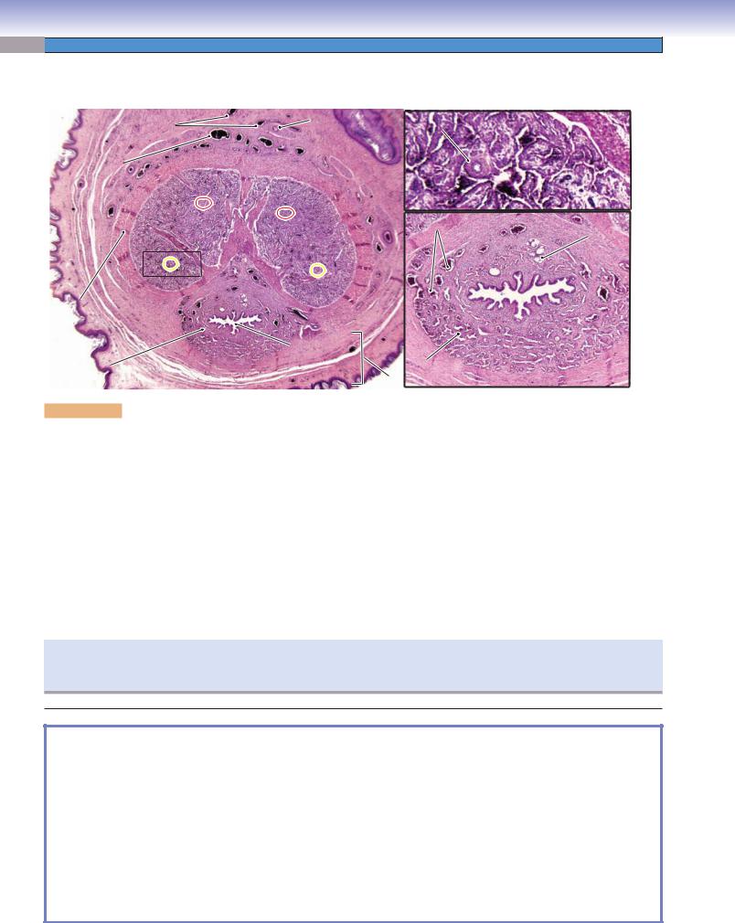
Figure 18-3A. Overview of the testis.
Each testis is composed of many convoluted seminiferous tubules. The anterior portion of the testis is covered by a closed sac of peritoneum called the tunica vaginalis (mesothelial sac). The tunica albuginea is a thick layer of capsule (dense connective tissue) that surrounds and divides the testis into small lobules; the connective tissue continues at the posterior part and becomes thicker and forms the vertically oriented connective tissue mass called the mediastinum. The mediastinum contains the rete testis, which consists of a labyrinth of small channels that collect sperm from the tubuli recti. The ductus epididymis is a single long, highly convoluted duct that receives sperm from the ductuli efferentes. The epididymis is divided into three parts: the head, body, and tail. The tail of the epididymis connects with the ductus deferens. The testes play important roles in the production of sperm and secretion of testosterone (sex hormone).
B
Interstitial connective
Septum
Lumen of the
tissue
Basement
seminiferous tubules
membrane
Tubuli recti
Figure 18-3B. Seminiferous tubules of the testis. H&E,
122
Seminiferous tubules are the main functional components of the testis. Each of the several hundred seminiferous tubules in each testis is a highly coiled tubule lined by a stratified germinal (seminiferous) epithelium containing various stages of spermatogenic cells. The seminiferous epithelium is supported by the basement membrane. Cross sections of some seminiferous tubules and a connective tissue septum are shown here. The tubuli recti (straight tubules) are located in the connective tissue septa. The connective tissue between neighboring tubules, which contains small vessels and endocrine cells, is called interstitial connective tissue.
Septum
C
Myoid
cells
Seminiferous
tubule
Basement membrane
Seminiferous tubule
Lumen
Seminiferous
epithelium
Fibroblast
Figure 18-3C. Seminiferous tubule. H&E, 281
This is an example of a single seminiferous tubule, consisting of germinal epithelium and its basement membrane. The seminiferous tubule is surrounded by a very thin connective tissue containing a few fibroblasts. Another type of cell, the myoid cell, has the appearance of smooth muscle cells, with flat and elongated nuclei. These cells surround the seminiferous tubules and contract to help the movement of the testicular fluid in which the spermatozoa are suspended. Neighboring seminiferous tubules are in close contact with one another. The various stages of the spermatogenic cells include spermatogonia, spermatocytes (primary and secondary), and spermatids (early, intermediate, and late). They are present in six different specific combinations of cell types that define the stages of the cycle of the seminiferous epithelium (Figs. 18-11A to 18-13B).
350 UNIT 3 ■ Organ Systems
Interstitial connective
Figure 18-4A.
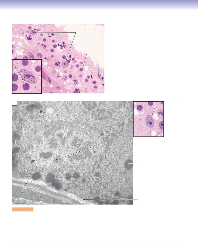
Cells in the seminiferous tubules. H&E, 458
tissue
Interstitial cell
Spermatogonia
of Leydig
Primary
Fibroblast
spermatocyte
Early
Myoid
Intermediate Spermatids
cell
Late
Sertoli
A
cells
There are two types of cells in the seminiferous tubules: spermatogenic (germ) cells and supporting (Sertoli) cells. The spermatogenic cells consist of
1.Spermatogonia: These cells have round or oval nuclei and are located near the basement membrane. They can be subdivided into type A and type B cells. Type A cells are stem cells that divide slowly and give rise to type B cells.
2.Spermatocytes: These derivatives of spermatogonia B cells undergo meiosis. They move toward the lumen and can be divided into primary (first meiotic division) and secondary (second meiotic division) spermatocytes. Primary spermatocytes in prophase are most commonly seen in sections. Their large nuclei contain strands of condensed chromosomes. Secondary spermatocytes complete the second meiotic division very quickly, so they are rarely seen.
3.Spermatids: These cells have small interphase nuclei that range from spherical to thin and elongated. They can be classified as early, intermediate, or late spermatids, based mainly on the appearance of the nucleus.
Late spermatids
Early spermatids
Primary spermatocytes
SpermatogoniumSertoli cells
Basement Sertoli cells membrane
Sertoli cell nuclei
B
Figure 18-4B. Seminiferous epithelium. EM, 4,600
This low magnification view shows almost the full thickness of the seminiferous epithelium. Sertoli cells are the nongerm cells that organize the epithelium and divide it into two compartments, basal and adluminal (Fig. 18-12A). The only cell type in the basal compartment is the spermatogonium, which, like the Sertoli cells, contacts the basement membrane. This patch of epithelium appears to be in stage one of the six stages (cell associations) of seminiferous epithelium. The adluminal compartment contains three cohorts of cells, each at a different stage of spermatogenesis. The least advanced cells are primary spermatocytes in prophase of meiosis I. The chromosomes of these diploid cells have begun condensing, and homologous pairs have aligned into synaptonemal complexes. Early spermatids, which appear as small, undifferentiated cells, predominate in the middle and superficial regions of the epithelium. The third and most advanced cell type here is the late spermatid. The heads of a few can be seen surrounded by the cytoplasm of a Sertoli cell.
CHAPTER 18 ■ Male Reproductive System
351
Nucleus of
Centrioles
early spermatid
Nucleus of
early spermatid Nucleus of early spermatid
Golgi complex
Figure 18-5. Seminiferous epithelium, early spermatid. EM, 17,000
Completion of meiosis II by secondary spermatocytes produces spermatids. These are haploid cells that do not divide but undergo spermiogenesis, that is, morphological differentiation into spermatozoa. The cells in this image are early spermatids that have not yet acquired many of the specializations of spermatozoa. The interphase nucleus in the center is still spherical, and the chromatin is not yet highly condensed. The Golgi complex, visible in the cell on the left, will be the site of development of the acrosomal vesicle and, ultimately the acrosome, which will form a cap on one side of the nucleus. Note that the plane of section happens to pass through the central cell’s centrosome with its pair of centrioles. One member of the pair will organize the development of the flagellum with its axoneme of microtubules. The other centriole will participate in the first cleavage division if the spermatozoan fertilizes a secondary oocyte. This spermatid is linked to its cohort spermatids by cytoplasmic bridges, although this is not evident in this image. Like all cells engaged in spermatogenesis, spermatids are embedded in cytoplasmic processes of Sertoli cells.
352
UNIT 3 ■
Organ Systems
A
Figure 18-6A.
Sertoli cells, seminiferous tubules.
Primary spermatocyte
Early spermatid
Sertoli cell
Sertoli cells
Early spermatid
Late spermatid
H&E, 732; inset 1,603
Sertoli cells have pale oval or irregularly shaped nuclei, and nucleoli are often present. They are irregular columnar cells with many folded cytoplasmic processes forming compartments for the spermatogenic cells. They form tight junctions (zonulae occludentes) with the neighboring Sertoli cells thereby providing a bloodtestis barrier to protect spermatogenic cells from being harmed by autoimmune reactions. Sertoli cells control hormones, nutrients, and other substances passing through the compartments and maintain the ideal environment for spermatogenesis. They play important roles in supporting, protecting, and nourishing spermatogenic cells as well as in secreting testicular fluid (rich in fructose) to help in transporting spermatozoa out of the seminiferous tubules. They also secrete
ABP, anti-Müllerian hormone, and inhibin and activin hormones.
Sertoli cells, the only somatic cells of the seminiferous epithelium, are dynamic cells with a long list of functions in support of spermatogenesis. Their processes envelope and support the germ cells throughout the many stages of meiosis and spermiogenesis. In this view, cytoplasmic extensions of the Sertoli cell (with its nucleus at the left edge) enshroud the primary spermatocyte that has entered prophase of meiosis I and isolate it from the basal compartment so that it is no longer accessible to the immune system. Note also the example of the junctional complexes (including tight junctions) that couple adjacent Sertoli cells and, thereby, establish a controlled and specialized environment in support of the cells that are undergoing spermatogenesis. Other functions of Sertoli cells are secretion of testicular fluid, concentration of androgens, and phagocytosis of the residual bodies jettisoned by late spermatids as they complete spermiogenesis.
CHAPTER 18 ■ Male Reproductive System
353
Seminiferous
tubule
Leydig
cells
Seminiferous
tubule
A
Basement membrane of the
seminiferous tubule
Leydig
cells
C
Lipid vacuoles of Leydig cell
Fibroblast
Lumen of
capillary
Leydig cell
B
Figure 18-7. Interstitial cells of Leydig. H&E, left 263; right
2,016; iron hematoxylin stain, 237
The interstitial cells of Leydig are located in the interstitial (connective) tissue near the blood capillaries and between the seminiferous tubules. These cells have round nuclei and pale-stained cytoplasm with lipid drops (lipid vacuoles) in the peripheral region of the cytoplasm, which give them a bubbly appearance (like many steroid-producing cells). These cells contain abundant smooth endoplasmic reticulum, which contributes to steroid hormone production. The interstitial cells of Leydig derive from the mesoderm and are usually large in size (about 20 μm in diameter) compared with other cells in the connective tissue. They are the endocrine cells that produce the important male sex hormone, testosterone. Testosterone plays important roles in developing and maintaining male sex characteristics, stimulating muscle and bone growth, and increasing bone density. A small amount of testosterone is also produced by the adrenal glands and ovaries in the female.
SYNOPSIS 18 - 1 Functions of Sertoli Cells
■Support: Provide physical support and nutrition for the different stages of spermatogenic cells.
■Protection: Form blood-testis barrier by tight junctions between adjacent Sertoli cells that protect spermatogenic cells from autoimmune destruction; also control hormones, nutrients, and other substances being transported in and out of the seminiferous tubules.
■Phagocytosis: Remove residual bodies after excess cytoplasm is shed from the spermatids during maturation of the spermatozoa.
■Secretion: (1) Secrete and release fructose-rich fluids (testicular fluid) to help nourish and move sperm from the seminiferous tubules to the epididymis; (2) secrete anti-Müllerian hormone to prevent oviducts from developing from the Müllerian duct in the early stages of the male embryo; (3) secrete androgen-binding protein (ABP) to maintain the concentration of testosterone in the seminiferous tubules, thereby promoting spermatogenesis; (4) secrete glial cell–derived neurotrophic factor (GDNF) to promote survival and differentiation of the spermatids (GDNF is better known for promoting development of neurons); and (5) produce inhibin and activin hormones to provide negative and positive feedback to the hypothalamus, thereby regulating follicle-stimulating hormone (FSH) secretion by the pituitary gland.
SYNOPSIS 18 - 2 Functions of Testosterone
The interstitial cells of Leydig secrete testosterone, which is the major male sex hormone. Its functions include:
■Promoting development of male sex organs in early fetal development.
■Promoting male sexual characteristics, such as growth of beard and axillary hair, enlargement of the larynx, and deepening the voice.
■Increasing muscle growth, thickness of the skin, and sebaceous gland secretion.
■Promoting bone growth and increasing bone density.
■Increasing basal metabolism and physical energy.
■Promoting spermatogenesis.
354 UNIT 3 ■ Organ Systems
Hypothalamus
(–)
GnRH(+) LH
GnRH(+) FSH
Pituitary gland
(–)
gonadotrophs
)
LH
(gonadotrophs
(+)
FSH
(+)
)
(
Testosterone
(+)
Interstitial (Leydig) cell
Anti-Müllerian
hormone
Supports development of
male sex organs and
maintains male sexual
characteristics;
Inhibits female organ
promotes spermatogenesis
development
( Inhibin
)–
Activin
(+)
Inhibin & activin hormones
ABP
Sertoli cell
(+)
Increases
concentration
of testosterone
Promotes
spermatogenesis
D. Cui
Figure 18-8. Hormone regulation involving the testicular cells (interstitial cells of Leydig and Sertoli cells). H&E, 1,005
The preoptic nucleus in the hypothalamus secretes gonadotropin-releasing hormone (GnRH), which stimulates the pituitary gland to produce and release luteinizing hormone (LH) and FSH. Secretion of testosterone by the interstitial cells of Leydig is stimulated by the LH produced by the gonadotrophs in the adenohypophysis of the pituitary gland. An excessive level of testosterone sends negative feedback to the hypothalamus to inhibit production of GnRH, resulting in decreased secretion of LH in the pituitary gland. Sertoli cells that release ABP are stimulated by FSH, which stimulates production of the ABP. Sertoli cells also secrete anti-Müllerian hormone as well as inhibin and activin hormones. The inhibin hormone suppresses and activin hormone stimulates the production of GnRH, which influences FSH production by the gonadotrophs in the adenohypophysis of the pituitary gland.
CHAPTER 18 ■ Male Reproductive System
355
1. Spermatogonium
2. Prepachytene primary
3. Pachytene primary
4. Metaphase primary or
spermatocyte
spermatocyte
secondary spermatocyte
J. Naftel
8. Detaching spermatozoan
7. Late spermatid
6. Intermediate
5. Early spermatid
spermatocyte
J. Naftel
Figure 18-9. Overview of readily identifiable spermatogenic cells in the seminiferous epithelium. H&E, 1,496
Spermatogenesis involves an orderly sequence of changes as cells proceed through mitosis (spermatogonia), meiosis (spermatocytes), and spermiogenesis (spermatids). Only the eight most readily identifiable stages are illustrated here owing to their distinctiveness and adequate abundance in ordinary sections. Spermatogonia can be identified by their position (in contact with the basement membrane) and their oval interphase nuclei with the long axis parallel to the basement membrane. Primary spermatocytes undergo several substages of prophase I, but, here, the earlier substages are denoted as prepachytene. The condensing chromosomes in prepachytene primary spermatocytes tend to be clumped at one edge of the nuclear envelope. By contrast, the paired chromosomes of pachytene primary spermatocytes form broad bands that fill the nucleus. Pachytene primary spermatocytes are abundant in sections, because this is the phase of meiosis with the longest duration. After a primary spermatocyte completes pachytene, the remaining steps of meiosis I and meiosis II (by the daughter secondary spermatocytes) are completed very rapidly, so examples of cells in these stages of meiosis are more difficult to find in sections. Cells in either metaphase I or metaphase II are most conspicuous and easiest to locate. The products of meiosis II are spermatids. These haploid cells begin as early spermatids, recognizable by their small spherical interphase nuclei. The first clear evidence that spermiogenesis is underway is a change in shape of the nucleus from spherical to a broad diamond shape; a cell with such a nucleus can be designated an intermediate spermatid. When the nucleus has assumed the sharply pointed shape and dense appearance of a spermatozoan but remains inserted deeply into the epithelium, the cell can be denoted a late spermatid. Finally, nuclei of detaching spermatozoa can readily be recognized, because they form a row at the surface of the epithelium and overlie a layer of residual bodies.
356 UNIT 3 ■ Organ Systems
I
II
III
IV
V
VI
I
Detaching spermatozoan with residual body
Intermediate
spermatid
Late spermatid
Early spermatid
Meiotic metaphase
spermatocyte
InterInterphaseaseprimaryprimary
Prepachytene
Pachytene primary
sperspermatocytetocyte
primary spermatocyte
spermatocyte
B
B
A
A
A
A
type B
type A
spermatogonium
spermatogonium
4.8 days’
3.1 days’
1.0 days’
1.2 days’
5.0 days’
0.8 days’
duration
duration
duration
duration
duration
duration
J. Naftel
Figure 18-10. Overview of spermatogenesis and the stages of the seminiferous epithelium.
If small patches of seminiferous epithelium are examined, it can be seen that there are six recurring groupings of stages of developing gametes that populate the epithelium. These groupings (associations) represent recurring stages that a patch of epithelium cycles through over time, and their existence indicates that the many steps in spermatogenesis proceed with a very rigid timetable. If the composition of a bit of epithelium could be monitored over time, it would be seen that, as cohorts of germ cells begin and complete spermatogenesis within the epithelium, the epithelium itself will change in appearance and contain the six different groupings of developing germ cells. The ability to recognize the stages in the cycle of the epithelium has made it possible to undertake simple experiments that have revealed some important dynamics of spermatogenesis. Therefore, the time required for a patch of epithelium to complete the six stages of one cycle is 16 days, and it can be inferred that with each 16-day cycle a single patch of epithelium will release a group of new spermatozoa for only a brief period when it reaches the end of stage II. Likewise, the end of stage II is the only time during the 16-day cycle that a new cohort or generation of primary spermatocytes will enter the adluminal compartment to begin meiosis I. The time required for a new type B spermatogonium to generate completed spermatozoa is 56 days.
CHAPTER 18 ■ Male Reproductive System
357
A
Late spermatid
Early spermatid
Pachytene
spermatocytes
in the epithelium
Sertoli cell
Spermatogonia
Basement
membrane
Figure 18-11A. Stage I seminiferous epithelium. H&E, left 207; right 747
Stage I seminiferous epithelium is shown at low magnification (left), and a detailed representation of the region is indicated by the box (right). Stage I seminiferous epithelium is characterized by the following: (1) Spermatogonia are located at the basement membrane; both type A and B spermatogonia are present, although the two types are not easily distinguished in H&E stained sections.
(2) A layer of pachytene primary spermatocytes is present. Although some of these cells are near the basement membrane, they are not in direct contact with it. (3) A prominent layer of round, undifferentiated early spermatids has moved toward the surface of the epithelium. (4) The late spermatids have their heads inserted deep into the epithelium.
B
DetachingLate spermatid spermatozoaon the surface onf theepitheliumthe surface of the epithelium
Pachytene primary spermatocyte
Residual
bodies
Sertoli
cell
Spermatogonium
Early spermatid
Figure 18-11B. Stage II seminiferous epithelium. H&E, left 207; right 747
Stage II seminiferous epithelium is shown at low magnification (left), and a detailed representation of the region is indicated by the box (right). The composition of the spermatogenic cell in stage II seminiferous epithelium is similar to that of stage I. The most characteristic features of this stage are the following: (1) Detaching spermatozoa (very late spermatids) have completed differentiation and are in the process of shedding their excess cytoplasm. (2) The small leftover cytoplasmic masses that have been shed are called residual bodies and are present in the luminal border of the epithelium, beneath the heads of the detaching spermatozoa.
(3) These detaching spermatozoa are positioned in a row on the surface of the epithelium ready to be released from the epithelium into the lumen. (4) Early spermatids with round nuclei and pachytene primary spermatocytes are present. (5) Both type A and B spermatogonia are present.
358 UNIT 3 ■ Organ Systems
A
Adluminal
compartment
Basement membrane
Basal compartment
Spermatogonium
Figure 18-12A. Stage III seminiferous epithelium. H&E, left 207; right 747
Lumen
Early sperspermatidatids
Pachytene primary sperspermatocyteatocytes
Sertoli cell
Stage III seminiferous epithelium is shown at low magnification (left), and a detailed representation of the region is indicated by the box (right). Stage III has a similar composition of spermatogenic cells as that of stage II, except for the absence of the detaching spermatozoa at the surface and the added presence of a new generation of primary spermatocytes near the basement membrane (not readily apparent). This stage is characterized by the following: (1) There is an absence of detaching spermatozoa, which have been released into the lumen to be suspended in testicular fluid and transported along the genital ducts. (2) Early spermatids with round nuclei have moved to the surface of the epithelium. (3) There is the continued presence of a generation (cohort) of primary spermatocytes in the pachytene stage of prophase I. (4) A new generation of primary spermatocytes has moved from the basal compartment into the adluminal compartment, although they are difficult to distinguish. Compartments are created by tight junctions of neighboring Sertoli cells. The basal compartment is situated on the basement membrane side of the tight junctions between Sertoli cells, and it contains spermatogonia. The adluminal compartment is situated above the junctions and toward the lumen and contains spermatocytes and spermatids. The tight junctions establish a blood-testis barrier that prevents immunoglobulins from entering the adluminal compartment. Newly formed primary spermatocytes are lifted from the basal compartment and into the adluminal compartment by outgrowth of processes extended from the local Sertoli cells.
B
Intermediate Lumen spermatids
Prepachytene primary spermatocyte
Pachytene
primary Spermatogonium spermatocytes
Sertoli cell
Figure 18-12B. Stage IV seminiferous epithelium. H&E, left 207; right 747
Stage IV seminiferous epithelium is shown at low magnification (left), and a detailed representation of the region is indicated by the box (right). Stage IV differs from stage III mainly in the progression of the generation of early spermatids (in stage III) to intermediate spermatids (in stage IV). Stage IV has the following composition: (1) Intermediate spermatids with diamond-shaped or irregular nuclei;
(2) one generation of primary spermatocytes in pachytene of prophase I; (3) a separate generation of primary spermatocytes at an early (prepachytene) stage of prophase I; and (4) Type A spermatogonia in the basal compartment.
CHAPTER 18 ■ Male Reproductive System
359
A
Late spermatids
Prepachytene primary spermatocyte
Pachytene
Basement
primary
Spermatogonium
membrane
spermatocyte
Figure 18-13A. Stage V seminiferous epithelium. H&E, left 207; right 747
Stage V seminiferous epithelium is shown at low magnification (left), and a detailed representation of the region is indicated by the box (right). In this example, stage V has newly developed late spermatids and two generations of primary spermatocytes. Stage V is characterized by the following: (1) There are no early or intermediate spermatids; only late spermatids are present. The cytoplasm of each late spermatid extends from the sharply pointed nucleus toward the lumen. (2) Two generations (both pachytene and prepachytene) of primary spermatocytes are present. (3) Type A spermatogonia are present, as always, in the basal compartment.
B
Spermatogonium
Secondary spermatocytes (in metaphases)
Lumen
Primary
spermatocytes
Figure 18-13B. Stage VI seminiferous epithelium. H&E, left 207; right 747
Stage VI seminiferous epithelium is shown at low magnification (left), and a detailed representation of the region is indicated by the box (right). The most characteristic feature of stage VI is the presence of metaphases of either primary or secondary spermatocytes, which are present only in this stage. Interphase secondary spermatocytes also occur in stage VI, but these are rarely apparent. The characteristics at this stage are as follows: (1) Late spermatids are present at the epithelium near the lumen. (2) Dividing spermatocytes with their chromosomes clumped in the center of the cytoplasm. (3) A separate generation of primary spermatocytes in prophase is present, although they are not easily distinguished. (4) Type A spermatogonia are present. This is a very active stage during which each member of the older cohort of primary spermatocytes completes the first meiotic division to produce two secondary spermatocytes, and, in a very short time, each secondary spermatocyte completes the second meiotic division to produce two spermatids.
The series of ducts which transport spermatozoa from the seminiferous tubules to the outside of the male body are called genital ducts. They include intratesticular genital ducts and extratesticular genital ducts. The intratesticular genital ducts refer to ducts within the testis. They are composed of the tubuli recti, rete testis, and ductuli efferentes. The extratesticular genital ducts include the ductus epididymis, ductus deferens, ejaculatory ducts, and urethra.
The tubuli recti are short (about 1 mm), straight tubules that carry the spermatozoa from the seminiferous tubules into the rete testis. Rectus means “straight.” Each tubulus rectus is lined by a single layer of cuboidal epithelial cells and is supported by a layer of dense connective tissue. It is located in the septum (connective tissue) and opens into the rete testis in the mediastinum. The terminal portion of the seminiferous tubules is lined by Sertoli cells. The germinal cells in the seminiferous tubules are gradually reduced in number near the tubuli recti and disappear at the terminal portion. The spermatozoa are pushed forward by the flow of the testicular fluid through the tubuli recti and into the rete testis. An example here shows two segments of a tubulus rectus.
C
Tubulus rectus
Rete testis
Lumen of rete testis
Mediastinum
Figure 18-14C. Rete testis. H&E, 35; inset 163
The rete testis is an interconnecting network of tubules located in the mediastinum. These tubules have an irregular lumen and are lined by simple cuboidal epithelium and supported by a dense connective tissue. This example shows the terminal portion of the tubulus rectus opening into the tubule of the rete testis.
CHAPTER 18 ■ Male Reproductive System
361
A
Figure 18-15A.
Ductuli efferentes. H&E, 68; inset 234
There are 15 to 20 tubules of ductuli efferentes leading from the
Ductuli
rete testis in the mediastinum to the head of the epididymis. They
efferentes
are also called efferent ducts. The ductuli efferentes are lined by two
types of epithelial cells: nonciliated cuboidal cells with microvilli
(absorptive cells) and ciliated columnar cells. They are alternately
distributed on the luminal surface of the ductuli efferentes. The epi-
Spermatozoa
thelium of the ductuli efferentes is supported by a thin layer of con-
nective tissue; beneath it is a thin layer of circular smooth muscle
that contracts to move testicular fluid and spermatozoa toward the
ductus epididymis (inset). Smooth muscle cells are interspersed with
some elastic fibers. The muscle layer becomes thicker as it nears the
Artery
ductus epididymis.
B
Ciliated columnar cells
Spermatozoa
Figure 18-15B. Ductuli efferentes. H&E, 463
An example of ductuli efferentes at high magnification is shown. The nonciliated cuboidal cells have an absorptive function, absorbing excess testicular fluid secreted by seminiferous tubules and increasing the concentration of spermatozoa in the lumen of the ductuli efferentes. The ciliated columnar cells of the ductuli efferentes have a motile function, sweeping spermatozoa and testicular fluid toward the epididymis.
Nonciliated cuboidal cells
CLINICAL CORRELATION
C
Lymphocytes
Seminoma
Figure 18-15C.Testis Seminoma. H&E, 192
Most tumors of the testes arise from the germinal epithelium of the seminiferous tubules; therefore, this group is termed germ cell tumors. These germ cell tumors can be divided into seminomas, and all other germ cell tumors are called nonseminomatous, examples of which include choriocarcinoma, embryonal carcinoma, yolk sac tumor, and teratoma. Seminomas are the most common germ cell tumor of the testis and are most likely to occur in the pure form, because germ cell tumors commonly contain multiple tumor types (mixed germ cell tumors). Germ cell tumors tend to arise from in situ lesions of the germinal epithelium called intratubular germ cell neoplasia (IGCN). The incidence of seminoma peaks in the fourth and fifth decades of life. Cryptorchidism and gonadal dysgenesis are risk factors for germ cell tumors. Patients present with painless enlargement of the testis. Grossly, classic seminomas are circumscribed and fleshy. Histologically, classic seminoma consists of sheets of large cells with distinct cell borders and nucleoli, infiltrated with lymphocytes. These cells are typically positive for placental alkaline phosphatase on immunohistochemical preparations. Because of the potential for seeding across fascial planes, biopsy is not recommended. Seminomas are very sensitive to radiation and chemotherapy.
This is an example of the ductus epididymis in the head region. The ductus epididymis is a single highly convoluted tube about 4 to 5 m in length. It connects the ductuli efferentes to the ductus deferens. The ductus epididymis is lined by pseudostratified columnar epithelium with long stereocilia and is supported by connective tissue (lamina propria) and a circular smooth muscle layer in its wall. The ductus epididymis can be divided into three regions: head, body, and tail. The head of the ductus epididymis, also called the initial region, receives spermatozoa from the ductuli efferentes. The spermatozoa in the head of ductus epididymis are weak and unable to function effectively in fertilization. They pass through the body region with the help of contraction of the smooth muscle in the wall of the ductus epididymis and are stored in the tail region, where the spermatozoa undergo maturation. The maturation of the spermatozoa is influenced by androgenic hormones. The final step of maturation is called capacitation and occurs only after the spermatozoa enter the female reproductive tract. Capacitation enables sperm to penetrate and fertilize an egg.
Epididymitis is an inflammation of the epididymis. Common causes include Chlamydia trachomatis, Escherichia coli, and Neisseria gonorrhoeae infections. In the male, urethritis (an inflammation of the urethra) is often associated with epididymitis. Chronic epididymitis may result in infertility, because spermatozoa are stored and mature in the tail of the ductus epididymis.
Pathway for Sperm Conduction through the Genital Ducts
Seminiferous
Tubuli
Rete
Ductus
Intratesticular
Ductuli
genital ducts
tubules
recti
testis
efferentes
epididymis
Extratesticular
genital ducts
Urethral
Penile
Ejaculatory
Ductus
Urethra
Penis
orifice
urethra
ducts
deferens
Bulbourethral
Prostate
Seminal
Accessory
D. Cui
gland
gland
vesicle
genital glands
CHAPTER 18 ■ Male Reproductive System
363
A
Basement
membrane
Pseudostratified columnar epithelium
Fig. 18-17B
Stereocilia
Connective
tissue
Figure 18-17A. Epithelium of the ductus epididymis. H&E, 305
The epithelium of the ductus epididymis is a pseudostratified columnar epithelium composed of basal cells and principal cells. The basal cells are short with round nuclei; they are stem cells and are capable of differentiating into principal cells. The principal cells are columnar cells with elongated nuclei and apical stereocilia (long microvilli, 10 μm or more in length, which function in absorption but have no motile function). They absorb large volumes of testicular fluid. The principal cells also play an important role in the secretion of glycerophosphocholine, which inhibits capacitation of the premature sperm in the male reproductive tract. Normally, capacitation happens only after the sperm enters the female reproductive tract.
B
Head of spermatozoan
Tail of spermatozoan
Middle piece of
spermatozoan
Stereocilia
Junctional complex
Figure 18-17B. Epithelium of the ductus epididymis. EM, 5,900
The view here is limited to part of the lumen and the apical ends of the columnar epithelial cells forming the wall of the ductus epididymis. Junctional complexes between neighboring epithelial cells seal the contents of the lumen from the interstitial compartment underlying the epithelium. The extremely long microvilli (stereocilia) are a reflection of the large amount of testicular fluid that is resorbed by the ductus epididymis. The lumen is the site of storage and final maturation of the spermatozoa that have been delivered to the epididymis by the flow of the testicular fluid. Note the randomly oriented sections through the heads, middle pieces, and tails of spermatozoa in the lumen.
364 UNIT 3 ■ Organ Systems
Ductus
Pampiniform venous
Figure 18-18A.
Ductus deferens, spermatic cord. H&E, 11
deferens
plexus
The ductus deferens (vas deferens) consists of bilateral tubes
that continue from the tails of the left and right ductus
epididymis. Each tube is about 30 to 40 cm long and is
surrounded by a thick wall of smooth muscle. Each ductus
deferens resides within a spermatic cord in its course from
the testis through the abdominal wall to the penis. The distal
portion of the ductus deferens becomes enlarged and is called
the ampulla (Fig. 18-18B). The spermatic cord contains the
The lining of the ductus deferens, like that of the epididymis, is pseudostratified columnar epithelium. The ductus deferens is characterized by fingerlike mucosal folds extending into the lumen. The thick muscularis contains inner longitudinal muscle, middle circular muscle, and outer longitudinal muscle, although these muscle layers are not easy to distinguish from one another. The outer longitudinal muscle is covered by an adventitia (connective tissue layer). The smooth muscle of the ductus deferens is richly innervated by postganglionic sympathetic nerve fibers, which initiate ejaculation when activated. The inset shows pseudostratified columnar epithelium with stereocilia of the ductus deferens.
The ampulla of the ductus deferens continues after it joins with the duct of the seminal vesicle to form the ejaculatory duct. The two ejaculatory ducts pass through the prostate gland to join with the urethra. Each ejaculatory duct is a short, straight tube (1–2 cm in length) and has a thin wall lined by pseudostratified (or simple) columnar epithelium and supported by connective tissue. Smooth muscle is present in the initial segment but disappears in most of the ejaculatory ducts. Here is an example of the two ejaculatory ducts within the prostate gland, surrounded by large amounts of connective tissue. The mucosa forms many folds extending into the lumen. The lumen may contain prostatic concretions (secretory material of the prostatic gland and often seen in older male patients).
The ejaculatory ducts penetrate the prostate gland and open into the prostatic urethra, at the seminal colliculus (also called the verumontanum), on the posterior wall of the prostatic urethra. This portion of the urethra has thick mucosa and shallow folds as shown here. The function of the ejaculatory ducts is to transport spermatozoa and seminal fluid into the prostatic urethra. The urethra includes three parts: the prostatic urethra (proximal part, near the bladder), the membranous urethra (intermediate part), and the penile (spongy) urethra (distal part). Prostatic concretions, also called corpora amylacea, are present in the lumen of the prostate gland shown here (Fig. 18-20B).
C
Transitional
epithelium
Prostatic
urethra
Duct
of
prostate
gland
Duct of prostate gland
Simple columnar epithelium
Figure 18-19C. Epithelium of the prostatic urethra. H&E,
272
Where the two ejaculatory ducts merge with the prostatic urethra, the epithelium changes from simple or pseudostratified columnar to the transitional epithelium that is characteristic of the urinary system. Here is an example of prostatic urethral epithelium at higher magnification; it is taken from the dashed box indicated in Figure 18-19B. The epithelium of the duct of the prostate gland is simple columnar epithelium with round nuclei. The prostatic secretions are delivered into the prostatic urethra through numerous small ducts of the prostate gland.
366 UNIT 3 ■ Organ Systems
Accessory Genital Glands
A
Prostatic
concretions
Lumen
Figure 18-20A. Prostate gland. H&E, 34
The prostate gland is similar to a chestnut in size and shape. It surrounds the initial portion of the urinary urethra (prostatic urethra) where the urethra exits the bladder. The prostate gland is penetrated by two ejaculatory ducts and the urethra (Fig. 18-19A–C). It contains many (about 30–50) highly branched tubuloalveolar glands (compound tubuloalveolar glands). Each gland has a duct that empties its
Stroma products into the prostatic urethra. The mucosa of the prostate gland is highly folded and is lined by simple columnar epithelium, which is
supported by a stroma (thin layer of connective tissue strands with many smooth muscle cells). Here is an example of the prostate gland with its characteristically irregular lumen that may contain prostatic concretions. These concretions are also known as corpora amylacea and are more prominent in older men; they are composed of material secreted by the prostate gland.
B
Lumen of the prostate gland
Connective
tissue Columnar epithelium
Prostatic concretion (corpora amylacea)
Figure 18-20B. Prostate gland. H&E, 272
A lumen in the prostate gland is shown housing a prostatic concretion
(corpora amylacea). This is composed of calcified prostatic secretions that typically display concentric rings. These structures increase in number with age. The secretion of the prostate gland contains proteolytic enzymes, acid phosphatase, citric acid, fibrinolysin, and lipids. The epithelial cells are columnar in shape with basally located round nuclei. The prostate gland produces secretions that empty into the urethra to mix with spermatozoa and seminal vesicle fluid to form semen. The prostatic secretion plays important roles in liquefying the coagulated semen, helping to expel the spermatozoa, and increasing their motility and survival rate after the semen has been transported into the female reproductive tract.
CLINICAL CORRELATION
Note the prominent nucleoli (malignant cells)
C
Prostate cancer
Figure 18-20C.Prostate Cancer. H&E, 96; inset 164
Prostate cancer is the most common cancer in men and typically affects men over the age of 50 years. It can be seen in younger men but is unusual before the age of 40. The etiology of prostate cancer is elusive, but known risk factors include a positive family history, African American race, androgenic hormonal influences, and environmental factors. The majority of prostate cancers are adenocarcinomas arising from the glandular component of the prostate. Patients may present with urinary symptoms, such as difficulty initiating or stopping the urine stream, or dysuria (pain on urination). Other patients may first present with bone pain due to advanced metastatic disease. Many patients are diagnosed with prostate cancer through screening programs utilizing the digital rectal exam and the serum prostate-specific antigen (PSA) test, and a needle biopsy if indicated. Histologically, the appearance of prostate cancer is highly varied, from well-formed tubular structures to individual infiltrating malignant cells. Prostate cancers are graded histologically on the Gleason system, from 1 (well differentiated) to 5 (poorly differentiated). Treatment of prostate cancer may involve chemotherapy, hormonal manipulation, radiation therapy (external beam and radioactive implants), or radical prostatectomy. For some patients, particularly the elderly, watchful waiting may be a reasonable alternative.
CHAPTER 18 ■ Male Reproductive System
367
A
Folded mucosa
Lumen
Lumen
Muscularis
Figure 18-21A. Seminal vesicles. H&E, 11
The seminal vesicles are paired glands that develop from the ductus deferens. Each seminal vesicle consists of a single highly convoluted tube with a duct that connects to the terminal portion (ampulla) of the ductus deferens. The ampulla of the ductus deferens is continuous with the ejaculatory duct. The mucosa of the seminal vesicles is extensively branched and folded and lined mostly by pseudostratified columnar epithelium. The epithelium is supported by a thin layer of connective tissue (lamina propria), and beneath it is the muscularis composed of inner circular and outer longitudinal smooth muscle. Contraction of the muscularis pushes the seminal secretion into the ejaculatory duct during ejaculation.
B
Epithelium
Lamina propria
Pseudostratified columnar epithelium
Smooth
muscle
Figure 18-21B. Seminal vesicle. H&E,
278; inset 635
This is an example of the mucosa of the seminal vesicle. The nonciliated, pseudostratified columnar epithelium (inset), underlying lamina propria, and some smooth muscle fibers are shown here. The epithelium of the seminal vesicle varies from simple to pseudostratified columnar epithelium. The mucosa appears branched and folded. The epithelium contains basal cells and secretory cells with abundant rough endoplasmic reticulum and well-developed Golgi complexes.
The seminal vesicles produce large volumes of seminal fluid, which contributes about 70% of the volume of semen. Seminal fluid contains fructose and other sugars, prostaglandins, flavins, phosphorylcholine, mucus, vitamin C, and proteins. The fructose provides an energy source for sperm motility; the flavins, also known as lipochrome pigment, add a yellowish color to the seminal fluid and have a strong fluorescent quality under the ultraviolet light. The inset shows seminal epithelium and seminal fluid (viscous material) filling the lumen of a seminal vesicle.
368 UNIT 3 ■ Organ Systems
Penis
Superficial
Dorsal
Helicine artery
artery
dorsal vein
Deep dorsal vein
Corpora
Sinuses (vein)
Urethral glands
cavernosa
(Littré glands)
Corpora
cavernosa
Tunica
Urethra
albuginea
Corpus
Urethra
Erectile
spongiosum
Skin
tissue
Figure 18-22. Overview of the penis. H&E, 13; inset (upper) 67; inset (lower) 26
The penis is composed of three cylinders of erectile tissue: the two corpora cavernosa and the corpus spongiosum as shown on the left. Each cavernosa is surrounded by a tunica albuginea (thick and dense connective tissue); the corpus spongiosum is surrounded by a thin layer of connective tissue with some smooth muscle fibers. The penile (spongy) urethra is enclosed in the center of the corpus spongiosum and extends into the terminal end (glans) of the penis. Each cylinder contains erectile tissue composed of a trabecular network of veins (sinuses) surrounded by collagen, elastic fibers, and smooth muscle cells. The three cylinders, with their tunica albuginea, are surrounded by hairless thin skin containing arteries, veins, nerves, and connective tissue and covered by stratified squamous epithelium. The two red circles indicate the position of the deep arteries; the yellow circles indicate the position of the helicine arteries in the corpora cavernosa.
The penis is supplied by the dorsal arteries, and blood drains into the dorsal veins. A special blood vessel arrangement called an arteriovenous (A-V) shunt allows blood to flow directly from arteries to veins. In the erect state, the arteriovenous shunt closes, which results in blood being forced from the helicine arteries into the sinuses (cavernous spaces) in the erectile tissue (right upper inset). Dilation of the sinuses produces penile erection. Erection is activated by parasympathetic stimulation through the spinal nerves and the sacral parasympathetic preganglionic motor neurons in the spinal cord (S2–S4). The right lower photomicrograph shows the penile urethra surrounded by erectile tissue and sinuses (veins) filled with blood. The urethral glands (Littré glands) are mucous glands in the submucosa of the penile urethra, also called paraurethral (periurethral) glands. The secretion of the urethral glands lubricates the urethra and contributes to the semen during ejaculation.
A clinical condition called erectile dysfunction is characterized by the inability to produce or maintain an erection of the penis. This happens due to insufficiency of dilation of the sinuses in the erectile tissue. The causes are varied, including hormonal disorders, neurological problems, hypertension, psychological factors, smoking, and alcohol use.
SYNOPSIS 18 - 3 Clinical and Pathological Terms for the Male Reproductive System
■IGCN: Noninvasive intratubular or in situ lesions within the seminiferous tubules that give rise to the majority of adult germ cell tumors of the testis; IGCN is often found adjacent to testicular germ cell tumors on histologic examination (Fig. 18-15C).
■Cryptorchidism: Lack of, or incomplete, descent of a testis, from the abdominal cavity into the scrotum; the testis may remain intra-abdominal or be found in the inguinal canal; an undescended testis is a risk factor for the development of testicular tumors (Fig. 18-15C).
■Gonadal dysgenesis: Abnormal development of the gonads with resultant alterations in sexual development; gonadal dysgenesis is a risk factor for the development of testicular tumors; the underdeveloped gonad is often referred to as a “streak gonad” (Fig. 18-15C).
■PSA: A protein synthesized by prostatic epithelial cells; elevated serum PSA levels are associated with benign processes such as benign prostatic hypertrophy as well as adenocarcinoma of the prostate; the PSA test is used as a screening test for prostate cancer as well as a tumor marker in patients with a history of prostate cancer who have received treatment (Fig. 18-20C).
19 Female Reproductive
System
Introduction and Key Concepts for the Female Reproductive System
Figure 19-1
Overview of the Female Reproductive System
Figure 19-2
Orientation of Detailed Female Reproductive Organ Illustrations


 Late spermatids
 Early spermatids
 Chromosomes in nucleus of primary spermatocyte
 Nucleus of Sertoli cell
 Cytoplasm of Sertoli cell
 capillary


 compartment


 C
C Lymphocytes
 Seminoma
 deferens
 Head of spermatozoan
 Tail of spermatozoan
 spermatozoan
 Stereocilia
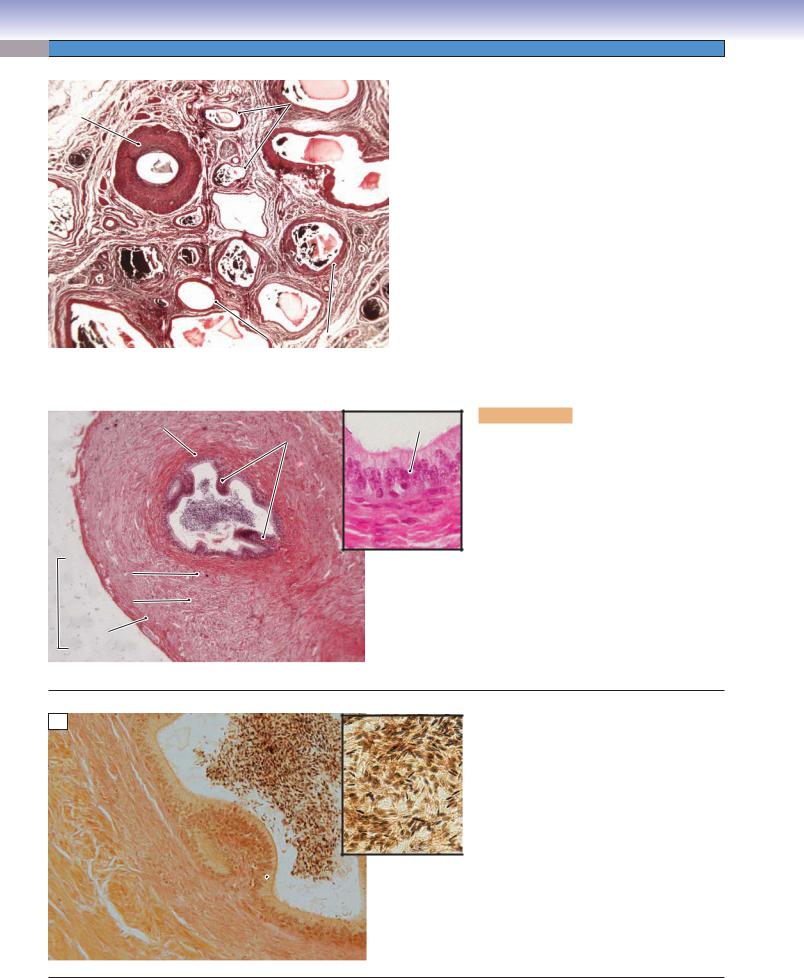
 Epithelium
 concretions
 nucleoli (malignant cells)
 C
C
 mucosa