


180 |
UNIT 3 ■ |
Organ Systems |
|
|
|
|
(Fig. 10-4A,B). They will die if they do not meet an antigen. |
and a Golgi apparatus in the cytoplasm (see Figs. 4-2 and 4-3). |
|||
|
Naive T cells migrate from the thymus to secondary lymphoid |
They actively produce antibodies known as immunoglobulins |
|||
|
organs where they encounter foreign antigens and become active |
(Igs), which are specific for each type of antigen (Fig. 10-3). |
|||
|
T cells. Once T cells are activated, they can boost the action of |
|
|
||
|
cytotoxic T cells and macrophages and help to expedite pro- |
Antigen-Presenting Cells |
|
||
|
liferation of B lymphocytes, which increase the production of |
These cells present antigens to lymphocytes. Most of them are |
|||
|
antibodies (see Fig. 10-5A). Activated T cells undergo cell divi- |
||||
|
MHC-II class, which have surface membrane molecules MHC-II |
||||
|
sion to become memory T cells or effector T cells. |
||||
|
(histocompatibility complex). These cells present antigen to |
||||
|
|
|
|
||
|
MEMORY T CELLS have a much longer life than naive (virgin) |
T cells (Fig.10-4B). Antigen-presenting cells include mac- |
|||
|
rophages, dendritic cells, Langerhans cells, and B cells. In gen- |
||||
|
T cells. They can survive for a long period in an inactive state |
||||
|
eral, B cells are both antigen-presenting and antigen-receiving |
||||
|
and can differentiate into effector T cells to participate in a stron- |
||||
|
cells. They present antigens to T cells and also receive antigens |
||||
|
ger and faster secondary immune response when they encounter |
||||
|
by either binding antigen to their receptors or through antigen- |
||||
|
the same antigen for the second time. Memory T cells include |
||||
|
presenting cells (follicle dendritic cells). Lymphocytes are acti- |
||||
|
central memory T cells and effector memory T cells. Central |
||||
|
vated after receiving an antigen. |
|
|||
|
memory T cells express CCR7 (chemokine receptor) surface |
|
|||
|
|
|
|||
|
molecules and secrete interleukin-2 (IL-2) that stimulates B cells |
Lymphatic Tissues and |
|
||
|
to proliferate. They reside in secondary lymphoid organs, such |
|
|||
|
as the paracortex of the lymph nodes, and are capable of dif- |
Lymphoid Organs |
|
||
|
ferentiating into effector memory T cells. Effector memory T |
|
|||
|
cells do not express CCR7 surface molecules but secrete IL-4 |
|
|
||
|
Mucosa-Associated Lymphatic Tissues |
|
|||
|
(to stimulate B cells and increase immunoglobulin G [IgG] and |
Diffuse lymphatic tissues or nodules are often located in the con- |
|||
|
IgM). They often migrate to an inflammatory site and develop |
||||
|
nective tissue, which support the wet epithelial membranes of |
||||
|
into effector T cells. |
|
|||
|
|
the body mucosae. The lymphatic tissues found in the mucosa |
|||
|
|
|
|
||
|
EFFECTOR T CELLS include helper T |
cells, cytotoxic T |
of the digestive, respiratory, and genitourinary tracts are called |
||
|
mucosa-associated lymphatic tissues (MALT). They can |
be |
|||
|
cells, and |
regulatory (suppressor) T cells. |
(1) Helper T cells |
||
|
subdivided into gut-associated lymphatic tissue (GALT) |
and |
|||
|
have the |
surface marker CD4, which |
restricts activation |
||
|
bronchus-associated lymphatic tissue (BALT), according to their |
||||
|
to antigens only if it is presented by another cell in associa- |
||||
|
locations. GALT is found in the digestive tract, such as Peyer |
||||
|
tion with major histocompatibility complex (MHC) class II. |
||||
|
patches in the ileum and lymphatic nodules in the appendix and |
||||
|
Helper T cells include Th0, Th1, and Th2 cells. Th0 cells can |
||||
|
large intestine. BALT is found in the respiratory tracts, mostly in |
||||
|
differentiate into Th1 and Th2 cells; Th1 cells secrete IL-2, |
||||
|
bronchi and bronchioles (see Chapter 11, “Respiratory System”). |
||||
|
interferon-g, and tumor necrosis factor and down regulate Th2 |
||||
|
Tonsils are covered by epithelium and have an incomplete cap- |
||||
|
cells’ response; Th2 cells secrete IL-4, IL-5, IL-6, and IL-10, |
||||
|
sule. Most tonsils contain lymphatic nodules but some of them |
||||
|
which help promote antibody production, stimulate prolifera- |
||||
|
have diffuse lymphatic tissues. Tonsils are located in the oral |
||||
|
tion of eosinophil and mast cells, and down regulate Th1 cells’ |
||||
|
cavity and posterior roof of the nasopharynx. Tonsils include |
||||
|
response. Helper T cells do not directly kill infected cells or |
||||
|
lingual tonsils, palatine tonsils, and a pharyngeal tonsil; they are |
||||
|
pathogens but function indirectly to promote and activate other |
||||
|
classified as MALT (Fig. 10-8A,B and Table 10-1). MALT traps |
||||
|
immune cells. (2) Cytotoxic (CD8) T cells have the CD8 surface |
||||
|
bacteria and viruses, defends against infection, and provides sites |
||||
|
marker, which restricts activation to antigen only if it is pre- |
||||
|
where lymphocytes meet antigens. Lymphatic nodules occur in |
||||
|
sented by another cell in association with MHC class I. They |
||||
|
most of the secondary lymphoid organs (MALT, lymph nodes, |
||||
|
kill target cells, such as virus-infected cells, tumor cells, and |
||||
|
and spleen). Lymphatic nodules with a germinal center are called |
||||
|
transplanted cells (grafts). (3) Regulatory T cells are also called |
||||
|
secondary nodules. The germinal center is evidence of prolifera- |
||||
|
suppressor |
T cells. They suppress the humoral and cellular |
|||
|
tion of lymphocytes after they encounter antigen and become |
||||
|
immune responses and are involved with immunological |
||||
|
activated. Lymphatic nodules contain various stages of B cells |
||||
|
tolerance. |
|
|
||
|
|
|
and most are lymphoblasts (enlarged and proliferated lympho- |
||
|
|
|
|
||
|
Null Cells |
|
cytes). The mantle zone (peripheral to the germinal center) of the |
||
|
|
lymphatic nodule contains tightly packed small lymphocytes. |
|||
|
Null cells resemble lymphocytes but do not have surface markers, |
The outside of the nodules is usually surrounded by T cells. A |
|||
|
which B and T cells have. They include pluripotential hemopoi- |
lymphatic nodule without a germinal center is called a primary |
|||
|
etic stem cells (PHSCs) and natural killer (NK) cells. PHSCs |
nodule, and it contains mostly inactivated (small) B cells. |
|
||
|
function as stem cells and can give rise to various types of blood |
Lymph Nodes |
|
||
|
cells. NK cells do not require exposure to antigens to become |
|
|||
|
activated. They function similarly to cytotoxic T cells but do |
Lymph nodes are bean-shaped organs that are covered by a layer |
|||
|
not have the surface markers CD8 or CD4. They kill invading |
||||
|
of connective tissue (capsule). They are distributed throughout |
||||
|
target cells, such as virus-infected cells and tumor cells. |
||||
|
the body. The regions that are associated with rich clusters of |
||||
|
|
|
|
||
|
Plasma Cells |
|
lymph nodes include the neck (cervical nodes and pericranial |
||
|
|
ring), axilla (axillary nodes), thorax (tracheal nodes), abdo- |
|||
|
Plasma cells differentiate from B cells. These activated large cells |
men (deep nodes), groin (inguinal nodes), and femoral (fem- |
|||
|
have clock-face nuclei, abundant rough endoplasmic reticulum, |
oral nodes) regions. They play important roles in circulating |
|||


CHAPTER 10 ■ Lymphoid System |
183 |
Hematopoietic stem cells (bone marrow)
Pro–B lymphocytes |
Pro–T lymphocytes |
Null cells |
(bone marrow, fetal liver) |
(bone marrow) |
(bone marrow, |
|
|
circulation) |
Virgin (inactive) B lymphocytes |
T lymphoblasts |
Pluripotential |
|
(circulation to lymphoid organs) |
(circulation to thymus) |
Natural killer |
|
|
|
hemopoietic |
(NK) cells |
|
|
stem cells (PHSCs) |
|
Memory B cells |
Plasma cells |
Virgin |
|
|
(inactive) |
|
|
||
(lymphoid organs to |
(lymphoid organs to |
T cells |
|
|
circulation) |
connective tissue) |
|
Effector |
|
|
|
|
|
|
|
|
|
T cells |
|
|
|
Memory |
|
|
|
|
T cells |
|
|
|
|
Helper T cells |
Cytotoxic |
Regulatory |
|
|
(Th0, Th1, Th2) |
(suppressor) |
|
|
|
T cells |
||
|
|
|
T cells |
|
|
|
|
|
Figure 10-2. A representation of types of lymphocytes.
B lymphocytes (B cells), T lymphocytes (T cells), and null cells are three major cell types in the immune system. Each of these cells originates from precursor cells in the bone marrow. B and T lymphocytes are the main cell types located in lymphoid organs.
(1) B cells mature and become naive (virgin) B cells (immunocompetent cells that have not been previously exposed to foreign antigen) in the bone marrow; they migrate to secondary lymph organs and may meet with antigens. B cells that become activated by exposure to antigens differentiate into memory B cells and effector B cells (plasma cells). (2) T cells differentiate from pro–T lymphocytes, which have migrated from the bone marrow into the thymus through the circulatory system. Thymocytes (developing lymphocytes) differentiate to naive (virgin) T cells in the thymus and then migrate to secondary lymphoid organs where they may be activated by exposure to foreign antigens. Activated T cells can differentiate into both memory T cells and effector T cells. Effector T cells include helper T cells, cytotoxic T cells, and regulatory (suppressor) T cells. B and T cells share some common features. Each B and T cell is programmed to respond to a particular antigenic determinant. Each naive B cell or T cell is relatively short lived unless it becomes activated by contact with the antigen it recognizes. Both types give rise to both memory cells and effector cells if they interact with an antigen (“antigen dependent”). Both B and T cells reside in specific regions in secondary lymphoid organs. However, there are some important differences between B and T cells. B-cell antigen recognition is mediated by Ig molecules in their surface membranes, whereas T-cell antigen recognition is mediated by the T-cell receptor (TCR), and activation requires presentation of the antigen in association with an MHC molecule on the surface of another cell. Finally, activated B cells function by differentiating into antibody-secreting plasma cells (humoral immune response), whereas activated T cells can differentiate into several functional forms: helper T cells, cytotoxic T cells, or suppressor T cells (cell-mediated immune responses). (3) Null cells are described in detail in the introduction.

184 UNIT 3 ■ Organ Systems
B Lymphocytes
IgD IgM |
First |
Activated |
Memory B cells in the |
|
|
encounter |
|
||
|
with an |
B cells |
circulation |
|
B lymphocytes in |
antigen |
|
and |
|
lymph nodes, spleen, |
|
|
lymph organs |
Second |
and other lymphoid organs |
|
|
|
encounter |
|
|
|
with same |
|
Pro–B lymphocytes |
|
|
|
|
|
|
M |
antigen |
|
in bone marrow |
|
|
|
|
Circulation |
|
|
|
IgG |
|
|
|
|
|
|
|
Plasma cell in the |
IgA |
|
|
|
|
||
|
|
lymphoid organs and |
|
|
|
|
connective tissue |
|
|
|
|
|
|
IgE |
Binding antigens to antibody; |
|
|
|
|
inactive B lymphocytes |
|
|
|
|
become activated B cells |
|
|
|
|
Antibodies secreted into blood, lymph, or connective tissue
Figure 10-3. A representation of B-lymphocyte maturation. H&E, 83
B lymphocytes (B cells) originate and mature in the bone marrow. Because naive (virgin) B lymphocytes differentiate from precursor cells (pro–B lymphocytes), they become randomly programmed to recognize a specific antigenic determinant. During the B-cell maturation process, they are subjected to negative selection, through which those B cells that happen to recognize self-antigens are induced to undergo apoptosis. Naive B lymphocytes are immunocompetent cells with specific antibodies (Igs) inserted into their plasma membrane as receptors. Each B lymphocyte has the ability to recognize and respond to a particular antigen. After newly matured B lymphocytes leave the bone marrow, they use the vasculature and their own motility to recirculate through the peripheral lymphoid organs (lymph nodes, spleen, MALT, etc.). This continual wandering increases the likelihood that a lymphocyte will encounter its antigen if the antigen has gained entry into the body. Naive B cells die in a few days or weeks if they do not meet their antigen, but those that encounter their specific antigen under favorable conditions will become activated. B cells that are activated by an encounter with antigens undergo cell division and differentiation. Some descendants of an activated B cell become memory B cells; others differentiate into effector B cells, that is, plasma cells, which are able to produce and secrete antibodies. Antibodies secreted by plasma cells become widely distributed throughout the body so that foreign antigens are unlikely to evade binding by antibodies and the defense mechanisms that are triggered by antibody binding. Memory B cells have a much longer life than naive B cells; they enter the blood circulation in an inactive state and may live and recirculate for decades. If there is a subsequent encounter with the same antigen, memory B cells rapidly divide and differentiate into plasma cells that secrete antibodies in great quantity, thereby producing a much quicker and more powerful secondary immune response.
SYNOPSIS 10 - 1 Characteristics of Types of Immunoglobulins
There are five types of Igs classified by their heavy chains:
■IgG: This is the most abundant type of Ig in blood serum and the only one that is able to cross the placenta. It is a major Ig during the secondary immune response and has high antigen-binding specificity.
■IgA: This is the major type of Ig in external secretions (milk, saliva, tears, sweat, and mucus) of epithelial cells, including gland epithelial cells. Its main function is to protect mucosal (epithelial) surfaces. It includes subclasses IgA1 and IgA2.
■IgM: This is the principal Ig in the primary immune response; it is most effective in activating a complement but with lower antigen-binding specificity. It activates macrophages and serves as an antigen receptor on the B cell surfaces.
■IgE: This is found only in small amounts in blood serum; it binds to Fc receptors of the mast cells and basophils and plays an important role in allergic reactions (see mast cell, Fig. 4-4B).
■IgD: This has a low concentration in blood serum; it serves along with IgM as an antigen receptor on the membranes of mature B cells.
CHAPTER 10 ■ Lymphoid System |
185 |
T Lymphocytes
A
|
Pro–T lymphocytes develop |
|
Migrate to T–cell regions |
||
|
|
(example of PALS in spleen) |
|||
|
into lymphoblasts in thymus |
|
|||
|
Memory T Cell |
|
|
||
|
|
|
|
||
|
|
(CD 4 and CD 8 cells) |
PALS |
||
Pro–T lymphocytes |
Pro–T lymphocytes |
|
|
||
|
|
|
|
||
in bone marrow |
|
|
|
|
|
|
Thymocytes |
|
|
|
|
|
(cortex) |
|
|
|
|
|
Circulation |
|
|
|
|
|
|
|
Activated effector T cells |
||
|
|
Helper |
Cytotoxic |
Regulatory |
|
|
|
T cells |
|
T cells |
(suppressor) T cells |
|
|
TCR |
|
TCR |
TCR |
|
Virgin/naive T cells |
CD4 |
CD8 |
|
CD4 |
|
|
|
|
||
(medulla)
Figure 10-4A. A representation of T-lymphocyte maturation. H&E, 19 (thymus); 200 (spleen)
T cells are derived from pro–T lymphocytes, which migrate from the bone marrow to the thymus where they undergo cell division to generate a large number of developing lymphocytes (thymocytes). As thymocytes undergo the differentiation process, they begin to express TCR and other cell-surface proteins. Some of the maturation markers of T cells help them to recognize and interact with MHC molecules. In order to survive and mature, thymocytes must negotiate both positive and negative selection processes. Positive selection involves promoting survival of only those thymocytes that are able to interact at an appropriate level with self-MHC molecules, a capacity essential to their ability to mount effective immune responses. Negative selection involves destruction of thymocytes that have too strong an interaction with self-MHC molecules; these cells have the potential to contribute to autoimmune disease, and they are removed by macrophages. Positive selection occurs in the cortex of the thymus and negative selection mainly in the medulla. It has been estimated that only 1% to 2% of thymocytes survive these selection processes and complete differentiation to become immunocompetent T cells (naive T cells). Naive T cells leave the medulla of the thymus through the circulation and migrate to the specific regions of the secondary lymphoid organs where they may encounter the foreign antigen that they are programmed to recognize. If antigen stimulation occurs, virgin T cells become active, undergo cell division, and give rise to clones composed of both memory T cells and effector T cells. Memory T cells can be found in the paracortex of the lymph nodes and may migrate to inflammatory sites and give rise to effector T cells. Effector cells include helper T cells, cytotoxic T cells, and regulatory (suppressor) T cells. Each effector cell has either CD4 or CD8 as a surface marker. Effector cells participate in cell-mediated immune responses.
B
Helper T cells |
|
Cytotoxic T cells |
Perforins |
|
TCR |
CD4 |
TCR |
|
|
CD4 |
CD8 |
CD8 |
||
|
||||
|
Peptide MHC II |
|
MHC I |
|
|
Antigen-presenting cell |
|
Virus-infected cell |
|
|
(macrophage) |
|
|
Figure 10-4B. A representation of helper T-cell and cytotoxic T-cell maturation markers.
Each T lymphocyte has in its plasmalemma numerous TCRs, each with the same antigen recognition site. Each T cell also has either CD4 or CD8 molecules that act as essential coreceptors with the TCR. In the early stages of T-cell development, each thymocyte has both CD4+ and CD8+ markers, and mature T cells have either CD4 or CD8 markers, but not both. CD8+ cells have the capacity to recognize and react to their specific antigen only if it is presented by another cell in association with MHC class I. All nucleated cells of the body express MHC class I and present fragments of internally synthesized peptides on their surface MHC class I molecules. If any cell in the body becomes infected by a virus and synthesizes viral proteins, fragments of these viral proteins are presented as foreign antigens by the cell’s surface MHC class I molecules. If such a virus-infected cell is encountered by a cytotoxic T cell (CD8+ cells) that bears TCRs that recognize one of the viral antigens, the cytotoxic T cell will become activated and destroy the virus-infected cell. CD4+ cells recognize their specific antigen only if it is presented by another cell in association with MHC class II. MHC class II is expressed by antigen-presenting cells. If an antigen-presenting cell presents antigen to a CD4+ (helper T cell) that recognizes the antigen, the helper T cell will become activated to provide signals that promote activation of other lymphocytes. The illustration on the left shows helper T cells with TCR and surface marker CD4. TCR is an antigen receptor that is specific to the peptide that is attached to the groove of the MHC II molecule on the macrophage. This peptide presents a foreign antigen to helper T cells. The illustration on the right shows TCR and CD8 markers on the cytotoxic T cells’ surface. TCR of the cytotoxic T cell responds to antigen presented in association with MHC I molecules of the infected cells. Once a cytotoxic T cell recognizes a nonself antigen, it releases perforins and enzymes from granules to kill the infected cells as well as some tumor cells, grafted cells, and virus-infected cells.


 M
M


 M
M


 Thoracic duct
 vein
vein Lymph node
 Spleen
 (ileum)
 Lymph node
 Lymphatic vessel
 Lymph node
188 UNIT 3 ■ Organ Systems
Fig. 10-8A
Fig. 10-8B
Fig. 10-13A,B,C
Fig. 10-14A,B,C
Fig. 10-15A,B
Fig. 10-16
Fig. 10-9A
Fig. 10-10
Fig.10-11 A,B,C,D
Fig. 10-12 A,B
Figure 10-7. Orientation of detailed lymphoid organ illustrations.
Structures of Lymphoid Organs with Figure Numbers
Pharyngeal tonsil |
Figure 10-12A |
|
Figure 10-8A |
Figure 10-12B |
|
Figure 10-12C |
||
|
||
Palatine tonsil |
Thymus |
|
Figure 10-8B |
||
Figure 10-13A |
||
|
||
Appendix |
Figure 10-13B |
|
Figure 10-13C |
||
Figure 10-9A |
||
|
||
Lymph node |
Spleen |
|
Figure 10-14A |
||
Figure 10-9B |
||
Figure 10-14B |
||
Figure 10-9C |
||
Figure 10-14C |
||
Figure 10-10 |
||
Figure 10-15A |
||
Figure 10-11A |
||
Figure 10-15B |
||
Figure 10-11B |
||
Figure 10-16 |
||
Figure 10-11C |
||
|
||
Figure 10-11D |
|
|
|
|


 Large irregular lymphoma cells
 Lymphoma cells with large nucleolus
 irregular-shaped lymphatic nodules
 lymphatic vessel
 Paracortex
 Paracortex
 Medulla
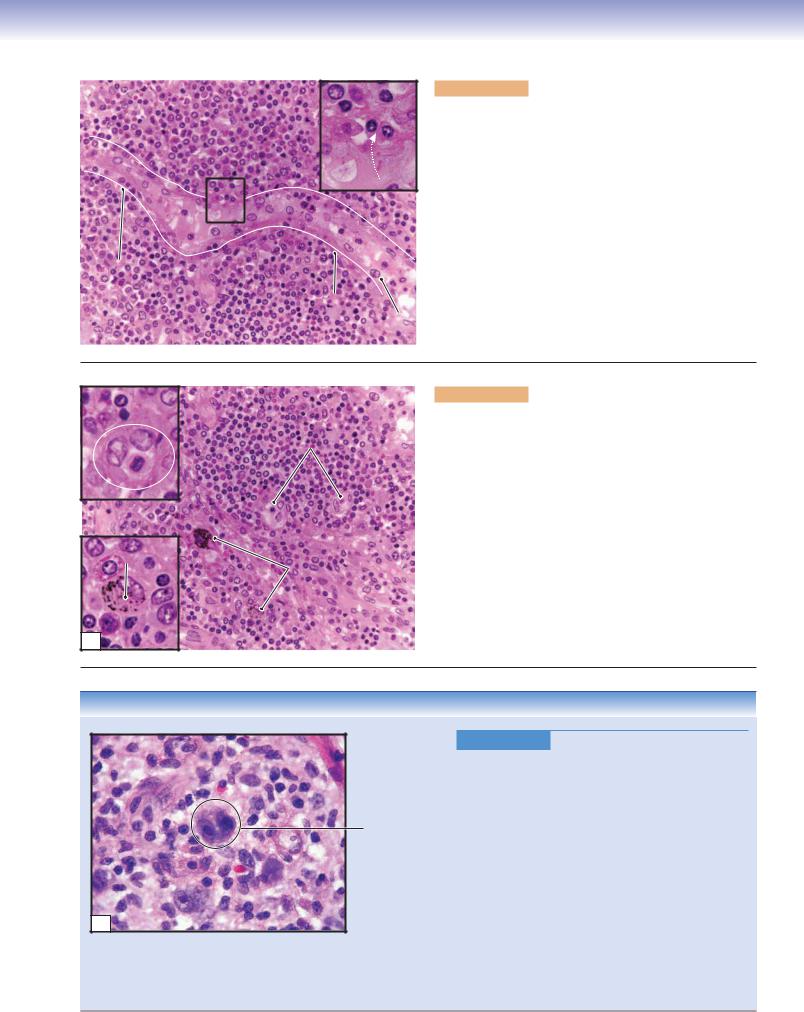
 Lymphocyte


 center
CHAPTER 10 ■ Lymphoid System |
197 |
Sinusoid
lumen
Plasma cell
Endothelial cells
 Macrophage
Basement membrane
Erythrocyte
Lymphocyte
 Platelet
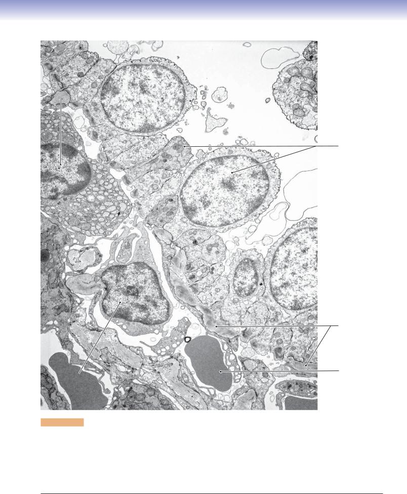
Figure 10-16. Red pulp of the spleen. TEM, 7,100.
Part of the wall and lumen of a red pulp sinusoid is shown here along with some adjacent red pulp cord (cord of Billroth) on the left. The plane of section through the sinusoid appears to be transverse to its long axis as indicated by the varying shapes and sizes of the many profiles of endothelial cells that make up this part of its wall. These cells have a fusiform three-dimensional shape, and their long axis parallels that of the vessel. A few endothelial cells are cut through the nucleus, but many more show only small profiles of cytoplasm. The basement membrane of the endothelium is incomplete, and only a couple of pieces are visible here. In life, the formed elements of blood squeeze between endothelial cells to move into and out of the red pulp cords. Macrophages, plasma cells, and all types of blood cells and platelets are suspended in the reticular framework of the red pulp cord tissue.

198 UNIT 3 ■ Organ Systems
TABLE 10 - 2 Lymphoid Organs
Organ |
Epithelium/ |
Cortex and |
Cords and |
B-cell Main |
T-cell Main |
Special Features (1) |
|
Capsule |
Medulla |
Sinuses |
Region |
Region |
and Functions (2) |
|
Covering |
|
|
|
|
|
|
|
|
|
|
|
|
Tonsils |
Incomplete |
No |
No |
Primary and |
Outside of |
1. Epithelial covering |
|
epithelium |
|
|
secondary nodules |
the lymphatic |
2. Promotes B cells to |
|
and capsule |
|
|
|
nodules |
proliferate and to |
|
|
|
|
|
|
produce IgA; immune |
|
|
|
|
|
|
defense against upper |
|
|
|
|
|
|
respiratory infections, |
|
|
|
|
|
|
where B and T cells |
|
|
|
|
|
|
encounter foreign |
|
|
|
|
|
|
antigens and initiate |
|
|
|
|
|
|
immune response |
Lymph |
Capsule |
Cortex, paracortex, |
Medullary |
Primary and |
Paracortex |
1. Afferent lymphatic |
nodes |
(thin) |
and medulla |
cords and |
secondary nodules |
|
vessels and |
|
|
|
medullary |
(most nodules |
|
subcapsular sinuses |
|
|
|
sinuses |
are secondary); |
|
2. Filter lymph and |
|
|
|
|
medullary cords |
|
recirculate both B |
|
|
|
|
|
|
and T cells; provide |
|
|
|
|
|
|
place for lymphocytes |
|
|
|
|
|
|
to meet antigens and |
|
|
|
|
|
|
start immune response |
Thymus |
Capsule |
Cortex (without |
No |
No |
Cortex and |
1. Epithelial reticular |
|
(thin) |
lymphatic nodules); |
|
|
medulla |
cells and Hassall |
|
|
medulla (with |
|
|
|
corpuscles; no |
|
|
Hassall corpuscles) |
|
|
|
lymphatic nodules |
|
|
|
|
|
|
2. Development and |
|
|
|
|
|
|
maturation of T cells |
Spleen |
Capsule |
No, arranged in |
Splenic cords |
Secondary nodules |
PALS |
1. Central arteries and |
|
(thick) |
white pulp and red |
and venous |
(splenic nodules) |
|
PALS |
|
|
pulp |
sinuses |
|
|
2. Red pulp filters |
|
|
|
|
|
|
blood, removes aged |
|
|
|
|
|
|
erythrocytes, and |
|
|
|
|
|
|
acts as a reservoir |
|
|
|
|
|
|
for erythrocytes and |
|
|
|
|
|
|
platelets; the white |
|
|
|
|
|
|
pulp hosts B and T |
|
|
|
|
|
|
lymphocytes, where |
|
|
|
|
|
|
they meet antigens, |
|
|
|
|
|
|
mature and prolif- |
|
|
|
|
|
|
erate, and initiate |
|
|
|
|
|
|
immune response |
SYNOPSIS 10 - 3 Pathological and Clinical Terms for the Lymphoid System
■Lymphadenopathy: Enlarged lymph nodes due to a variety of causes including lymphoma, infection, autoimmune disease, medications, and metastatic disease (Fig. 10-9C).
■Myalgia: Muscle pain that may be caused by a variety of conditions including exercise, autoimmune disease, medications, infections, and neoplasms (Fig. 10-9C).
■Lymphoid hyperplasia: A reactive proliferative process of lymphoid tissues, particularly lymph nodes, characterized by enlarged follicles with abundant macrophages within the germinal center (Fig. 10-9C).
■Reed-Sternberg cell: Characteristic cell of classical Hodgkin lymphoma containing two nuclei or nuclear lobes, each with a prominent nucleolus (Fig. 10-12C).
■Waldeyer ring: Lymphoid tissues of the nasopharynx including the palatine tonsils and pharyngeal tonsils (adenoids) that may be an extranodal site of lymphoma development (Fig. 10-9B).
