


Brainstem 65
Lesions of the Brainstem
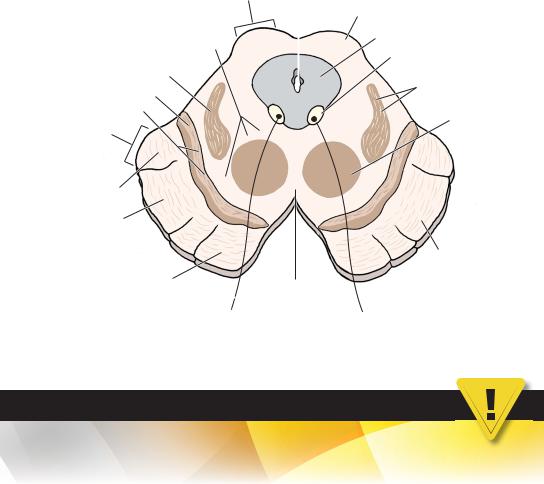
ILesions of the Medulla (Figure 7-6)
A.Medial Medullary Syndrome (Anterior Spinal Artery Syndrome). Affected structures include the following:
1.Corticospinal tract (medullary pyramid). Lesions result in contralateral spastic hemiparesis;
2.Medial lemniscus. Lesions result in contralateral loss of tactile and vibration sensation from the trunk and extremities;
3.Hypoglossal nucleus and its intra-axial fibers. Lesions result in ipsilateral flaccid hemiparalysis of the tongue. When protruded, the tongue points to the side of the lesion (i.e., the weak side). See Figure 9.8.
B.Lateral Medullary (Wallenberg; Posterior Inferior Cerebellar Artery [PICA]) Syndrome is characterized by dissociated sensory loss. Affected structures include the following:
1.Vestibular nuclei. Lesions result in nystagmus, nausea, vomiting, and vertigo;
2.Inferior cerebellar peduncle. Lesions result in ipsilateral cerebellar signs (e.g., dystaxia, dysmetria [past pointing], and dysdiadochokinesia);
3.Nucleus ambiguus. Lesions result in ipsilateral laryngeal, pharyngeal, and palatal hemiparalysis (i.e., loss of the gag reflex [efferent limb], dysarthria, dysphagia, and dysphonia [hoarseness]);
4.Glossopharyngeal nerve roots. Lesions result in loss of the gag reflex (afferent limb);
5.Vagal nerve roots. Lesions result in the same deficits as seen in lesions involving the nucleus ambiguus;
6.Spinothalamic tracts (spinal lemniscus). Lesions result in contralateral loss of pain and temperature sensation;
7.Spinal trigeminal nucleus and tract. Lesions result in ipsilateral loss of pain and temperature sensation from the face (facial hemianesthesia);
8.Descending sympathetic tract. Lesions result in ipsilateral Horner syndrome (i.e., ptosis, miosis, hemianhidrosis, and apparent enophthalmos).
IILesions of the Pons (Figure 7-7A)
A.Medial Inferior Pontine Syndrome results from occlusion of the paramedian branches of the basilar artery. Affected structures include the following:
1.Corticospinal tract. Lesions result in contralateral spastic hemiparesis.
2.Medial lemniscus. Lesions result in contralateral loss of tactile sensation from the trunk and extremities.
3.Abducent nerve roots. Lesions result in ipsilateral lateral rectus paralysis.
B.Lateral Inferior Pontine Syndrome (anterior inferior cerebellar artery syndrome; Figure 7-7B). Affected structures include the following:
1.Facial nucleus and intra-axial nerve fibers. Lesions result in:
a.Ipsilateral facial paralysis;
b.Ipsilateral loss of taste from the anterior two-thirds of the tongue;
c.Ipsilateral loss of lacrimation and reduced salivation;
d.Loss of the efferent limb of the corneal blink and stapedial reflexes (hyperacusis).
2.Cochlear nuclei and intra-axial nerve fibers. Lesions result in ipsilateral deafness;
3.Vestibular nuclei and intra-axial nerve fibers. Lesions result in nystagmus, nausea, vomiting, and vertigo;

66Chapter 7
4.Spinal nucleus and tract of the trigeminal nerve. Lesions result in ipsilateral loss of pain and temperature sensation from the face (facial hemianesthesia);
5.Middle and inferior cerebellar peduncles. Lesions result in ipsilateral limb and gait dystaxia;
6.Spinothalamic tracts (spinal lemniscus). Lesions result in contralateral loss of pain and temperature sensation from the trunk and extremities;
7.Descending sympathetic tract. Lesions result in ipsilateral Horner syndrome.
C.MLF Syndrome (Internuclear Ophthalmoplegia) (Figure 7-7C) interrupts fibers
from the contralateral abducent nucleus that project through the MLF to the ipsilateral medial rectus subnucleus of CN III. It causes medial rectus palsy on attempted lateral conjugate gaze and nystagmus in the abducting eye. Convergence remains intact. This syndrome is often seen in patients with multiple sclerosis.
D.Facial Colliculus Syndrome usually results from a pontine glioma or a vascular accident. The internal genu of CN VII and the abducent nucleus underlie the facial colliculus.
1.Lesions of the internal genu of the facial nerve cause:
a.Ipsilateral facial paralysis;
b.Ipsilateral loss of the corneal blink reflex (efferent limb).
2.Lesions of the abducent nucleus cause:
a.Lateral rectus paralysis (medial strabismus);
b.Horizontal diplopia.
IIILesions of the Midbrain (Figure 7-8)
A.Posterior Midbrain (Parinaud) Syndrome (see Figure 7-8A) is often the result of a pinealoma or germinoma of the pineal region. Affected structures include the following:
1.Superior colliculus and pretectal area. Lesions cause paralysis of upward and downward gaze, pupillary disturbances, and absence of convergence;
2.Cerebral aqueduct. Compression causes noncommunicating hydrocephalus.
B.Paramedian Midbrain (Benedikt) Syndrome (see Figure 7-8B). Affected structures include the following:
1.Oculomotor nerve roots (intra-axial fibers). Lesions cause complete ipsilateral oculomotor paralysis. Eye abduction and depression is caused by the intact lateral rectus (CN VI) and superior oblique (CN IV). Ptosis (paralysis of the levator palpebrae superioris) and fixation and dilation of the ipsilateral pupil (complete internal ophthalmoplegia) also occur;
2.Dentatothalamic fibers. Lesions cause contralateral cerebellar dystaxia with intention tremor;
3.Medial lemniscus. Lesions result in contralateral loss of tactile sensation from the trunk and extremities.
C.Medial Midbrain (Weber) Syndrome (see Figure 7-8C). Affected structures and resultant deficits include the following:
1.Oculomotor nerve roots (intra-axial fibers). Lesions cause complete ipsilateral oculomotor paralysis. Eye abduction and depression are caused by intact lateral rectus (CN VI) and superior oblique (CN IV). Ptosis, fixation, and dilation of the ipsilateral pupil also occur;
2.Corticospinal tracts. Lesions result in contralateral spastic hemiparesis;
3.Corticonuclear fibers. Lesions cause contralateral weakness of the lower face (CN VII), tongue (CN XII), and palate (CN X). The upper face division of the facial nucleus receives bilateral corticonuclear input. The uvula and pharyngeal wall are pulled toward the normal side (CN X), and the protruded tongue points to the weak side.
Brainstem 67
IV Acoustic Neuroma (Schwannoma) (Figure 7-9) is a benign
tumor of Schwann cells that affects the vestibulocochlear nerve (CN VIII). It accounts for 8% of all intracranial tumors. It is a posterior fossa tumor near the internal auditory meatus and cerebellopontine angle. The neuroma often compresses the facial nerve (CN VII), which
accompanies CN VIII in the cerebellopontine angle and internal auditory meatus. It may impinge on the pons and affect the spinal trigeminal tract (CN V). Schwannomas occur twice
as often in women as in men. Affected structures and resultant deficits include the following:
A.Cochlear Division of CN VIII. Damage results in tinnitus and unilateral nerve deafness;
B.Vestibular Division of CN VIII. Damage results in vertigo, nystagmus, nausea, vomiting, and unsteadiness of gait;
C.Facial Nerve (CN VII). Damage results in facial weakness and loss of the corneal blink reflex (efferent limb);
D.Spinal Tract of Trigeminal Nerve (CN V). Damage results in paresthesia, anesthesia of
the ipsilateral face, and loss of the corneal blink reflex (afferent limb). NEUROFIBROMATOSIS TYPE 2. This disorder often occurs with bilateral acoustic neuromas.
VJugular Foramen Syndrome usually results from a posterior fossa tumor
(e.g., glomus jugulare tumor, the most common inner ear tumor) that compresses CNs IX, X, and XI. Affected structures and resultant deficits include the following:
A.Glossopharyngeal Nerve (CN IX). Damage results in:
1.Ipsilateral loss of the gag reflex;
2.Ipsilateral loss of pain, temperature, and taste in the tongue.
|
|
Cerebral |
|
|
Tectum |
aqueduct |
|
|
|
|
Inferior |
Lateral lemniscus |
|
|
colliculus |
Trigeminal thalamic fibers |
|
|
|
Spinothalamic fibers |
|
|
Periaqueductal gray |
|
|
|
Trochlear nucleus |
Medial |
|
|
|
|
|
|
|
lemniscus |
|
|
|
Cerebral peduncle
Superior |
|
Crus cerebri |
|
|
|
cerebellar |
|
|
peduncle |
|
|
|
Tegmentum |
Substantia nigra |
|
|
|
|
(reticular formation) |
|
Figure 7-9 Caudal midbrain.

68 |
Chapter 7 |
B.Vagal Nerve (CN X). Damage results in:
1.Ipsilateral paralysis of the soft palate and larynx;
2.Ipsilateral loss of the gag reflex.
C.Accessory Nerve (CN XI). Damage results in:
1.Paralysis of sternocleidomastoid, which results in the inability to turn the head to the opposite side;
2.Paralysis of trapezius, which causes shoulder droop and inability to shrug the shoulder.
VI |
“Locked-in” Syndrome is a lesion of the base of the pons as the result of |
|
infarction, trauma, tumor, or demyelination. The corticospinal and corticonuclear tracts are |
|
affected bilaterally. The oculomotor and trochlear nerves are not injured. Patients are conscious |
|
and may communicate through vertical eye movements. |
VII |
Central Pontine Myelinolysis is a lesion of the base of the pons that |
|
affects the corticospinal and corticonuclear tracts. More than 75% of cases are associated with |
|
alcoholism or rapid correction of hyponatremia. Symptoms include spastic quadriparesis, |
|
pseudobulbar palsy, and mental changes. This condition may become the locked-in syndrome. |
VIII |
“Top of the Basilar” Syndrome results from embolic occlusion of the |
|
rostral basilar artery. Neurologic signs include optic ataxia and psychic paralysis of fixation of |
|
gaze (Balint syndrome), ectopic pupils, somnolence, and cortical blindness, with or without |
|
visual anosognosia (Anton syndrome). |
IX |
Subclavian Steal Syndrome (Figure 7-10) results from thrombosis of the |
|
left subclavian artery proximal to the vertebral artery. Blood is shunted in the left vertebral artery |
|
and into the left subclavian artery. Clinical signs include transient weakness and claudication of |
|
the left upper limb on exercise and vertebrobasilar insufficiency (i.e., vertigo, dizziness). |
XThe Cerebellopontine Angle is the junction of the medulla, pons, and
cerebellum. CNs VII and VIII are found there. Five brain tumors, including a cyst, are often
located in the cerebellopontine angle cistern. Remember the acronym SAME: schwannoma (75%), arachnoid cyst (1%), meningioma (10%), and ependymoma (1%) and epidermoid
(5%). The percentages refer to cerebellopontine angle tumors.
|
|
|
|
Brainstem |
69 |
|
Tectum |
Cerebral |
Superior |
|
|
|
aqueduct |
|
|||
|
|
|
|||
|
|
colliculus |
|
||
Tegmentum |
|
|
|
|
|
|
|
|
|
||
|
|
|
Periaqueductal gray |
|
|
(reticular formation) |
|
|
|
|
|
|
|
|
|
|
|
|
|
|
|
Oculomotor nucleus |
|
Medial lemniscus |
|
|
|
Spinothalamic and |
|
|
|
|
|
|
|
Substantia nigra |
|
|
|
trigeminothalamic tracts |
|
|
|
|
|||
|
|
|
|
|
|
Pars compacta |
|
|
|
|
|
Cerebral |
|
|
|
Red nucleus |
|
|
|
|
|
|
|
peduncle |
|
|
|
|
|
Occipito, parieto, temporopontine fibers
Corticospinal fibers
Corticonuclear fibers 
Crus cerebri
Frontopontine fibers
CN III |
|
Ventral tegmental |
|
area |
|
|
|
Figure 7-10 Rostral midbrain.
CASE 7-1
For several weeks, a 60-year-old hypertensive, diabetic man has experienced sudden dizziness, facial pain, double vision, and difficulty in walking. He is also having problems in swallowing and speaking. What is the most likely diagnosis?
Relevant Physical Exam Findings
●Decreased temperature and pain sense below the left T-4 level
●Horner syndrome on the right side
●Decreased position sensation in the right fingers and toes
●Ataxia and mild weakness in the right limbs
Relevant Lab Finding
●An infarct involving the right lateral part of the lower medulla and the cerebellum seen on brain magnetic resonance image.
Diagnosis
●Wallenberg’s syndrome is an infarction involving the lateral or medial branches of the posterior inferior cerebellar artery.