


58 |
Chapter 6 |
C.Unilateral muscle atrophy and absent quadriceps (L3) and ankle jerk (S1) reflex activity.
D.Unremarkable incontinence and sexual function.
E.Gradual and unilateral onset.
VII Conus Medullaris Syndrome (Cord Segments S3 to C0)
usually results from an intramedullary tumor (e.g., ependymoma). It is characterized by:
A.Pain, usually bilateral and not severe.
B.Sensory distribution in a bilateral saddle-shaped area.
C.Unremarkable muscle changes; normal quadriceps and ankle jerk reflexes.
D.Severely impaired incontinence and sexual function.
E.Sudden and bilateral onset.
C H A P T E R 7
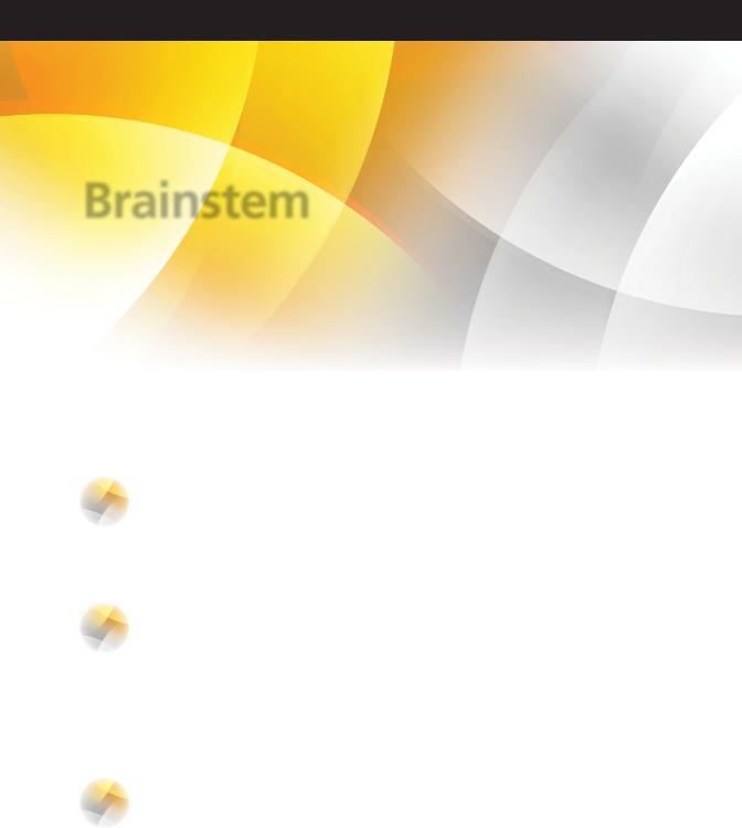
Brainstem
Objectives
1.Identify the brainstem nuclei associated with the cranial nerves and be able to locate them on a brainstem cross section.
2.Identify the cranial nerves where they connect to the brainstem.
3.Describe the reticular formation—connections, functions, and structure.
4.Describe the result of occlusion of the anterior spinal artery and the posterior inferior cerebellar artery, include all brainstem nuclei and pathways affected.
5.Describe medial longitudinal fasciculus syndrome and Weber syndrome.
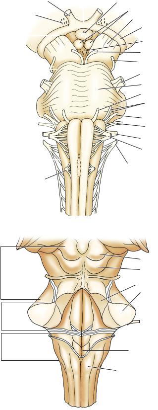
IIntroduction. The brainstem includes the medulla, pons, and midbrain. It extends
from the pyramidal decussation inferiorly to the posterior commissure superiorly. The brainstem receives its blood supply from the vertebrobasilar system. It gives rise to CNs III to X and XII (Figures 7-1 and 7-2).
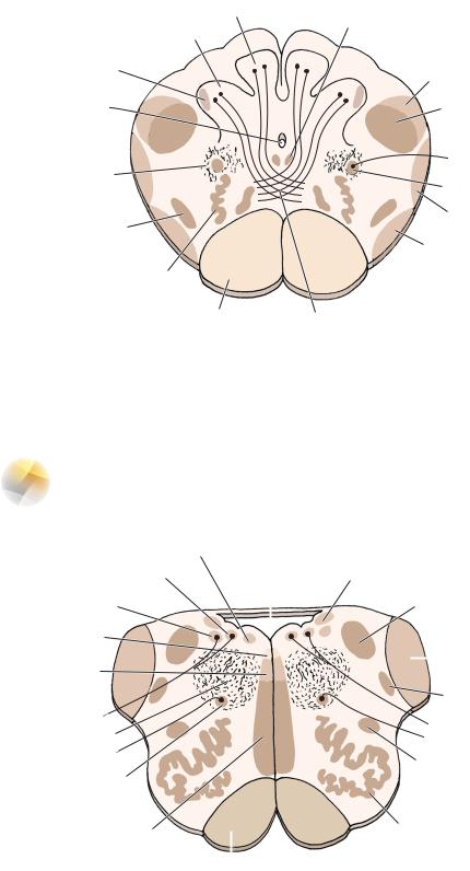
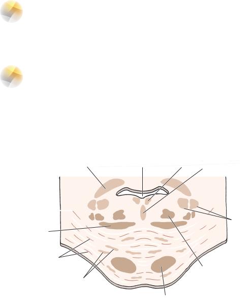
IICross Section Through the Caudal Medulla (Figure 7-3)
1.Pyramid (corticospinal fibers)
2.Nucleus gracilis and nucleus cuneatus—give rise to arcuate fibers that cross the midline to form the medial lemniscus
3.Anterior and posterior spinocerebellar tracts
4.Inferior olivary nucleus
5.Accessory cuneate nucleus
IIICross Section Through the Mid-Medulla (Figure 7-4)
1.Hypoglossal nucleus
2.Dorsal motor nucleus of the vagus—preganglionic parasympathetic cell bodies that send fibers into CN X
3.Solitary nucleus—special sense nucleus
4.Medial longitudinal fasciculus—yolks together cranial nerve nuclei from opposite sides of the brainstem
5.Tectospinal tract—fibers descending from the midbrain colliculi to lower motor neurons of the cervical spinal cord
59
|
|
Optic nerve |
|
Optic chiasm |
||||
|
|
|
|
Infundibulum |
||||
|
|
|
|
|||||
Anterior perforated substance |
|
|
Mamillary bodies |
|||||
|
|
Cerebral peduncle |
||||||
|
|
|
|
|
|
|
|
|
|
|
|
|
|
|
|
|
Optic tract |
|
|
|
|
|
|
|
|
|
|
|
|
|
|
|
|
|
Posterior perforated substance |
|
|
|
|
|
|
|
|
|
Midbrain |
|
|
|
|
|
|
|
Oculomotor nerve |
|
|
|
|
|
|
|||
|
|
|
|
|
|
|
|
Trochlear nerve |
|
|
|
|
|
|
|
|
Trigeminal nerve |
|
|
|
|
|
|
|
|
|
|
|
|
|
|
|
|
|
|
|
|
|
|
|
|
|
|
Pons |
Pons |
|
|
|
|
|
|
|
Abducent nerve |
|
|
|
|
|
|
|
Facial nerve and |
|
|
|
|
|
|
|
|
|
|
|
|
|
|
|
|
|
|
nervus intermedius |
|
|
|
|
|
|
|
|
Vestibulocochlear nerve |
|
|
|
|
|
|
|
|
Glossopharyngeal nerve |
|
|
|
|
|
|
|
|
|
|
|
|
|
|
|
|
||
Medulla |
|
|
|
|
|
|
|
Vagus nerve |
|
|
|
|
|
|
|
Hypoglossal nerve |
|
|
|
|
|
|
|
|
|
|
|
|
|
|
|
|
|
|
Spinal accessory nerve |
|
|
|
|
|
|
|
|
|
Anterior root C1 |
|
|
|
|||||
Figure 7-1 The anterior or ventral surface of the brainstem and the attached cranial nerves.
Superior colliculus
Inferior colliculus
Midbrain 
Superior cerebellar peduncle
Middle cerebellar peduncle
 CN V
CN V
Pons 
 CN VII
 CN VIII
Medulla 
Area postrema
 Tuberculum cuneatus
Tuberculum gracilis
Figure 7-2 The posterior or dorsal surface of the brainstem.
60
|
Brainstem |
61 |
Nucleus gracilis |
Medial longitutinal |
|
|
|
|
Nucleus cuneatus |
fasciculus |
|
|
|
|
Accessory cuneate |
|
|
nucleus |
Spinal tract (CN V) |
|
|
|
|
Central canal |
Spinal nucleus (CN V) |
|
|
Fibers of CN X |
|
Reticular |
|
|
formation |
Nucleus ambiguus |
|
Posterior Spinothalamic spinocerebellar tract
tracts
Anterior spinocerebellar tract
Inferior olivary nucleus
Pyramid |
Decussation of |
|
medial lemniscus |
Figure 7-3 Caudal medulla.
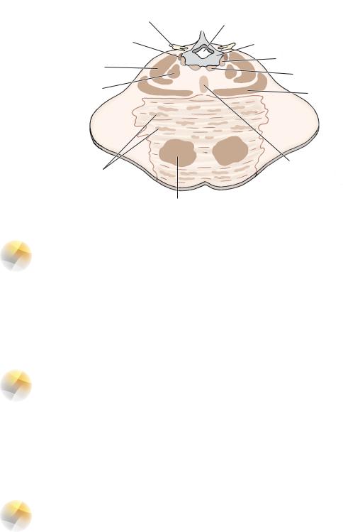
6.Nucleus ambiguus—lower motor neuron nucleus that sends fibers into CNs IX and X
7.Medial lemniscus
8.Inferior cerebellar peduncle
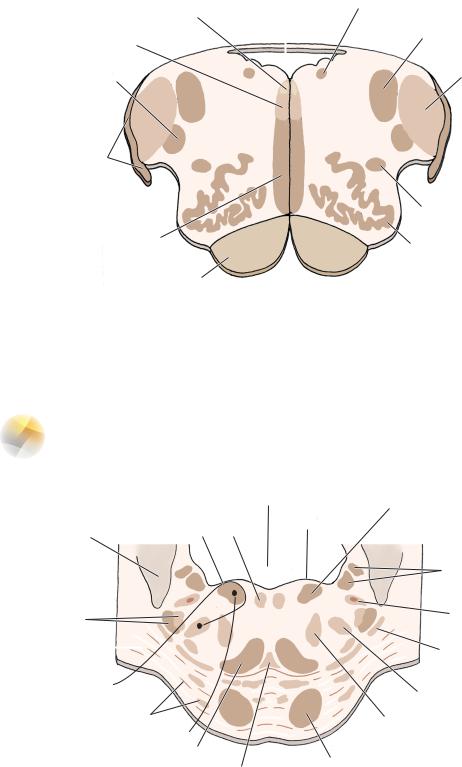
IV Cross Section Through the Rostral Medulla (Figure 7-5)
Hypoglossal nucleus |
|
|
|
Medial vestibular |
|||
Dorsal motor nucleus |
Fourth ventricle |
||||||
nucleus |
|||||||
|
|
|
|||||
|
|
|
|||||
of CN X |
|
|
|
|
|
|
|
Solitary nucleus |
|
|
|
|
Inferior vestibular |
||
|
|
|
|
|
|
nucleus |
|
Medial longitudinal |
|
|
|
|
|
|
|
fasciculus |
|
|
|
|
|
Inferior cerebellar |
|
|
|
|
|
|
|
||
|
|
|
|
|
|
||
Tectospinal tract |
|
|
|
|
|
peduncle |
|
|
|
|
|
|
|
Spinal nucleus and |
|
|
|
|
|
|
|
tract of CN V |
|
Reticular formation |
|
|
|
|
|
|
|
|
|
|
|
|
Lateral spinothalamic |
||
Nucleus ambiguus |
|
|
|
|
|
tract |
|
Medial lemniscus |
|
|
|
Inferior olivary nucleus |
|||
|
|
|
|
|
|
||
|
|
|
|
|
|
||
|
Pyramid |
|
|
|
|||
Figure 7-4 Mid-medulla. |
|
|
|
|
|
|
|
62 |
Chapter 7 |
|
||
|
Fourth ventricle |
Solitary nucleus |
||
|
Medial longitudinal |
and tract |
||
|
fasciculus |
|
|
|
|
|
|
Inferior vestibular |
|
|
Tectospinal |
|
|
|
|
|
|
nucleus |
|
|
tract |
|
|
|
|
Spinal nucleus |
|
|
Inferior cerebellar |
|
|
|||
|
and tract of CN V |
peduncle |
||
Dorsal and ventral cochlear nuclei
|
Spinothalamic |
Medial |
tracts |
|
|
lemniscus |
Inferior olivary |
|
nucleus |
Pyramid
Figure 7-5 Rostral medulla.
1.Spinothalamic tracts (spinal lemniscus)
2.Spinal nucleus and tract of trigeminal nerve
3.Inferior cerebellar peduncle—contains olivocerebellar, cuneocerebellar, and posterior spinocerebellar tracts
VCross Section Through the Caudal Pons (Figure 7-6). The pons
has a posterior tegmentum and an anterior base.
|
|
|
|
Fourth ventricle |
Abducens nucleus |
|
|
|
|
Genu of |
|
Facial colliculus |
of CN VI |
Inferior cerebellar |
MLF |
|
||||
facial nerve |
|
|||||
|
|
|||||
peduncle |
|
|
|
|
||
|
|
|
|
|
|
Vestibular nuclei |
|
|
|
|
|
|
(lateral and superior) |
Spinal nucleus |
|
|
|
Solitary nucleus |
||
and tract |
|
|
|
|||
|
|
|
and tract |
|||
|
|
|
|
|
|
|
Middle cerebellar |
|
|
|
|
|
Spinothalamic tracts |
|
|
|
|
|||
peduncle |
|
|
|
|||
|
|
|
|
|||
|
|
|
|
|
|
Facial motor |
Transverse pontine fibers |
|
|
|
nucleus |
||
|
|
|
Central tegmental |
|||
and deep pontine nuclei |
|
|
|
|||
|
|
|
|
|
|
tract |
|
Medial lemniscus |
|
Corticonuclear and |
|||
|
|
|
Trapezoid body |
|||
|
|
|
corticospinal tracts |
|||
Figure 7-6 Caudal pons.
Brainstem 63
1.Medial longitudinal fasciculus (MLF)
2.Abducent nucleus of CN VI (underlies facial colliculus)
3.Genu (internal) of CN VII (underlies facial nerve; facial colliculus)
4.Medial lemniscus
5.Corticospinal and corticonuclear tracts (in the base of the pons)
6.Facial motor nucleus (CN VII)
7.Spinal nucleus and tract of trigeminal nerve (CN V)
8.Spinothalamic tracts (spinal lemniscus)
9.Vestibular nuclei of CN VIII
10.Inferior and middle cerebellar peduncle
11.Central tegmental tract—fiber pathway traversing the reticular formation
VI Cross Section Through the Mid-Pons (Figure 7-7)
1.Raphe nuclei
2.Deep pontine nuclei and transverse pontine fibers
3.Superior cerebellar peduncle—main cerebellar outflow pathway, also contains decussating anterior spinocerebellar fibers
VII Cross Section Through the Rostral Pons (Figure 7-8)
1.Mesencephalic nucleus—unconscious proprioception for the head
2.Locus ceruleus—source of norepinephrine for the brain
3.Cerebral aqueduct—connects the third and fourth ventricles
4.Periaqueductal gray—involved in pain modulation, source of serotonin
Superior cerebellar |
Fourth ventricle |
MLF |
|
peduncle |
Raphe nuclei |
||
|
|
|
Middle cerebellar |
|
M |
S |
|
|
|
|
|
|
||
peduncle |
Sensory (S) and |
||
|
|
|
|
Medial lemniscus |
motor (M) |
||
nuclei of CN V |
|||
Transverse pontine fibers
Central tegmental
tract
Deep pontine nuclei
Corticonuclear and
corticospinal tracts
Figure 7-7 Mid-pons.
64 |
Chapter 7 |
Superior medullary velum
Trochlear nerve (CN IV) |
Cerebral aqueduct |
|
|
||
Mesencephalic |
|
|
|
||
nucleus |
Periaqueductal gray |
|
|
||
Superior cerebellar |
Locus ceruleus |
|
peduncle |
MLF |
|
|
||
Central tegmental |
Medial lemniscus |
|
tract |
||
|
Middle cerebellar  peduncle
Raphe nucleus
Deep pontine nuclei
Corticonuclear and
corticospinal tracts
Figure 7-8 Rostral pons.
VIII Cross Section Through the Caudal Midbrain (Figure 7-9).
The midbrain has a posterior tectum, an intermediate tegmentum, and a base. The cerebral aqueduct lies between the tectum and the tegmentum.
Medial lemniscus
1.Inferior colliculus—auditory relay nucleus
2.Trochlear nucleus—lower motor neuron nucleus that innervates the superior oblique
3.Cerebral aqueduct
4.Crus cerebri (basis pedunculi cerebri or cerebral peduncle)—composed of descending fibers.
IX Cross Section Through the Rostral Medulla (Figure 7-10)
1.Superior colliculus—visual relay nucleus
2.Oculomotor nucleus—lower motor neuron nucleus that innervates the majority of the extraocular musculature
3.Red nucleus—part of the primitive motor system, involved in coordination
4.Ventral tegmental area—source of dopamine, involved in the reward system
5.Spinothalamic and trigeminothalamic tracts
6.Medial lemniscus
XCorticonuclear Fibers project bilaterally to all motor cranial nerve nuclei except
the facial nucleus. The division of the facial nerve nucleus that innervates the upper face (the orbicularis oculi and above) receives bilateral corticonuclear input. The division of the facial nerve nucleus that innervates the lower face receives only contralateral corticonuclear input.