Добавил:
Опубликованный материал нарушает ваши авторские права? Сообщите нам.
Вуз: Предмет: Файл:

KAPLAN_USMLE_STEP_1_LECTURE_NOTES_2018_BIOCHEMISTRY_and_GENETICS

.pdf
Скачиваний:
8
Добавлен:
29.01.2024
Размер:
10.81 Mб
Скачать

Part II Medical Genetics

Recombination Frequencies and

Gene Mapping

High-Yield

The closer together two linked loci are—for instance, a gene and a marker—the lower the recombination frequency will be between them. Therefore, recombination frequency can be used to estimate proximity between a gene and a linked marker.

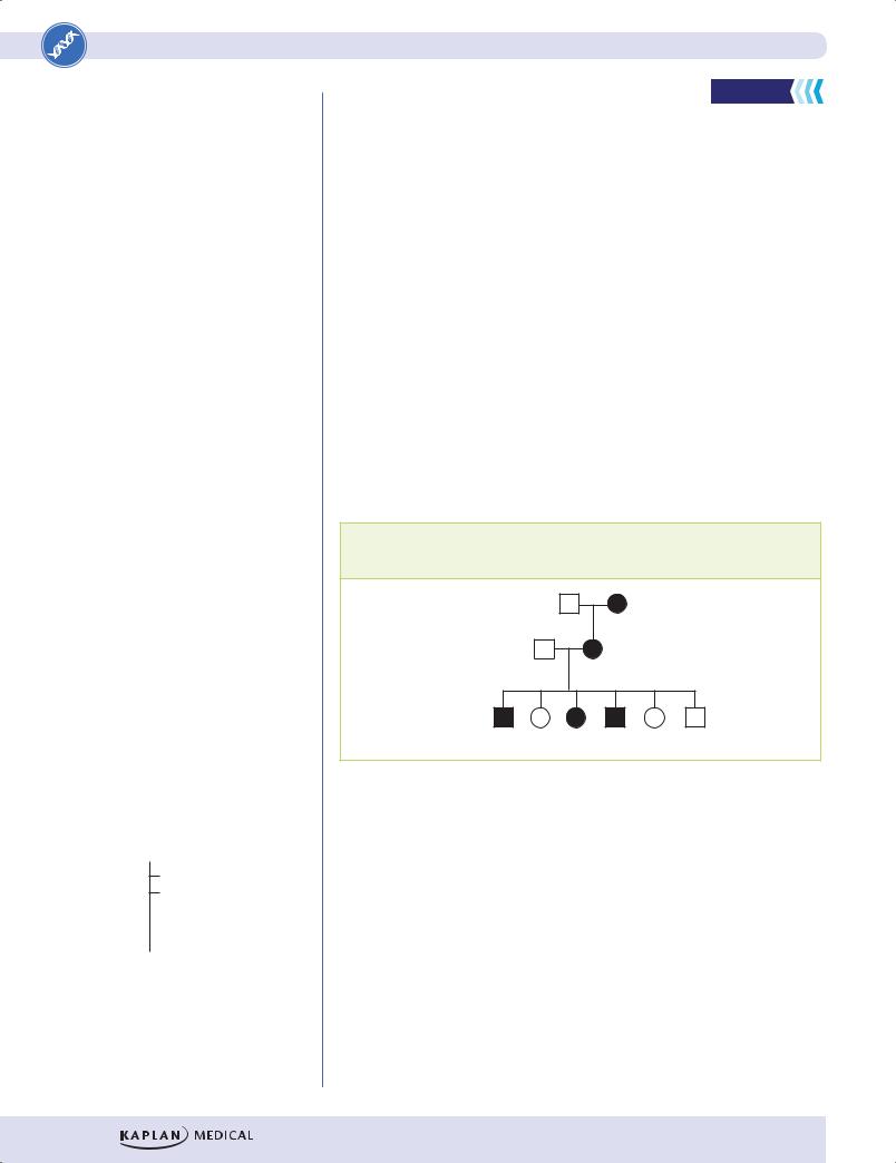
The following example of a family with neurofibromatosis type 1, an autosomal dominant disorder with complete penetrance, illustrates the concept of recombination frequency. Some members of the family have the disease-producing allele of the gene (indicated by phenotype in the pedigree) whose location is to be determined. Other individuals have the normal allele of the gene.

Each individual has also been typed for his or her allele(s) of a 2-allele marker (1 or 2). Three steps are involved in determining whether linkage exists and, if so, estimating the distance between the gene and the known marker.

1.Establish linkage phase between the disease-producing allele of the gene and an allele of the marker in the family.

2.Determine if linkage exists between the 2 alleles.

3.If linkage exists, estimate the recombination frequency.

Note

A haplotype is the combination of alleles on a single chromosome. With respect to the gene and the marker in Figure II-5-3, individual II-2 has two haplotypes, AM1 and aM2, depicted below, where A and a are alleles of the gene causing the disease. The marker has alleles 1 and 2.

A


a


M1


M1


384

A family in which a mutation causing neurofibromatosis type 1 is transmitted in 3 generations. The genotype of a marker locus is shown for each pedigree member.

I

2,2 1,2

II

1,1 1,2

III

1,1 1,2 1,1 1,1 1,2 1,1

Figure II-5-4. Pedigree for Neurofibromatosis Type 1

Linkage Phase. The pedigree indicates that the grandmother (I-2) had the dis- ease-producing allele (A) of the gene, which she passed to her daughter (II-2). Is it also possible to determine which allele of the marker was passed from the grandmother to her daughter? Yes, allele 1. If linkage is present, the diseaseproducing allele (A) is linked to allele 1 of the marker. We can then designate the daughter’s 2 haplotypes as AM1/aM2, indicating the chromosomes from her mother/father respectively.

Determine If Linkage Exists. Are the gene and the marker actually linked as we hypothesize? Looking at the children in generation III (each representing a meiotic event in their mother, II-2), we would expect a child who inherited marker allele 1 from the mother to have the disease. The children who inherited allele 2 from the mother should not have the disease. Examination of the 6 children’s haplotypes shows that this assumption is true in all but one case (III-6). Because

P (linkage at recombination frequency, θ)

Chapter 5 Recombination Frequency

the AM1 and aM2 haplotypes remain together more than 50% of the time (or, conversely, are separated by recombination less than 50% of the time), our hypothesis of linkage is correct.

Estimate the Recombination Frequency. Out of 6 children, there is only one recombinant (III-6). The estimated recombination frequency in this family is 1/6, or 17%.

Recombination frequencies can be related to physical distance by the centimorgan (cM)

The recombination frequency provides a measure of genetic distance between any pair of linked loci. This distance is expressed in centimorgans. The centimorgan is equal to 1% recombination frequency. For example, if two loci show a recombination frequency of 2%, they are said to be 2 centimorgans apart. Physically, 1 cM is approximately equal to 1 million base pairs of DNA (1 Mb). This relationship is only approximate, however, because crossover frequencies are somewhat different throughout the genome, e.g., they are less common near centromeres and more common near telomeres.

Determining Recombination Frequency Accurately: LOD Scores

In the previous example, a very small population (6 children, or 6 meiotic events) was used to determine and calculate linkage, allowing only a very rough estimate of linkage distance. In fact, there is some small chance that the gene and the marker are not actually linked at all and the data were obtained by chance. We could be more confident that our conclusions were correct if we had used a much larger population. Because families don’t have 100 or 200 children, the next best approach is to combine data from different families with this same disease to increase the number of meioses examined. These data can be combined by using LOD (log of the odds) calculations.

A LOD score, calculated by computer, compares the probability (P) that the data resulted from actual linkage with a recombination frequency of theta (θ) versus the probability that the gene and the marker are unlinked (θ = 50%) and that the data were obtained by chance alone. In the example calculation:

Probability of observing pedigree data if θ = 0.17

Odds = Probability of observing pedigree data if θ = 0.5

In practice, because 17% might not be the correct number, the computer calculates these probabilities assuming a variety of recombination frequencies from θ = 0 (gene and marker are in the same location) to θ = 0.5 (gene and marker are unlinked). The “odds of linkage” is simply the probability that each recombination frequency (θ) is consistent with the family data. If data from multiple families are combined, the numbers can be added by using the log10 of these odds.

Log of the Odds (LOD) = log10 P (unlinked, recom bination frequency, 50%)

This equation need not be memorized. These calculations are done by computer and are displayed as a LOD table that gives the LOD score for each recombination frequency, θ.

Note

For Linked Loci:

1 centimorgan (cM) = 1% recombination frequency

1 cM ≈ 1 million base pairs

385

Part II Medical Genetics

Note

LOD score >3 indicates linkage, while score <–2 indicates no linkage.

To determine the recombination frequency:

First examine the row of LOD scores.

The highest LOD score >3 is the estimate of the recombination frequency.

Then examine the row of recombination frequencies.

The value directly above the LOD score >3 is the recombination frequency.

Table II-5-1. LOD Scores for a Gene and a Marker

Recombination

0.01

0.05

0.10

0.20

0.30

0.40

frequency (θ)


LOD score

0.58

1.89

3.47

2.03

–0.44

–1.20


When interpreting LOD scores, the following rules apply:

LOD score >3.00 shows statistical evidence of linkage. (It is 1,000 times more likely that the gene and the marker are linked at that distance than unlinked.)

LOD score <–2.00 shows statistical evidence of nonlinkage. (It is 100 times more likely that the gene and the marker are unlinked than linked at that distance.)

LOD score between –2.00 and 3.00 is indeterminate.

An examination of Table II-5-1 shows that in only one case is there convincing evidence for linkage and that score has a recombination frequency of 0.10. Therefore, the most likely distance between the gene and the marker is a recombination frequency of 10%, or 10 cM.

If no LOD score on the table is >3.00, the data may be suggestive of linkage, but results from additional families with the disease would need to be gathered.

Gene mapping by linkage analysis serves several important functions:

It can define the approximate location of a disease-causing gene.

Linked markers can be used along with family pedigree information for genetic testing (see Chapter 6). In practice, markers that are useful for genetic testing must show less than 1% recombination with the gene involved (be <1 cM distant from the gene).

Linkage analysis can identify locus heterogeneity.

386

Chapter 5 Recombination Frequency

Review Questions

Select the ONE best answer.

1.A family with an autosomal dominant disorder is typed for a 2 allele marker, which is closely linked to the disease locus. Based on the individuals in Generation III, what is the recombination rate between the disease locus and the marker locus?

I

1,1 2,2

II

1,2 2,2

III

1,2 2,2 2,2 1,2

A.0

B.0.25

C.0.50

D.0.75

E.1.0

F.The marker is uninformative

2.A man who has alkaptonuria marries a woman who has hereditary sucrose intolerance. Both are autosomal recessive diseases and both map to 3q with a distance of 10 cM separating the two loci. What is the chance they will have a child with alkaptonuria and sucrose intolerance?

A.0%

B.12.5%

C.25%

D.50%

E.100%

387

Part II Medical Genetics

3.In a family study following an autosomal dominant trait through 3 generations, two loci are compared for their potential linkage to the disease locus. In the following 3-generation pedigree, shaded symbols indicate the presence of the disease phenotype, and the expression of ABO blood type and MN alleles are shown beneath each individual symbol.

AO OO

MN NN

AO OO

MN NN

AO AO OO AO OO OO AO

MN MN NN NN MN NN NN

Which of the following conclusions can be made about the linkage of the disease allele, ABO blood group locus, and MN locus?

A.The ABO and MN alleles are linked, but assort independently from the disease allele

B.The ABO, MN, and disease alleles all assort independently

C.The disease allele is linked to the ABO locus

D.The disease allele is linked to the ABO and MN loci

E.The disease allele is linked to the MN locus

388

Chapter 5 Recombination Frequency

Answers

1.Answer: A. In this pedigree, the disease allele is consistently transmitted with the 1 allele. There is no case in this small number of individuals where recombination between these two loci has occurred. Therefore, in Generation III, there is no recombination seen in any of the 4 individuals. Receiving the one allele always goes together with receiving the disease gene. Linked markers can be “uninformative” (choice E) in some pedigrees if, for example, the same alleles are expressed in all family members. In such a case, it would be impossible to determine any recombination frequency.

2.Answer: A. A child will inherit a gene for alkaptonuria from the father and the normal allele of this gene from the mother. Conversely, the child will inherit a gene for hereditary sucrose intolerance from the mother and a normal allele of this gene from the father. The child will therefore be a carrier for each disease but will not be affected with either one.

3.Answer: C. In this pedigree, the disease locus alleles are segregating with the ABO blood locus alleles. In each case, individuals who receive the A allele also receive the disease allele. The MN locus is not linked to the AO locus because individuals III-4, -5, and -7 are each recombinants between these loci. The MN locus is not linked to the disease allele because individuals III-4, -5, and -7 are each recombinants between these loci.

389

Genetic Diagnosis

6

Learning Objectives

Understand concepts of direct and indirect genetic diagnosis

Understand concepts of allele-specific oligonucelotides and dot blots

GENETIC DIAGNOSIS

Once a gene is identified, the associated genetic disease in at-risk individuals can be diagnosed.

The goal of genetic diagnosis is to determine whether an at-risk individual has inherited a disease-causing gene. Genetic diagnosis can be distinguished into

2types:

Direct diagnosis: the mutation itself is examined

Indirect diagnosis: linked markers are used to infer whether the individual has inherited the chromosome segment containing the disease-causing mutation

Direct Diagnosis

High-Yield


PCR and allele-specific oligonucleotide (ASO) probes

ASO probes are short nucleotide sequences that bind specifically to a single allele of the gene. For example, the most common mutation causing hemochromatosis is the C282Y mutation that results from a G to A substitution in codon 282.

Codon:

280

281

282

283

284

Normal HFE allele:

TAT

ACG

TGC

CAG

GTG


C282Y allele:

TAT

ACG

TAC

CAG

GTG


The ASO for the normal allele would have the sequence


3

ATA

TGC

ACG

GTC

CAC 5

The ASO for the C282Y allele would have the sequence


3

ATA

TGC

ATG

GTC

CAC 5

391

Part II Medical Genetics

The 2 ASOs could be used to probe the PCR-amplified material on a dot blot.

1

2

3

Normal ASO

C282Y ASO

Probe does not react with sample

Probe reacts with sample

Figure II-6-1. Allele-Specific Oligonucleotide Probes in Hemochromatosis

The results show that individual 1 is homozygous for the normal HFE allele. Individual 2 is heterozygous for the normal and C282Y alleles. Individual 3 is homozygous for the C282Y allele. Only individual 3 would be expected to have symptoms. Note that this test merely determines genotype, and many considerations must be taken into account before predictions about phenotype could be made. Hemochromatosis has only about 15% penetrance, and in those who do have symptoms, variable expression is seen.

DNA chips

This approach involves embedding thousands of different oligonucleotides, representing various mutations and normal sequences, on a silicone chip. Patient DNA from specific regions is amplified by PCR, tagged with a fluorescent label, and exposed to the oligonucleotides on the chip. The sites of hybridization on the chip are recorded by a computer. This approach has the advantages of ready computerization and miniaturization (hundreds of thousands of oligonucleotides can be embedded on a single 2-cm2 chip).

392

Chapter 6 Genetic Diagnosis

Restriction fragment length polymorphism (RFLP) analysis of PCR products (RFLP-PCR)

Occasionally a mutation that creates a disease-producing allele also destroys (or creates in some instances) a restriction enzyme site, as illustrated by the following case:

A 14-year-old girl has been diagnosed with Gaucher disease (glucocerebrosidase A deficiency), an autosomal recessive disorder of sphingolipid catabolism. The mutation, T1448C, in this family also affects an HphI restriction site. PCR amplification of the area containing the mutation yields a 150-bp product. The PCR product from the normal allele of the gene is not cut by HphI. The PCR product of the mutant allele T1448C is cut by HphI to yield 114and 36-bp fragments. The PCR product(s) is visualized directly by gel electrophoresis. Based on the results shown below in Figure II-6-3 using this assay on DNA samples from this family, what is the most likely conclusion about sibling 2?

Affected

Sibling 1 Sibling 2

Mother Father female

150 bp

114 bp

36 bp

Figure II-6-2. PCR and RFLP for Gaucher Disease

(Ans.: Sibling 2 is also affected)

RFLP diagnosis of myotonic dystrophy

RFLP analysis is also useful in a few cases in which polymorphisms are too large to conveniently amplify with a PCR. One such case is myotonic dystrophy, in which the expanded sequence is within the gene region itself (a CTG in the 3untranslated region). This disease shows anticipation, and family members with a severe form of myotonic dystrophy may have several thousand copies of this repeat. As shown in Figure II-6-3, when EcoRI digests are analyzed by Southern blotting, a probe reveals 9- to 10-kb fragments in unaffected individuals. The size of the fragment can reach 20 kb in severely affected individuals.

393