

KAPLAN_USMLE_STEP_1_LECTURE_NOTES_2018_BIOCHEMISTRY_and_GENETICS
.pdfPart II ● Medical Genetics
Note
Major types of single-gene mutations are:
•Missense
•Nonsense
•Deletion
•Insertion
•Frameshift
304
For example, the β-globin gene encodes a protein (β-globin). It has been mapped to chromosome 11p15.5 indicating its locus, a specific location on chromosome 11. Throughout human history there have been many mutations in the β-globin gene, and each mutation has created a new allele in the population. The β-globin locus is therefore polymorphic. Some alleles cause no clinical disease, but others, like the sickle cell allele, are associated with significant disease. Included among the disease-causing alleles are those associated with sickle cell anemia and several associated with β-thalassemia.
Genotype
The specific DNA sequence at a locus is termed a genotype. In diploid somatic cells a genotype may be:
•Homozygous if the individual has the same allele on both homologs (homologous chromosomes) at that locus.
•Heterozygous if the individual has different alleles on the two homologs (homologous chromosomes) at that locus.
Phenotype
The phenotype is generally understood as the expression of the genotype in terms of observable characteristics.
Mutations
A mutation is an alteration in DNA sequence (thus, mutations produce new alleles). When mutations occur in cells giving rise to gametes, the mutations can be transmitted to future generations. Missense mutations result in the substitution of a single amino acid in the polypeptide chain (e.g., sickle cell disease is caused by a missense mutation that produces a substitution of valine for glutamic acid in the β-globin polypeptide). Nonsense mutations produce a stop codon, resulting in premature termination of translation and a truncated protein. Nucleotide bases may be inserted or deleted. When the number of inserted or deleted bases is a multiple of 3, the mutation is said to be in-frame. If not a multiple of 3, the mutation is a frameshift, which alters all codons downstream of the mutation, typically producing a truncated or severely altered protein product. Mutations can occur in promoter and other regulatory regions or in genes for transcription factors that bind to these regions. This can decrease or increase the amount of gene product produced in the cell. (For a complete description of these and other mutations, see Section I, Chapter 4: Translation; Mutations.)
Mutations can also be classified according to their phenotypic effects. Mutations that cause a missing protein product or cause decreased activity of the protein are termed loss-of-function. Those that produce a protein product with a new function or increased activity are termed gain-of-function.
Recurrence risk
The recurrence risk is the probability that the offspring of a couple will express a genetic disease. For example, in the mating of a normal homozygote with a heterozygote who has a dominant disease-causing allele, the recurrence risk for each offspring is 1/2, or 50%. It is important to remember that each reproductive event is statistically independent of all previous events. Therefore, the recurrence risk remains the same regardless of the number of previously affected or unaffected offspring. Determining the mode of inheritance of a disease

Chapter 1 ● Single-Gene Disorders
(e.g., autosomal dominant versus autosomal recessive) enables one to assign an appropriate recurrence risk for a family.
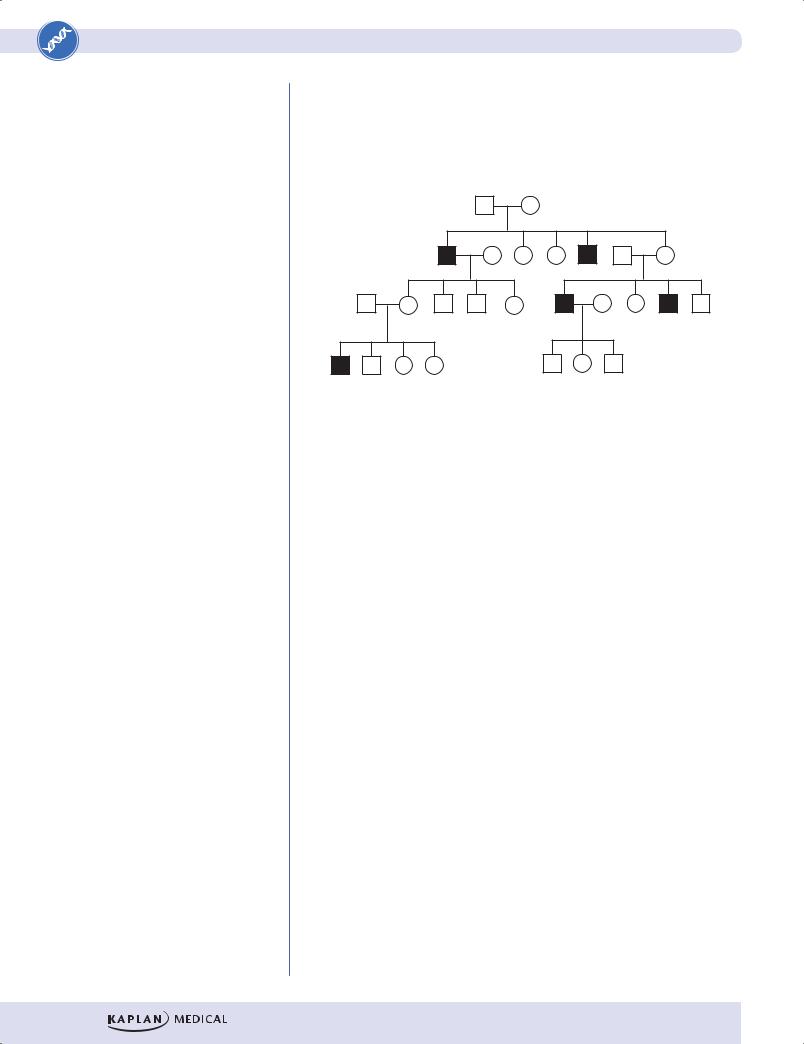
Pedigrees
A patient’s family history is diagrammed in a pedigree. The first affected individual to be identified in the family is termed the proband.
Generation |
|
|
|
|
|
|
|
I |
|
1 |
2 |
|
|
|
|
|
|
|
|
|
|
||
II |
2 |
3 |
4 |
5 |
6 |
|
|
1 |
|
|
|||||
III |
|
|
|
|
|
|
|
1 |
2 |
3 |
4 |
5 |
6 |
7 |
SB |
IV |
|
1 |
2 |
|
3 |
|
|
|
|
|
|
|
|||
Male |
|
|
|
|
Dead |
|
|
Female |
|
|
|
|
Mating |
|
|
Unknown sex |
|
|
|
Consanguineous or |
|||
|
|
|
|
|
incestuous mating |
||
Affected |
|
|
|
|
Sibship |
|
|
Carrier of an autosomal |
|
|
Dizygotic twins |
|
|||
recessive (Optional) |
|
|
|
||||
Carrier of an X-linked |
|
|
|
|
|
||
recessive (Optional) |
|
|
Monozygotic twins |
||||
Stillborn |
|
|
|
|
|||
|
|
|
|
|
|
|
|
SB |
|
|
|
|
|
|
|
Figure II-1-1. Pedigree Nomenclature
MAJOR MODES OF INHERITANCE
Autosomal Dominant Inheritance
High-Yield
A number of features in a pedigree help identify autosomal dominant inheritance:
•Because affected individuals must receive a disease-causing gene from an affected parent, the disease is typically observed in multiple generations of a pedigree.
•Skipped generations are not typically seen because two unaffected parents cannot transmit a disease-causing allele to their offspring (an exception occurs when there is reduced penetrance).
•Because these genes are located on autosomes, males and females are affected in roughly equal frequencies.
305

Part II ● Medical Genetics
Autosomal dominant alleles are relatively rare in populations, so the typical mating pattern is a heterozygous affected individual (Aa genotype) mating with a homozygous normal individual (aa genotype). Note that, by convention, the dominant allele is shown in uppercase (A) and the recessive allele is shown in lowercase (a). The recurrence risk is thus 50%, and half the children, on average, will be affected with the disease. If both parents are heterozygous, the recurrence risk is 75%.
Note
Autosomal Dominant Diseases
•Familial hypercholesterolemia (LDL receptor deficiency)
•Huntington disease
•Neurofibromatosis type 1
•Marfan syndrome
•Acute intermittent porphyria
Figure II-1-2. Autosomal Dominant Inheritance
|
|
A |
a |
|
|
|
|
|
|
a |
Aa |
aa |
|
|
|
|
|
|
|
a |
Aa |
aa |
||
A Punnett square: Affected offspring (Aa) are shaded.
Figure II-1-3. Recurrence Risk for the Mating of Affected Individual (Aa) with a Homozygous Unaffected Individual (aa) using a Punnett Square
Autosomal Recessive Inheritance |
High-Yield |
|
Important features that distinguish autosomal recessive inheritance:
•Because autosomal recessive alleles are clinically expressed only in the homozygous state, the offspring must inherit one copy of the diseasecausing allele from each parent.
•In contrast to autosomal dominant diseases, autosomal recessive diseases are typically seen in only one generation of a pedigree.
•Because these genes are located on autosomes, males and females are affected in roughly equal frequencies.
Most commonly, a homozygote is produced by the union of two heterozygous (carrier) parents. The recurrence risk for offspring of such matings is 25%.
306

Chapter 1 ● Single-Gene Disorders
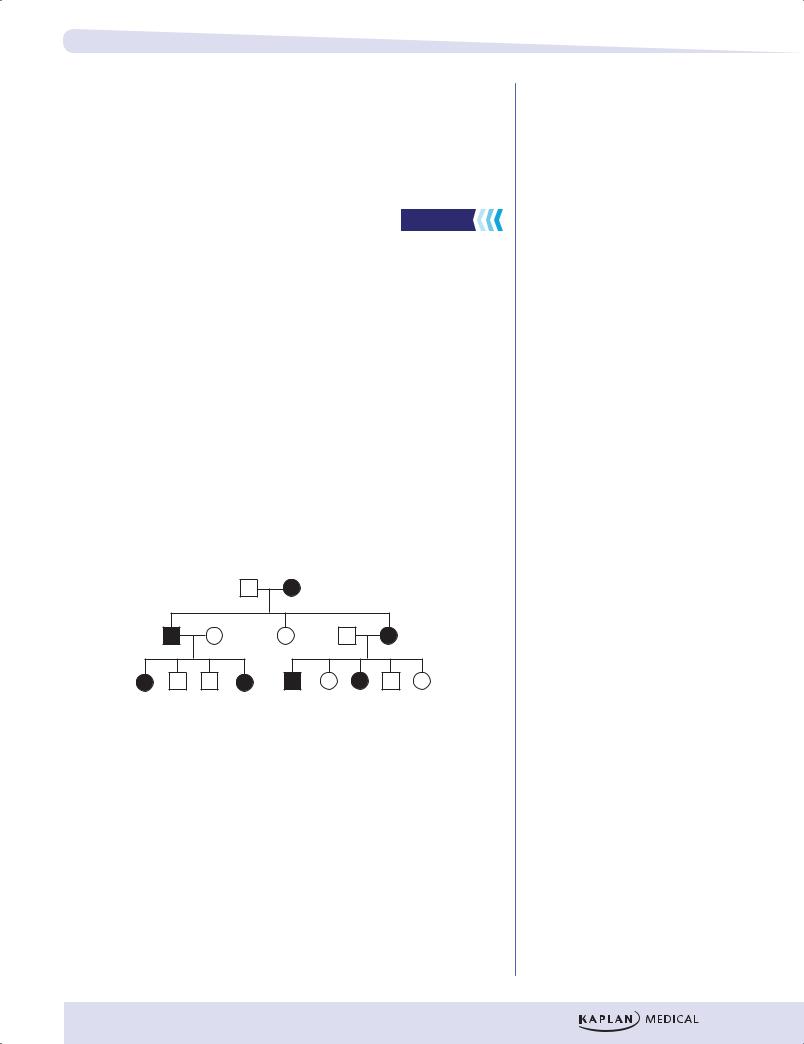
Consanguinity (the mating of related individuals) is sometimes seen in recessive pedigrees because individuals who share common ancestors are more likely to carry the same recessive disease-causing alleles.
A consanguineous mating has produced two affected offspring.
Figure II-1-4. Pedigree for an Autosomal Recessive Disease
Determining the Recurrence Risk for an Individual Whose Phenotype Is Known. In Figure II-1-4, individual IV-1 may wish to know his risk of being a carrier. Because his phenotype is known, there are only 3 possible genotypes he can have, assuming complete penetrance of the disease-producing allele. He cannot be homozygous for the recessive allele (aa). Two of the remaining 3 possibilities are carriers (Aa and aA), and one is homozygous normal (AA). Thus, his risk of being a carrier is 2/3, or 0.67 (67%).
|
A |
a |
A |
AA |
Aa |
a |
Aa |
aa |
The affected genotype (aa) is shaded.
Figure II-1-5. Recurrence Risk for the Mating of
Two Heterozygous Carriers (Aa) of a Recessive Mutation
X-Linked Recessive Inheritance |
High-Yield |
|
Properties of X-linked recessive inheritance
Because males have only one copy of the X chromosome, they are said to be hemizygous (hemi = “half”) for the X chromosome. If a recessive diseasecausing mutation occurs on the X chromosome, a male will be affected with the disease.
•Because males require only one copy of the mutation to express the disease and females require 2 copies, X-linked recessive diseases are seen much more commonly in males than in females.
Note
Autosomal Recessive Diseases
•Sickle cell anemia
•Cystic fibrosis
•Phenylketonuria (PKU)
•Tay-Sachs disease (hexosaminidase A deficiency)
Note
Cystic fibrosis is an important topic on the exam.
307
Part II ● Medical Genetics
Note
X-Linked Recessive Diseases
•Duchenne muscular dystrophy
•Lesch-Nyhan syndrome (hypoxanthine-guanine phosphoribosyltransferase [HGPRT] deficiency)
•Glucose-6-phosphate dehydrogenase deficiency
•Hemophilia A and B
•Red-green color blindness
•Menke’s disease
•Ornithine transcarbamoylase (OTC) deficiency
•SCID (IL-receptor γ-chain deficiency)
•Skipped generations are commonly seen because an affected male can transmit the disease-causing mutation to a heterozygous daughter, who is unaffected but who can transmit the disease-causing allele to her sons.
•Male-to-male transmission is not seen in X-linked inheritance; this helps distinguish it from autosomal inheritance.
Figure II-1-6. X-Linked Recessive Inheritance
Recurrence risks
Figure II-1-7 shows the recurrence risks for X-linked recessive diseases.
•Affected male–homozygous normal female: All of the daughters will be heterozygous carriers; all of the sons will be homozygous normal.
•Normal male–carrier female: On average, half of the sons will be affected and half of the daughters will be carriers. Note that in this case, the recurrence rate is different depending on the sex of the child. If the fetal sex is known, the recurrence rate for a daughter is 0, and that for a son is 50%. If the sex of the fetus is not known, then the recurrence rate is multiplied by 1/2, the probability that the fetus is a male versus a female. Therefore if the sex is unknown, the recurrence risk is 25%.
|
x |
|
Y |
|
X |
|
Y |
X |
Xx |
|
XY |
X |
XX |
|
XY |
|
|
|
|
|
|
|
|
X |
Xx |
|
XY |
x |
Xx |
|
xY |
|
|
|
|
|
|
|
|
|
|
A |
|
|
B |
||
A.Affected male–homozygous normal female
(X chromosome with mutation is in lower case)
B.Normal male–carrier female
Figure II-1-7. Recurrence Risks for X-Linked Recessive Diseases
308

X inactivation
Normal males inherit an X chromosome from their mother and a Y chromosome from their father, whereas normal females inherit an X chromosome from each parent. Because the Y chromosome carries only about 50 protein-coding genes and the X chromosome carries hundreds of protein-coding genes, a mechanism must exist to equalize the amount of protein encoded by X chromosomes in males and females. This mechanism, termed X inactivation, occurs in the blastocyst (~100 cells) during the development of female embryos. When an X chromosome is inactivated, its DNA is not transcribed into mRNA, and the chromosome is visualized under the microscope as a highly condensed Barr body in the nuclei of interphase cells. X inactivation has several important characteristics:
•It is random—in some cells of the female embryo, the X chromosome inherited from the father is inactivated, and in others the X chromosome inherited from the mother is inactivated. Like coin tossing, this is a random process. As shown in Figure II-1-6, most women have their paternal X chromosome active in approximately 50% of their cells and the maternal X chromosome active in approximately 50% of their cells. Thus, females are said to be mosaics with respect to the active X chromosome.
•It is fixed—once inactivation of an X chromosome occurs in a cell, the same X chromosome is inactivated in all descendants of the cell.
•It is incomplete—there are regions throughout the X chromosome, including the tips of both the long and short arms, that are not inactivated.
•X-chromosome inactivation is permanent in somatic cells and reversible in developing germ line cells. Both X chromosomes are active during oogenesis.
•All X chromosomes in a cell are inactivated except one. For example, females with 3 X chromosomes in each cell (see Chapter 3) have two X chromosomes inactivated in each cell (thus, two Barr bodies can be visualized in an interphase cell). X-chromosome inactivation is thought to be mediated by >1 mechanism.
•A gene called XIST has been identified as the primary gene that causes X inactivation. XIST produces an RNA product that coats the chromosome, helping produce its inactivation.
•Condensation into heterochromatin
•Methylation of gene regions on the X chromosome
Chapter 1 ● Single-Gene Disorders
Note
X inactivation occurs early in the female embryo and is random, fixed, and incomplete. In a cell, all X chromosomes but one are inactivated.
Note
Genetic mosaicism is the presence of 2 or more cell lines with different karyotypes in an individual. It arises from mitotic nondisjunction. The number of cell lines that develop and their relative proportions are influenced by the timing of nondisjunction during embryogenesis and the viability of the aneuploid cells produced.
309

Part II ● Medical Genetics
Paternal X |
Maternal |
Barr body |
X active |
Blastocyst ~100 cells
Paternal |
Maternal X |
X active |
Barr body |
Females
Number of
5/95 50/50 95/5
Percentage of cells with paternal/maternal X active
Figure II-1-8. Inactivation of the X Chromosome during
Embryogenesis Is a Random Process
Manifesting (female) heterozygotes
Normal females have two copies of the X chromosome, so they usually require two copies of the mutation to express the disease. However, because X inactivation is a random process, a heterozygous female will occasionally express an X-linked recessive mutation because, by random chance, most of the X chromosomes carrying the normal allele have been inactivated. Such females are termed manifesting heterozygotes. Because they usually have at least a small population of active X chromosomes carrying the normal allele, their disease expression is typically milder than that of hemizygous males.
Recall Question
Which of the following is an X-linked recessive disease?
A.Acute intermittent porphyria
B.Duchenne muscular dystrophy
C.Huntington disease
D.Marfan syndrome
Answer: B
310
Chapter 1 ● Single-Gene Disorders
Y Chromosome Highlights
•The SRY (sex determining region) gene is a transcription factor that initiates male development.
•The q arm of Y chromosomes contains a large block of heterochromatin.
•Microdeletions of Yq in males result in nonobstructive azoospermia.
X-Linked Dominant Inheritance |
High-Yield |
|
There are relatively few diseases whose inheritance is classified as X-linked dominant. Fragile X syndrome is an important example. In this condition, females are differently affected than males, and whereas penetrance in males is 100%, that in females is approximately 60%. The typical fragile X patient described is male.
As in X-linked recessive inheritance, male–male transmission of the diseasecausing mutation is not seen.
•Heterozygous females are affected. Because females have 2 X chromosomes (and thus 2 chances to inherit an X-linked diseasecausing mutation) and males have only one, X-linked dominant diseases are seen about twice as often in females as in males.
•As in autosomal dominant inheritance, the disease phenotype is seen in multiple generations of a pedigree; skipped generations are relatively unusual.
•Examine the children of an affected male (II-1 in the figure below). None of his sons will be affected, but all of his daughters have the disease (assuming complete penetrance).
Figure II-1-9. X-Linked Dominant Inheritance
Clinical Correlate
Fragile X Syndrome
Males: 100% penetrance
•Mental retardation
•Large ears
•Prominent jaw
•Macro-orchidism (usually postpubertal)
Females: 60% penetrance
• Mental retardation
Note
The penetrance of a disease-causing mutation is the percentage of individuals who are known to have the disease-causing genotype who display the disease phenotype (develop symptoms).
Recurrence Risks
Figure II-1-10 shows the recurrence risks for X-linked dominant inheritance.
•Affected male–homozygous normal female: None of the sons are affected; all of the daughters are affected. Note that in this case, the recurrence rate is different depending on the sex of the child. If the fetal sex is known, the recurrence rate for a daughter is 100%, and that for a son is 0%. If the sex of the fetus is not known, then the recurrence rate is multiplied by 1/2, the probability that the fetus is a male versus a female. Therefore if the sex is unknown, the recurrence risk is 50%.
•Normal male–heterozygous affected female: on average, 50% of sons are affected and 50% of daughters are affected.
311
Part II ● Medical Genetics
Note
X-Linked Dominant Diseases
•Hypophosphatemic rickets
•Fragile X syndrome
Affected male–homozygous normal female (the mutation-carrying chromosome is upper case)
|
X |
Y |
|
|
|
x |
Xx |
xY |
|
|
|
x |
Xx |
xY |
|
|
|
Normal male–heterozygous affected female
x Y
X |
Xx |
XY |
x |
xx |
xY |
Figure II-1-10. Recurrence Risks for X-Linked Dominant Inheritance
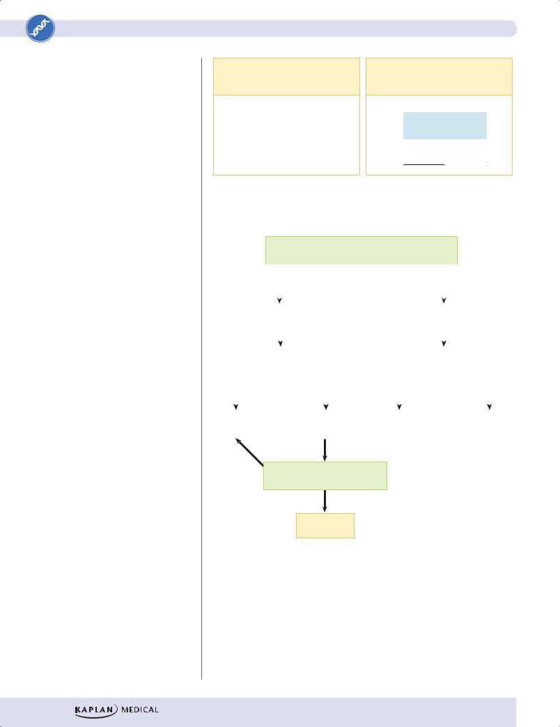
Affected individuals have an affected parent? (Multiple generations affected?)
|
|
|
|
|
|
|
|
|
|
|
|
|
|
|
|
|
|
|
|
|
|
|
|
|
|
|
|
Yes |
|
|
|
|
|
|
|
|
|
|
|
|
|
|
No |
||||||
|
|
|
|
|
|
|
|
|
|
|
|
|
|
|
|
|
||||||||
|
|
|
|
|
|
|
|
|
|
|
|
|
|
|
|
|
|
|
|
|
|
|
|
|
|
|
|
|
|
|
|
|
|
|
|
|
|
|
|
|
|
|
|
||||||
|
|
|
Dominant |
|
|
|
|
|
|
|
|
|
|
Recessive |
|
|||||||||
|
|
|
|
|
|
|
|
|
|
|
|
|
|
|
|
|
|
|
|
|
|
|
|
|
|
|
|
|
|
|
|
|
|
|
|
|
|
|
|
|
|
|
|
|
|
|
|
|
|
|
|
|
Male-male |
|
|
|
|
|
|
|
All (or almost all) affected |
|
||||||||||||
|
|
|
transmission? |
|
|
|
|
|
|
|
are males? |
|
||||||||||||
Yes |
|
|
|
|
|
|
|
|
No |
|
|
Yes |
|
|
|
|
|
|
|
No |
||||
|
|
|
|
|
|
|
|
|
|
|
|
|
|
|
|
|
||||||||
|
|
|
|
|
|
|
|
|
|
|
|
|
|
|
|
|
|
|
|
|
|
|
|
|
|
|
|
|
|
|
|
|
|
|
|
|
|
|
|
|
|
||||||||
Autosomal |
|
|
|
|
May be X- |
|
|
X-linked |
|
|
|
Autosomal |
||||||||||||
dominant |
|
|
|
|
dominant |
|
|
recessive |
|
|
|
recessive |
||||||||||||
No
Are all daughters of an affected male also affected?
Yes
X-dominant
Note: If transmission occurs only through affected mothers and never through affected sons, the pedigree is likely to reflect mitochondrial inheritance.
Figure II-1-11. A Basic Decision Tree for Determining the Mode of Inheritance in a Pedigree
312
Chapter 1 ● Single-Gene Disorders
Mitochondrial Inheritance
High-Yield
Mitochondria, which are cytoplasmic organelles involved in cellular respiration, have their own chromosomes, each of which contains 16,569 DNA base pairs (bp) arranged in a circular molecule. This DNA encodes 13 proteins that are subunits of complexes in the electron transport and oxidative phosphorylation processes (see Part I, Chapter 13). In addition, mitochondrial DNA encodes 22 transfer RNAs and 2 ribosomal RNAs.
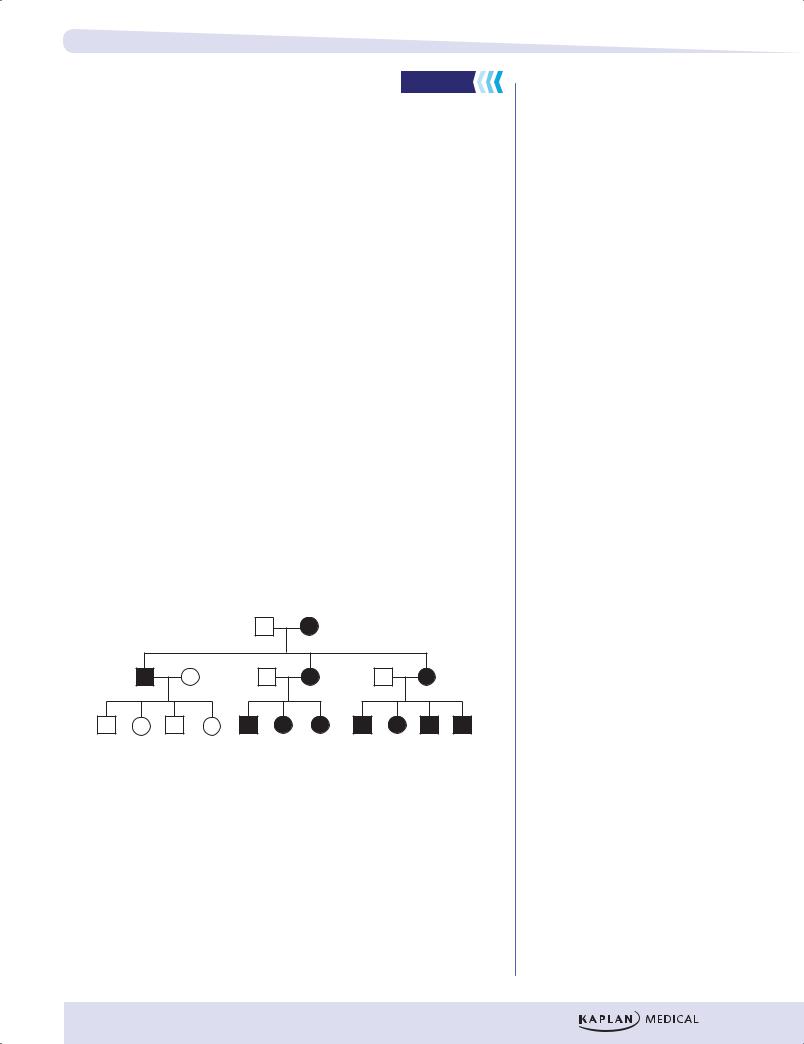
Because a sperm cell contributes no mitochondria to the egg cell during fertilization, mitochondrial DNA is inherited exclusively through females. Pedigrees for mitochondrial diseases thus display a distinct mode of inheritance: Diseases are transmitted only from affected females to their offspring.
•Both males and females are affected.
•Transmission of the disease is only from a female.
•All offspring of an affected female are affected.
•None of the offspring of an affected male is affected.
•Diseases are typically neuropathies and/or myopathies.
Heteroplasmy
A typical cell contains hundreds of mitochondria in its cytoplasm, and each mitochondrion has its own copy of the mitochondrial genome. When a specific mutation occurs in some of the mitochondria, this mutation can be unevenly distributed into daughter cells during cell division: Some cells may inherit more mitochondria in which the normal DNA sequence predominates, while others inherit mostly mitochondria with the mutated, disease-causing gene. This condition is known as heteroplasmy. Variations in heteroplasmy account for substantial variation in the severity of expression of mitochondrial diseases.
Note
Mitochondrial Diseases
• Leber hereditary optic neuropathy
• |
MELAS: mitochondrial |
|
encephalomyopathy, lactic acidosis, |
|
and stroke-like episodes |
• |
Myoclonic epilepsy with ragged red |
Figure II-1-12. Pedigree for a Mitochondrial Disease |
muscle fibers |
|
313