
- •Preface
- •Foreword
- •Contents
- •Contributors
- •1. Medical History
- •1.1 Congestive Heart Failure
- •1.2 Angina Pectoris
- •1.3 Myocardial Infarction
- •1.4 Rheumatic Heart Disease
- •1.5 Heart Murmur
- •1.6 Congenital Heart Disease
- •1.7 Cardiac Arrhythmia
- •1.8 Prosthetic Heart Valve
- •1.9 Surgically Corrected Heart Disease
- •1.10 Heart Pacemaker
- •1.11 Hypertension
- •1.12 Orthostatic Hypotension
- •1.13 Cerebrovascular Accident
- •1.14 Anemia and Other Blood Diseases
- •1.15 Leukemia
- •1.16 Hemorrhagic Diatheses
- •1.17 Patients Receiving Anticoagulants
- •1.18 Hyperthyroidism
- •1.19 Diabetes Mellitus
- •1.20 Renal Disease
- •1.21 Patients Receiving Corticosteroids
- •1.22 Cushing’s Syndrome
- •1.23 Asthma
- •1.24 Tuberculosis
- •1.25 Infectious Diseases (Hepatitis B, C, and AIDS)
- •1.26 Epilepsy
- •1.27 Diseases of the Skeletal System
- •1.28 Radiotherapy Patients
- •1.29 Allergy
- •1.30 Fainting
- •1.31 Pregnancy
- •Bibliography
- •2.1 Radiographic Assessment
- •2.2 Magnification Technique
- •2.4 Tube Shift Principle
- •2.5 Vertical Transversal Tomography of the Jaw
- •Bibliography
- •3. Principles of Surgery
- •3.1 Sterilization of Instruments
- •3.2 Preparation of Patient
- •3.3 Preparation of Surgeon
- •3.4 Surgical Incisions and Flaps
- •3.5 Types of Flaps
- •3.6 Reflection of the Mucoperiosteum
- •3.7 Suturing
- •Bibliography
- •4.1 Surgical Unit and Handpiece
- •4.2 Bone Burs
- •4.3 Scalpel (Handle and Blade)
- •4.4 Periosteal Elevator
- •4.5 Hemostats
- •4.6 Surgical – Anatomic Forceps
- •4.7 Rongeur Forceps
- •4.8 Bone File
- •4.9 Chisel and Mallet
- •4.10 Needle Holders
- •4.11 Scissors
- •4.12 Towel Clamps
- •4.13 Retractors
- •4.14 Bite Blocks and Mouth Props
- •4.15 Surgical Suction
- •4.16 Irrigation Instruments
- •4.17 Electrosurgical Unit
- •4.18 Binocular Loupes with Light Source
- •4.19 Extraction Forceps
- •4.20 Elevators
- •4.21 Other Types of Elevators
- •4.22 Special Instrument for Removal of Roots
- •4.23 Periapical Curettes
- •4.24 Desmotomes
- •4.25 Sets of Necessary Instruments
- •4.26 Sutures
- •4.27 Needles
- •4.28 Local Hemostatic Drugs
- •4.30 Materials for Tissue Regeneration
- •Bibliography
- •5. Simple Tooth Extraction
- •5.1 Patient Position
- •5.2 Separation of Tooth from Soft Tissues
- •5.3 Extraction Technique Using Tooth Forceps
- •5.4 Extraction Technique Using Root Tip Forceps
- •5.5 Extraction Technique Using Elevator
- •5.6 Postextraction Care of Tooth Socket
- •5.7 Postoperative Instructions
- •Bibliography
- •6. Surgical Tooth Extraction
- •6.1 Indications
- •6.2 Contraindications
- •6.3 Steps of Surgical Extraction
- •6.4 Surgical Extraction of Teeth with Intact Crown
- •6.5 Surgical Extraction of Roots
- •6.6 Surgical Extraction of Root Tips
- •Bibliography
- •7.1 Medical History
- •7.2 Clinical Examination
- •7.3 Radiographic Examination
- •7.4 Indications for Extraction
- •7.5 Appropriate Timing for Removal of Impacted Teeth
- •7.6 Steps of Surgical Procedure
- •7.7 Extraction of Impacted Mandibular Teeth
- •7.8 Extraction of Impacted Maxillary Teeth
- •7.9 Exposure of Impacted Teeth for Orthodontic Treatment
- •Bibliography
- •8.1 Perioperative Complications
- •8.2 Postoperative Complications
- •Bibliography
- •9. Odontogenic Infections
- •9.1 Infections of the Orofacial Region
- •Bibliography
- •10. Preprosthetic Surgery
- •10.1 Hard Tissue Lesions or Abnormalities
- •10.2 Soft Tissue Lesions or Abnormalities
- •Bibliography
- •11.1 Principles for Successful Outcome of Biopsy
- •11.2 Instruments and Materials
- •11.3 Excisional Biopsy
- •11.4 Incisional Biopsy
- •11.5 Aspiration Biopsy
- •11.6 Specimen Care
- •11.7 Exfoliative Cytology
- •11.8 Tolouidine Blue Staining
- •Bibliography
- •12.1 Clinical Presentation
- •12.2 Radiographic Examination
- •12.3 Aspiration of Contents of Cystic Sac
- •12.4 Surgical Technique
- •Bibliography
- •13. Apicoectomy
- •13.1 Indications
- •13.2 Contraindications
- •13.3 Armamentarium
- •13.4 Surgical Technique
- •13.5 Complications
- •Bibliography
- •14.1 Removal of Sialolith from Duct of Submandibular Gland
- •14.2 Removal of Mucus Cysts
- •Bibliography
- •15. Osseointegrated Implants
- •15.1 Indications
- •15.2 Contraindications
- •15.3 Instruments
- •15.4 Surgical Procedure
- •15.5 Complications
- •15.6 Bone Augmentation Procedures
- •Bibliography
- •16.1 Treatment of Odontogenic Infections
- •16.2 Prophylactic Use of Antibiotics
- •16.3 Osteomyelitis
- •16.4 Actinomycosis
- •Bibliography
- •Subject Index
64 F. D. Fragiskos
4.25
Sets of Necessary Instruments
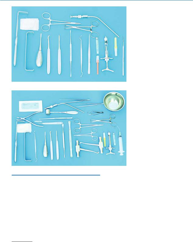
For practical reasons, sterilized and packaged full sets of instruments for the most common surgical procedures must always be available. These sets include:
a.Set for simple tooth extraction (Fig. 4.57)1):
1.Local anesthesia syringe, needle, and ampule.
2.Desmotome or Freer elevator.
3.Retractor or mouth mirror.
4.Extraction forceps
(depending on the tooth to be removed).
1)Disposable materials (e.g., needles for anesthesia, gauze, sutures, etc.) shown in Figs. 4.57–4.60 are not included in the set at the time of sterilization. These are usually placed on the surgery tray afterwards together with the rest of the instruments.
Fig. 4.57. Set of instruments necessary for simple tooth extraction
Fig. 4.58. Set of instruments necessary for surgical tooth extraction
5.Surgical or anatomic forceps.
6.Elevators.
7.Sterile gauze.
8.Periapical curette.
9.Suction tip.
10.Towel clamp.
11.Needle holder.
b. Set for surgical tooth extraction (Fig. 4.58):
1.Local anesthesia syringe, needle, and ampule.
2.Scalpel and blade.
3.Periosteal elevators.
4.Elevators.
5.Bone chisel.
6.Mallet.
7.Rongeur forceps.
8.Bone file.
9.Periapical curette.
10.Bone burs.
11.Hemostat.
Chapter 4 Equipment, Instruments, and Materials |
65 |
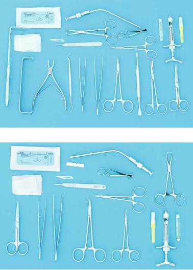
Fig. 4.59. Set of instruments necessary for soft tissue specimen sampling by biopsy
Fig. 4.60. Set of instruments necessary for incision and drainage of abscesses
12.Retractors.
13.Needle holder.
14.Surgical forceps and anatomic forceps.
15.Scissors.
16.Towel clamps.
17.Disposable plastic syringe.
18.Suction tip.
19.Straight handpiece.
20.Bowl for saline solution.
21.Sutures.
22.Sterile gauze.
c.Set of instruments for surgical biopsy
(bone and soft tissue) (Fig. 4.59):
1.Local anesthesia syringe, needle, and ampule.
2.Scalpel and blade.
3.Periosteal elevator.
4.Scissors.
5.Surgical forceps and anatomic forceps.
6.Periapical curette.
7.Needle holder.
8.Hemostats.
9.Rongeur forceps.
10.Towel clamps.
11.Suction tip.
12.Sutures.
13.Sterile gauze.
14.Retractors.
d.Set of instruments for incision and drainage of abscess (Fig. 4.60):
1.Local anesthesia syringe, needle, and ampule.
2.Scalpel and blade.
3.Hemostats.
4.Surgical and anatomic forceps.
5.Scissors.
6.Needle holder.
7.Suction tip.
8.Towel clamps.
9.Sutures.
10.Sterilized Penrose rubber drain 1/4 in.
11.Sterile gauze.
66 F. D. Fragiskos
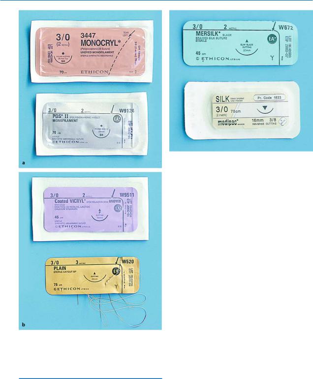
Fig. 4.62. Nonresorbable surgical sutures made of silk
Fig. 4.61 a, b. Different types of resorbable sutures made from gut tissue and synthetic material
4.26 Sutures
Great progress in sutures has been made since 1865, when disinfection and sterilization first started being used in surgery. There is a big variety in the size of
surgical sutures available today, and two basic categories: (1) resorbable, and (2) nonresorbable sutures.
Resorbable Sutures. These sutures are resorbed after a certain time, which usually coincides with healing of the wound. These sutures are made of gut or vital tissue (catgut, collagen, fascia, etc.) and are plain or chromic, or of synthetic material, e.g., polyglycolic acid
(Dexon) (Fig. 4.61). Plain catgut sutures are resorbed postsurgically over 8 days, chromic sutures in 12– 15 days, and synthetic (Dexon) sutures in approximately 30 days. These types of sutures are used for flaps with little tension, children, mentally handicapped patients, and generally for patients who cannot return to the clinic to have the sutures removed.
Nonresorbable Sutures. These sutures remain in the tissues and are not resorbed, but have to be cut and removed about 7 days after their placement. They are fabricated of various natural materials, mainly surgical silk (monofilamentous or multifilamentous, in many diameters and lengths) and surgical cotton suture. Silk sutures are the easiest to use and the most economical, and have a satisfactory ability to hold a knot (Fig. 4.62).
The most commonly used suture sizes are 4–0 and 3–0 for resorbable sutures, and 3–0 and 2–0 for nonresorbable sutures. These kinds of sutures are sold in sterilized packages with pre-attached atraumatic needles or in bundles without needles.
Chapter 4 Equipment, Instruments, and Materials |
67 |
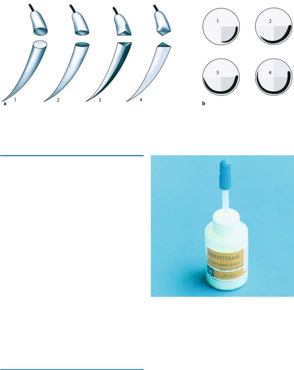
Fig. 4.63 a, b. Cross-sectional view of needles. a Round tapered (1), oval tapered (2), cutting (3, triangular with one of the three cutting edges on the inside of the semicircle), reverse-cutting (4, triangular with two cutting edges on the
inside of the semi-circle). b Size of needle compared to regular circle: one-quarter of a circle (1), three-eighths of a circle (2), half a circle (3), three-quarters of a circle (4)
4.27 Needles
A variety of needles are available in oral surgery, and they may differ in shape, diameter, cross-sectional view, and size (Fig. 4.63). They are usually made of stainless steel, which is a strong and flexible material. The needles preferred by surgeons today are atraumatic disposable needles with pre-attached sutures on their posterior ends. Needles that may be used and sterilized many times are also available, with an eye or groove in the needle, through which the suture is passed.
Needles with Round or Oval Cross-Sectional View.
These are considered atraumatic and are mainly used for suturing thin mucosa. Their disadvantage is that great pressure is required when passing through the tissues, which may make suturing the wound harder.
Triangular Needles. These needles have sharp cutting edges and are preferred for suturing thicker tissues. When they are used for thin mucosa, care is required because they may tear the tissues. The most suitable needles are semicircular or three-eighths of a circle and 19–20 mm long, in both cases.
4.28
Local Hemostatic Drugs
These drugs are suitable only for local use and can stop heavy bleeding, which is due to injury of capillaries or arterioles. The main hemostatic drugs are listed below.
Fig. 4.64. Hemostatic powder suitable for stopping capillary bleeding
Alginic Acid. This is sold in powder form in special
5-mg packages (Fig. 4.64). It is placed on the bleeding surface, creating a protective membrane that applies pressure to the capillaries and helps hold the blood clot in place.
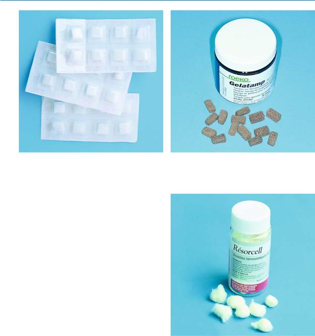
Natural Collagen Sponge. This is a white sponge material, nonantigenic and fully absorbable (Fig. 4.65). Its hemostatic ability is due to promotion of platelet aggregation. Also, it activates coagulation factors XI and XIII. It is used for patients who are prone to hemorrhage after dental surgical procedures.
68 F. D. Fragiskos
Fig. 4.65. Absorbable hemostatic natural collagen sponges. These are indicated in cases of postextraction bleeding
Fibrin Sponge. The fibrin sponge is nonantigenic, and is prepared from bovine material that has been processed in order to avoid allergic reactions. It is used locally in the bleeding area and especially in the postextraction socket. It promotes coagulation, creating a normal hemostatic blood clot, but it also functions as a plug over the edges of the bleeding area. The fibrin sponge is fully absorbed by the tissues within
4–6 weeks.
Gelatin Sponge. This is a relatively spongy material, nonantigenic and fully absorbable (Fig. 4.66). Its hemostatic action and application are the same as that of the fibrin sponge.
Oxidized Cellulose. This is an absorbable hemostatic material, which is manufactured by controlled oxidation of cellulose by nitrous dioxide. It is available in gauze form or pellet form (Fig. 4.67). It is used topically as a hemostatic material, because it releases cytotoxic acid, which has significant affinity for hemoglobin. Its attachment to the walls of the postextraction socket for the treatment of bleeding is quite satisfactory and therefore it is considered superior to various other hemostatic sponges, which have a tendency to expel the material from the socket.
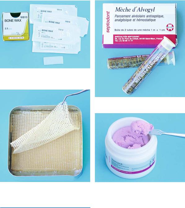
Bone Wax. Bone wax is a sterilized, nonabsorbable mix of waxes, and is composed of white beeswax, paraffin wax, and an isopropyl ester of palmitic acid
Fig. 4.66. Gelatin sponges. These are used to treat postextraction bleeding
Fig. 4.67. Oxidized cellulose in pellet form
(Fig. 4.68). It is white and available as a solid rectangular plate weighing 2.5 g. It is used to control bleeding that originates in bone or chipped edges of bone. Before its application, bone wax is first warmed with the fingers, so that the desirable consistency is reached. Its hemostatic action is brought about through mechanical obstruction of the osseous cavity, which contains the bleeding vessels.
Chapter 4 Equipment, Instruments, and Materials |
69 |
Fig. 4.68. Surgical bone wax for treatment of bone hemorrhage
Fig. 4.69. Petrolatum (Vaseline®) gauze in a sterile container
4.29
Materials for Covering
or Filling a Surgical Wound
Petrolatum Gauze. Petrolatum (Vaseline®) gauze is available in sterilized packages and is used mainly for covering exposed wounds, for tamponade of bone cavities after marsupialization of cysts, for surgical procedures in the maxillary sinus, etc. Before its application, the excess petrolatum must be removed and the gauze saturated with antibiotic ointment (oxytetracycline)
(Fig. 4.69), if deemed necessary.
Fig. 4.70. Iodoform gauze for the treatment of fibrinolytic alveolitis (dry socket)
Fig. 4.71. Surgical gingival dressing for protection of an exposed postoperative field, until healing by secondary intention occurs
Iodoform Gauze. This gauze has antiseptic, analgesic and hemostatic properties. Its indications for use are the same as for petrolatum gauze, although it may remain in place for longer. The iodoform gauze is also available in small-sized packages (Fig. 4.70), for the treatment of dry socket.
Surgical Dressing. This is an autopolymerized puttylike paste, available in sterilized packaging. It is used in periodontology and oral surgery as a temporary protective covering of intraoral wounds after surgical procedures (Figs. 4.71, 4.72).
