- •Preface
- •Foreword
- •Contents
- •Contributors
- •1. Medical History
- •1.1 Congestive Heart Failure
- •1.2 Angina Pectoris
- •1.3 Myocardial Infarction
- •1.4 Rheumatic Heart Disease
- •1.5 Heart Murmur
- •1.6 Congenital Heart Disease
- •1.7 Cardiac Arrhythmia
- •1.8 Prosthetic Heart Valve
- •1.9 Surgically Corrected Heart Disease
- •1.10 Heart Pacemaker
- •1.11 Hypertension
- •1.12 Orthostatic Hypotension
- •1.13 Cerebrovascular Accident
- •1.14 Anemia and Other Blood Diseases
- •1.15 Leukemia
- •1.16 Hemorrhagic Diatheses
- •1.17 Patients Receiving Anticoagulants
- •1.18 Hyperthyroidism
- •1.19 Diabetes Mellitus
- •1.20 Renal Disease
- •1.21 Patients Receiving Corticosteroids
- •1.22 Cushing’s Syndrome
- •1.23 Asthma
- •1.24 Tuberculosis
- •1.25 Infectious Diseases (Hepatitis B, C, and AIDS)
- •1.26 Epilepsy
- •1.27 Diseases of the Skeletal System
- •1.28 Radiotherapy Patients
- •1.29 Allergy
- •1.30 Fainting
- •1.31 Pregnancy
- •Bibliography
- •2.1 Radiographic Assessment
- •2.2 Magnification Technique
- •2.4 Tube Shift Principle
- •2.5 Vertical Transversal Tomography of the Jaw
- •Bibliography
- •3. Principles of Surgery
- •3.1 Sterilization of Instruments
- •3.2 Preparation of Patient
- •3.3 Preparation of Surgeon
- •3.4 Surgical Incisions and Flaps
- •3.5 Types of Flaps
- •3.6 Reflection of the Mucoperiosteum
- •3.7 Suturing
- •Bibliography
- •4.1 Surgical Unit and Handpiece
- •4.2 Bone Burs
- •4.3 Scalpel (Handle and Blade)
- •4.4 Periosteal Elevator
- •4.5 Hemostats
- •4.6 Surgical – Anatomic Forceps
- •4.7 Rongeur Forceps
- •4.8 Bone File
- •4.9 Chisel and Mallet
- •4.10 Needle Holders
- •4.11 Scissors
- •4.12 Towel Clamps
- •4.13 Retractors
- •4.14 Bite Blocks and Mouth Props
- •4.15 Surgical Suction
- •4.16 Irrigation Instruments
- •4.17 Electrosurgical Unit
- •4.18 Binocular Loupes with Light Source
- •4.19 Extraction Forceps
- •4.20 Elevators
- •4.21 Other Types of Elevators
- •4.22 Special Instrument for Removal of Roots
- •4.23 Periapical Curettes
- •4.24 Desmotomes
- •4.25 Sets of Necessary Instruments
- •4.26 Sutures
- •4.27 Needles
- •4.28 Local Hemostatic Drugs
- •4.30 Materials for Tissue Regeneration
- •Bibliography
- •5. Simple Tooth Extraction
- •5.1 Patient Position
- •5.2 Separation of Tooth from Soft Tissues
- •5.3 Extraction Technique Using Tooth Forceps
- •5.4 Extraction Technique Using Root Tip Forceps
- •5.5 Extraction Technique Using Elevator
- •5.6 Postextraction Care of Tooth Socket
- •5.7 Postoperative Instructions
- •Bibliography
- •6. Surgical Tooth Extraction
- •6.1 Indications
- •6.2 Contraindications
- •6.3 Steps of Surgical Extraction
- •6.4 Surgical Extraction of Teeth with Intact Crown
- •6.5 Surgical Extraction of Roots
- •6.6 Surgical Extraction of Root Tips
- •Bibliography
- •7.1 Medical History
- •7.2 Clinical Examination
- •7.3 Radiographic Examination
- •7.4 Indications for Extraction
- •7.5 Appropriate Timing for Removal of Impacted Teeth
- •7.6 Steps of Surgical Procedure
- •7.7 Extraction of Impacted Mandibular Teeth
- •7.8 Extraction of Impacted Maxillary Teeth
- •7.9 Exposure of Impacted Teeth for Orthodontic Treatment
- •Bibliography
- •8.1 Perioperative Complications
- •8.2 Postoperative Complications
- •Bibliography
- •9. Odontogenic Infections
- •9.1 Infections of the Orofacial Region
- •Bibliography
- •10. Preprosthetic Surgery
- •10.1 Hard Tissue Lesions or Abnormalities
- •10.2 Soft Tissue Lesions or Abnormalities
- •Bibliography
- •11.1 Principles for Successful Outcome of Biopsy
- •11.2 Instruments and Materials
- •11.3 Excisional Biopsy
- •11.4 Incisional Biopsy
- •11.5 Aspiration Biopsy
- •11.6 Specimen Care
- •11.7 Exfoliative Cytology
- •11.8 Tolouidine Blue Staining
- •Bibliography
- •12.1 Clinical Presentation
- •12.2 Radiographic Examination
- •12.3 Aspiration of Contents of Cystic Sac
- •12.4 Surgical Technique
- •Bibliography
- •13. Apicoectomy
- •13.1 Indications
- •13.2 Contraindications
- •13.3 Armamentarium
- •13.4 Surgical Technique
- •13.5 Complications
- •Bibliography
- •14.1 Removal of Sialolith from Duct of Submandibular Gland
- •14.2 Removal of Mucus Cysts
- •Bibliography
- •15. Osseointegrated Implants
- •15.1 Indications
- •15.2 Contraindications
- •15.3 Instruments
- •15.4 Surgical Procedure
- •15.5 Complications
- •15.6 Bone Augmentation Procedures
- •Bibliography
- •16.1 Treatment of Odontogenic Infections
- •16.2 Prophylactic Use of Antibiotics
- •16.3 Osteomyelitis
- •16.4 Actinomycosis
- •Bibliography
- •Subject Index

Chapter 4 Equipment, Instruments, and Materials |
47 |
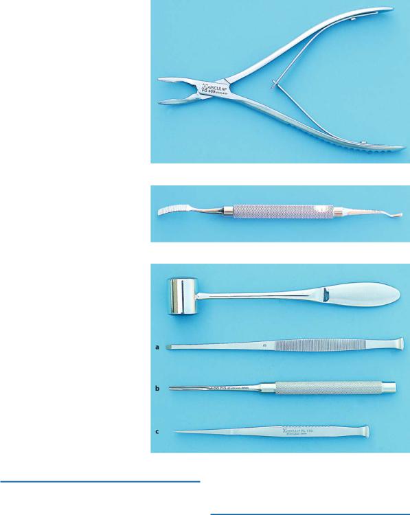
Fig. 4.12. Luer–Friedmann rongeur forceps with side-cutting/end-cutting edge
Fig. 4.13. Double-ended bone file with small and large ends
Fig. 4.14 a–c. Surgical mallet and chisels. a Partsch monobevel chisel. b Lucas chisel with concave end.
c Lambotte bibevel chisel
4.9
Chisel and Mallet
Mallets are instruments with heavy-weighted ends.
The surfaces of the ends are made of lead or of plastic so that some of the shock is absorbed when the mallet strikes the chisel.
The chisels used in oral surgery have different shapes and sizes. Their cutting edges are concave,
monobeveled or bibeveled (Fig. 4.14). The bibevel chisel is used for sectioning multi-rooted teeth.
4.10
Needle Holders
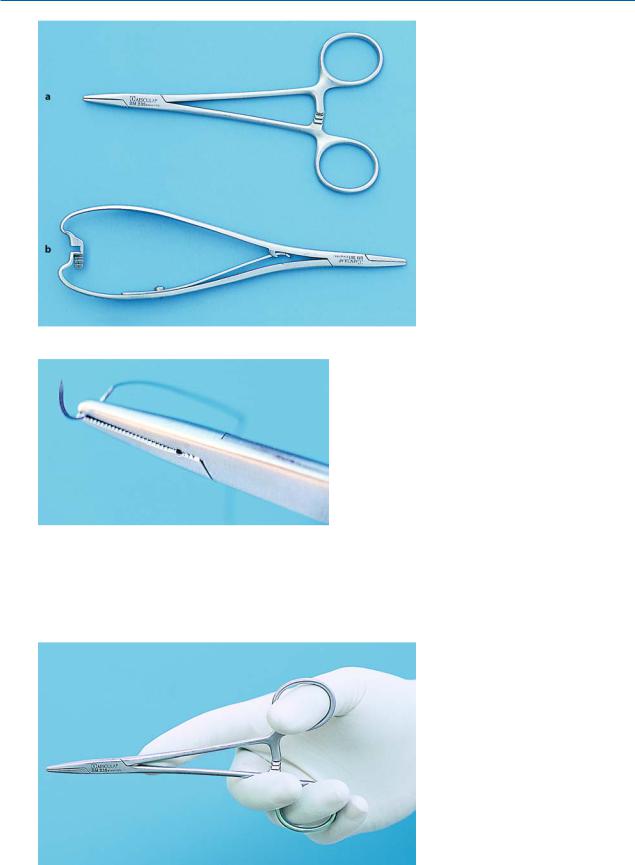
Needle holders are used for suturing the wound. The Mayo–Hegar and Mathieu needle holders are considered suitable for this purpose (Fig. 4.15). The first type

48 F. D. Fragiskos
Fig. 4.16. Beak of the needle holder grasps a suture needle. The needle holder’s beak face is crosshatched, ensuring stability of the needle during tissue penetration
Fig. 4.15 a, b. Needle holders. a Mayo–Hegar needle holder. b Mathieu needle holder
looks similar to a hemostat and is preferred mainly for intraoral placement of sutures. The hemostat and needle holder have the following differences:
ΟThe short beaks of the hemostat are thinner and longer compared to those of the needle holder.
ΟOn the needle holder, the internal surface of the short beaks is grooved and crosshatched, permitting a firm and stable grasp of the needle (Fig. 4.16), while the short beaks of the hemostat have parallel grooves which are perpendicular to the long axis of the instrument.
ΟThe needle holder can release the needle with simple pressure, because of the gap in the last step of the locking handle, whereas the hemostat requires a special maneuver, because it does not have that gap in the last step of the locking handle.
Fig. 4.17. Correct position of the fingers for holding the needle holder

Chapter 4 Equipment, Instruments, and Materials |
49 |
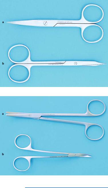
Fig. 4.18 a, b. a Standard suture scissors. b Goldman–Fox soft tissue scissors
Fig. 4.19 a, b. a Blunt-nosed Metzenbaum soft tissue scissors. b Lagrange soft tissue scissors
The correct way to hold the needle holder is to place the thumb in one ring of the handle and the ring finger in the other. The rest of the fingers are curved around the outside of the rings, while the fingertip of the index finger is placed on the hinge or a little further up, for better control of the instrument (Fig. 4.17).
4.11 Scissors
Various types of scissors are used in oral surgery, depending on the surgical procedure. They belong to the following categories: suture scissors and soft tissue scissors (Figs. 4.18, 4.19). The most commonly used scissors for cutting sutures have sharp cutting edges, while Goldman–Fox, Lagrange (which have slightly upward curved blades), and Metzenbaum are used for soft tissue. Lagrange scissors are narrow scissors with sharp blades and are mainly used for removing excess

50 F. D. Fragiskos
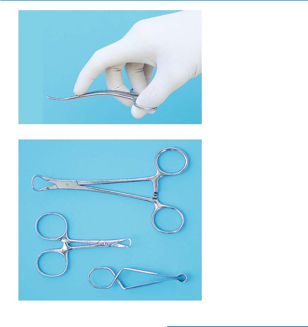
Fig. 4.20. Correct way to hold scissors
Fig. 4.21. Towel clamps
gingival tissue, while the Metzenbaum are blunt-nosed scissors and are suitable for dissecting and undermining the mucosa from the underlying soft tissues. Scissors are held the same way as needle holders (Fig. 4.20).
4.12
Towel Clamps
Towel clamps are mainly used for fastening sterile towels and drapes placed on the patient’s head and chest, as well as for securing the surgical suction tube and the tube connected to the handpiece with the sterile drape covering the patient’s chest (Fig. 4.21).
