

Budras_Анатомия лошади
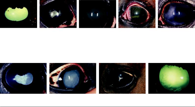
.pdfhumor into the cornea (Fig. 42.1.2a). Even if the fluid would be absorbed again from the cornea and it was possible to successfully lower the elevated internal ocular pressure, so-called “band-like opacities” may persist (Fig. 42.1.2b).
After injuries, superficial and medicamentally well treatable defects within the cornea may develop. Some of these defects are visible with the naked eye, others can be easily identified only after staining with fluorescein. With any injury to the corneal epithelium (positive fluorescein test; that is, the primary stain remains adherent to the cornea), it is important to abstain from the use of cortisone containing ointments (Fig. 42.1.3). If an initially harmless defect is treated with cortisone, then the painfulness of the eye subsides rapidly; however, the danger of an infection of the injury is increased considerably. If an infection of the cornea develops, which leads to lysis of corneal tissue, one speaks of a corneal ulcer (Fig. 42.1.4). Certain, very aggressive microorgaisms (collagenase producing Pseudomonadae) can breakdown the corneal parenchyma within a short time and lead to a rupture of the cornea. Some ulcers for that reason must be cleaned up surgically (Fig. 42.1.5) and stabilized by a sutured “conjunctival flap.” In these cases it is advisable to bring in a subpalpebral catheter into the dorsal fornix in order to prevent the loss of the flap by the blinking of the eye and by rubbing of the eye (to be able to apply medicaments from the neck via a catheter) and to install a temporary ankyloblepharon (temporary artificial closure of the palpebral fissure), until the cornea is stabilized (about 12 days duration) (Fig. 42.1.6a, b, and c).
Perforating corneal injuries are recognizable with the bulging of iris tissue within the corneal lesion (Fig. 42.1.7). In uncomplicated cases the cornea can be stabilized by suture, installation of a subpalpebral catheter and a temporary ankyloblepharon (see corneal ulcer), and often even visibility can be retained. If there is considerable intraocular hemorrhage, the lens dislocated and the retina detached (if necessary, ultrasound examination), it is sometimes advisable to remove the eyeball. If there is a bacterial or mycotic infection of the interior of the eye (septic endophthalmitis) after corneal injuries (ulcer or perforation), the danger exists that it may lead to an ascending infection by way of the optic nerve and finally to an encephalitis. The distance to the brain via the optic nerve is only a few centimeters. For this reason, often eyes that are involved in a severe ophthalmitis have to be removed (Fig. 42.1.8).
42.2. Anterior Eye Chamber and Iridocorneal Angle. The anterior chamber of the eye is the space between the cornea and the iris. In adult warmblood horses it contains about 2.4 ml aqueous humor and, in the center of the anterior chamber of the eye, the distance from the corneal endothelium to the iris is about 5 mm. The posterior chamber of the eye is located between iris and lens. It is distinctly smaller than the anterior chamber and not completely observable (see the upper figure on page 43). With different diseases deposits can be found in the anterior chamber. The most frequent finding is fibrin in the anterior eye chamber, which develops after traumata or inflammation of the iris (“iritis”). It may be in the form of very fine and tiny coagula, which are only visible with a very precise inspection of the anterior chamber (Fig. 42.2.1a). But there may also be a great deal of fibrin in the anterior chamber (Fig. 42.2.1b) and sometimes the entire anterior eye chamber is filled by fibrin. More rarely blood is found in the anterior chamber (“hyphema”, Fig. 42.2.1c). Blood may reach the anterior chamber following traumata or severe inflammation of the iris. Leukocytes may reach the anterior chamber as a result of severe corneal inflammation, infectious processes of the cornea or in the interior of the eye, as well as in rare cases in the course of uveitis (hypopyon). Very rarely, foreign bodies (for example, glass, metal, or wood splinters) can also be found in the anterior chamber. Fibrin coagula in the anterior chamber usually dissolve by themselves, larger amounts of blood must be dissolved with the use of medication or be surgically removed, and foreign bodies are removed surgically.
The ligamentum pectinatum is located circularly in the periphery and meets the sclera at a right-angle. The ligamentum pectinatum in the horse is directly observable and gonioscopy of the iridocorneal angle applied in other species is unnecessary in the horse (Fig.
42.2.2). The iridocorneal angle of the horse is wide open and a
“narrow-angle glaucoma” (clogging of the iridocorneal angle by iris-tissue, especially with a dilated pupil) does not exist. The predominant drainage of the aqueous humor in horses is realized via the suprachoroidal space, which exhibits a low resistance to drainage (“uveoscleral drainage of aqueous humor”). Glaucoma of the horse is almost always an “open-angle glaucoma.” This is associated with a diminished drainage of the aqueous humor owing to a clogging of the trabecular meshwork at the chamber angle by products of inflammation and with that an increase in intraocular pres-
a |
b |
c |
Fig. 42.2.1: Nonphysiological content within the anterior eye chamber: (a) very small and scarcely perceptible coagulum of fibrin; (b) large and easily recognizable coagulum of fibrin; (c) hyphema (blood within the anterior eye chamber).
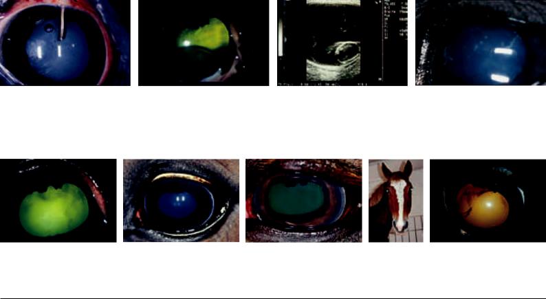
Fig. 42.2.2 (left): Open and visible iridocorneal angle of the horse. The grey strip at the temporal corneal border is the pectinate ligament, which meets the cornea at a right angle. Nasally and temporally it is normally easily seen, dorsally and ventrally it can be hidden by the eyelids or by pigmentation of the cornea near the limbus. Fig. 42.2.3 (2nd left): Endoscopic view of the lens (L), zonule fibers (Zo), ciliary body (Zi) and ora serrata (OS) (beginning of the retina) from the vitreous body out. Aboveleft, the pupil is visible though the lens.
Fig. 42.3.1 (3rd left): Remnant of the pupillary membrane circularly on the anterior surface of the iris.
Fig. 42.3.2 (4th left): Iris coloboma: Nasal and temporal of the pupil. Temporally, the border of the lens and the folded part (pars plicata) of the ciliary body are visible. Fig. 42.3.3 (2nd right): Iris hypoplasia and “stromal cyst” ventral (cyst-like projection of the thin iris tissue owing to the flow of aqueous humor). Additionally, the consequences of posterior synechiae and a cloudy lenticular nucleus are present (the lens has a normal size, the cortex is however normally transparent and therefore not visible).
Fig. 42.3.4 (right): Iridic granular cysts on the ventral border of the pupil.
155
sure (“secondary glaucoma”). In individual cases the iridocorneal angle may be clogged even by blood or connective tissue (after hyphema), iris melanomas, iris cysts or a lens loosened by a partial rupture of zonular fibers, which may also be a reason for increase in the intraocular pressure (Fig. 42.2.3).
Because narrow-angle glaucoma does not play a role in horses and uveoscleral drainage of aqueous humor is fostered by atropine, the administration of atropine is indicated in horses with increased intraocular pressure (in contrast to other species in which glaucoma must be regarded as a contraindicaton for the dispensing of atropine). In addition, elevated intraocular pressure is treated with antiinflammatory drugs as also with medications, which lower the production of aqueous humor.
42.3. Iris, Granula iridica and Pupil. The stroma of the iris is normally brownish pigmented and well vascularized. Frequently, in the middle of the iris, there can be recognized a somewhat irregular prominent ring of dark iris tissue, which represents a remnant of the embryonal pupillary membrane (Fig. 42.3.1). Congenital malformations are rare such as a persisting pupillary membrane or iris coloboma (Fig. 42.3.2). More often there appears a deficiency of pigment of the iris tissue, which may involve one or both eyes, partially (heterochromia) or completely (“wall-eye”; albinismus iridis externum). Often these color variants go along with a broad white blaze or another white marking extended onto the orbital region. Depending on the degree of pigment deficiency, the iris tissue appears then blue, white—very seldom—reddish (albinism). In connection with a deficiency of pigment the iris stroma can be very thin to transparent (iris hypoplasia). The concerned tissue will then be pushed anteriorly by the aqueous humor formed behind the iris, and looks similar to a cyst (stromal cyst; Fig. 42.3.3). Within the iris is located, the sphincter pupillae muscle which is innervated by parasympathetic fibers, as well as the dilatator pupillae muscle, which is innervated by sympathetic fibers. In very excited horses the pupil is then sometimes very wide. A wide pupil can be obtained by medication, either by sympathomimetics (less effective) or by parasympatholytics (for example, atropine, very effective).
In any case, the reaction of the pupil to the incident light (pupillary constriction, pupillary reflex) should be checked, before the pupil is enlarged by medication. In the case of nervous horses the constriction of the pupil may be somewhat delayed and incomplete.
Movement should, however, always be observed. Also, in the presence of cataract, the reaction of the pupil is maintained, provided there is no retinal damage. If the eye has been treated with a mydriatic prior to the examination and, for this reason, the pupil is unre-
sponsive to the incident light, in a doubtful case (if, for example, no response or only a weak response to a menace gesture results) the “consensual pupillary reflex” can also be checked. The consensual pupillary reflex arises by the fact that in the optic chiasm a partial decussation of nerve fibers of the left and right eyes takes place and the sphincter pupillae muscle of the second eye is activated via the reflex arc. This means that in the case of light directed into the eye with the pupil widened by medication, the pupil of the partner eye, which must not be brightly illuminated, must constrict. The movement of the pupil of the partner eye should be observed with minimal tangential lighting of the anterior eye chamber from temporal.
If the consensual pupillary reflex does not appear, then there is retinal damage of the brightly illuminated eye or a pathological process in the area of the visual pathways. If a pupillary reaction to light results, then as a rule vision is present.
Iridic granules (granula iridica) are found at the dorsal margin of the pupil. These have in each horse a variable size and shape and should not be confused with a tumor. Relatively often iridic granular cysts (fluid-filled, vesicularly enlarged iridic granules) are found, which as a rule are an incidental finding and have no pathological importance (Fig. 42.3.4). Only rarely a problem may appear, if the pupil is altered by large cysts and vision is correspondingly limited by this or if the iridic granular cysts reach the corneal endothelium, where they irritate. In an individual case therefore it may be necessary to remove the iridic granules surgically. Adhesions between the iris and the lens (posterior synechiae) are a frequent result of painful inflammations of the iris, accompanied by miosis (constriction of the pupil) and fibrin accumulation in the anterior eye chamber. Sometimes these synechiae can be recognized only if the pupil is dilated. The synechiae may concern small portions of the iris tissue.
In some eyes, however, a larger area of adhesion occurs between iris and lens (Fig. 42.3.5a and b). With posterior synechiae there often arises a deformation of the normally transverse-oval pupil (dyscoria; Fig. 42.3.5b). Sometimes, with enlargement of the pupil, some iris tissue rips off, remains sticking on the anterior surface of the lens (iris residuum), and is the only indication of a former iritis (Fig.
42.3.6). If the border of the pupil is circularly connected to the lens, then one speaks of a secclusio pupillae. If then the pupil is still covered by fibrin, we are dealing with an occlusio pupillae (Fig. 42.3.7). In both cases, flow of aqueous humor from the posterior chamber through the pupil into the anterior chamber is impaired, in which case the iris tissue that is not adherent to the lens may bulge anteriorly (“Bundt-cake iris”). In white horses, rarely also in differently pigmented horses, intraocular melanomas (darkly pigmented tumors), which originate from iris tissue, can arise (Fig. 42.3.8). In
a |
b |
Fig. 42.3.5: (a) Small posterior synechiae (adhesions between iris and lens) at “10 o’clock”; (b) areolar posterior synechiae and dyscoria. Additionally a cataract is present. Fig. 42.3.6 (3rd left): Iris residuum in the center of the anterior surface of the lens.
Fig. 42.3.7 (2nd right): Pupillary occlusion (pupil closed by fibrin) and “Bundt-cake iris” (Bundt-cake-like projection of the iris, the border of the pupil is adherent to the lens).
Fig. 42.3.8 (right): Melanoma of the iris (pigmented tumor, which starts from the iris). Dorsonasally a brown pigment can be seen in the cornea. Additionally there is a smoky-milky cloudiness of the cornea and individual delicate ribbon-like opacities. In the left half of the pupil there is irregular brown tumor tissue.
a |
b |
c |
d |
Fig. 42.4.1: (a) Mature cataract (complete milky cloudiness of the lens); (b) Nuclear cataract (cloudiness of the nucleus of the lens); (c) “Suture-star cataract” (cataract in 156 the area of the lenticular sutures); (d) Vacuolar cataract in the region of the posterior surface of the lens.
contrast to the iridic granular cysts, the melanomas have an irregular structure and prefer to grow at the iridocorneal angle. With these, corneal opacities often develop. With continuing growth a brown pigment can be observed within the cloudy cornea. If, with expanding cloudiness, sight in the eye is no longer possible, ultrasound examination can be helpful to assess the extent of the tumor in the eye.
42.4. Lens. The most frequent alteration of the lens is a cloudiness (cataract; Fig. 42.4.1a–d). The cataract may be congenital, in young horses genetically conditioned in the first months or years of life (juvenile) or as a consequence of inflammation, posterior synechiae or traumata (cataracta complicata). In old horses a senile cataract can develop. The cataract may involve the entire lens or only parts of it. According to the location of the cataract, one speaks of a “suture star” (turbidity or cloudiness in the region of the former lens sutures), a nuclear cataract (concerning the lens nucleus), a cortical cataract (concerning the cortex of the lens), subcapsular cataract (lying beneath the capsule of the lens) or capsular cataract (concerning the lens capsule) cataract (Fig. 42.4.1a–c). In a normally transparent cortex, the cloudiness of the nucleus of the lens may simulate a microphakia. In these cases an examination by slitlamp is helpful, because the slit-lamp makes the normally invisible cortex visible. Often, as a consequence of an uveitis, a vacuolar cataract is present in the region of the posterior surface of the lens (Fig. 42.4.1d). Lens operations by phacoemulsification are now technically possible (Fig. 42.4.2). (Phacoemulsification is a minimally invasive operative technique by which the lens material is shattered by ultrasound and aspirated by way of a small incision in the cornea. Simultaneously, the internal pressure of the eye is maintained by infusion.)
Subluxations (partial tearing out of the zonule fibers) of the lens may be inherited, a consequence of traumata, a chronic uveitis or a chronic glaucoma with gradual enlargement of the ocular bulb.
With a widely dilated pupil, they can be recognized because the border of the lens and possibly torn out zonular fibers become visible in the periphery of the lens (Fig. 42.2.3). Complete luxations of the lens can take place into the anterior eye chamber or into the vitreous chamber. The lens, which is usually cataractous, is then often well visible (Fig. 42.4.3a) or, in case of considerable cloudiness of the cornea, may be easily localized by ultrasound (Fig. 42.4.3b).
Concentric rings within the lens (Berlin rings), striking in appearance, are to be regarded as normal variants. Refraction anomalies of the lens appear in connection with its deformation (for example, lenticonus, bulging of the posterior surface of the lens into the
vitreous chamber), but may show up first in the examination of the fundus of the eye, in that uncommon dioptric values are needed for focusing. Rarely, also colobomas of the lens can be observed with a more dilated pupil (indentation of the lens border in the periphery) (Fig. 42.4.4).
42.5.Vitreous Body. Direct ophthalmoscopy is imperative for examination of the vitreous body. A frequent finding, which becomes visible in the course of time in every horse older than perhaps 8 years, is liquefaction of the vitreous body. This liquefaction presents itself ophthalmoscopically as two mixed clear phases of fluids (for example, egg-white in water). Sometimes also filamentous structures of the framework of the vitreous body can be recognized. A diffuse turbidity of the vitreous body as well as cloudy inclusions, which present themselves as dark inclusions in direct ophthalmoscopy, are as a rule the result of an uveitis (Fig. 42.5.1). Since these inclusions are not resorbed as fibrin is from the anterior eye chamber, in the inflammation-free interval that follows the abatement of an acute ocular inflammation they are also of importance as an indication of the existence of a recidivistic equine uveitis (formerly “periodic ophthalmia”). The first changes are always observable entirely in the periphery of the vitreous body, near the ciliary body. In the further course of the disease, the cloudinesses may be present in the entire vitreous chamber. A diffuse yellowish cloudiness of the vitreous body, a result of inflammation in the posterior portion of the eye, is best seen by shining a light into each pupil in a bilateral comparison (Fig. 42.4.2a–c). By superimposition of the yellowish color in the vitreous chamber with the blue to greenish color of the fundus of the eye, the color that is observed in the pupil with lateral lighting appears strikingly green. Rarely the diffuse cloudinesses of the vitreous body or inclusions in the vitreous body are the result of hemorrhage and then, with illumination, reddish in color (Fig. 42.5.3).
42.6.Fundus of the Eye and Retina. The fundus of the eye bears the tapetum lucidum in its upper 2/3. The tapetum lucidum has a greenish to bluish color and is set through with small brown dots (WINSLOW’s stars = transverse sections of small vessels running from the choroidea to supply the retina). The ventral part of the ocular fundus is brown in color. The optic disc (papilla of the optic nerve) is found just ventral to the transition from the tapetum lucidum (Fig. 42.6.1a–d). Only a few short blood vessels extend from the entrance of the optic nerve into the periphery.
For ophthalmoscopic examination of the fundus, it is advisable to focus first on the WINSLOW’s stars to find the proper ophthal-
a |
b |
Fig. 42.4.2 (left): Phacoemulsification: Using a metal cannula the lens material is shattered by ultrasound and aspirated. At the same time, fluid is injected by means of the surrounding plastic cannula and the metal cannula is cooled.
Fig. 42.4.3 (middle): Luxation of the lens: (a) into the vitreous chamber (luxatio lentis posterior); (b) ultrasound image of posterior luxation of the lens.
Fig. 42.4.4 (right): Coloboma of the lens.
a |
b |
c |
Fig. 42.5.1 (left): Cloudy, dark grey-brown inflammatory inclusions in the vitreous chamber as viewed by direct ophthalmoscopy.
Fig. 42.5.2 (middle): Color of the pupil: (a) normal eye; (b) diffuse cloudiness of the vitreous body in uveitis; (c) color of the pupil in a comparison of both sides: the right (normal) eye glimmers bluish, the left eye yellowish-green owing to cloudiness of the vitreous body in uveitis.
Fig. 42.5.3 (right): Hemorrhage of the vitreous body after trauma.
157
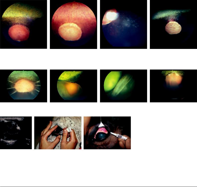
moscopic setting. After that, the fundus is examined in a meandering manner and checked for alterations. The clinically important changes are normally found in the region of the optic disc. Early stages of retinal detachment (Fig. 42.6.2a) as well as signs of choroiditis (Fig. 42.6.2b–d) and formations (structural changes) as a rule are revealed first in this region. Atrophy of the optic nerve is recognizable as a strikingly pale color of the optic disc as well as an absence of the few blood vessels that are normally present.
The retina consists of the layers of the neurosensory retina and the pigment layer (stratum pigmentosum). In retinal detachment (amotio or ablatio retinae), the neurosensory retina separates from the pigment layer of the retina, so that there is only a light, curtainlike structure in folds, vesicles or in extended areas loosened from the ocular fundus (Fig. 42.6.3); however, the pigmentation of the fundus is entirely maintained (Fig. 42.6.2d).
After detachment of the retina, the pigment of the fundus appears still lighter and more iridescent, since the otherwise anteriorly located retinal layers are lacking (hyperreflection). A red color of the fundus (fundus albinismus) has no pathological significance and is a normal finding in human beings and swine. The red background does not represent a hyperemia in connection with an inflammation, but shows only the absence of pigment, so that the view of the choroidal blood vessels becomes possible (Fig. 42.6.1b). If the pigment is absent, then in horses an impairment of vision in dim light
and darkness can be assumed, since the tapetum lucidum otherwise reflects the light and enables a weaker intensity of light to be utilized.
Application of Ophthalmic Ointments and Ophthalmic Drops
For the application of ophthalmic ointments, the upper eyelid is pushed dorsally with the index finger in the palpebral sulcus and the lower eyelid somewhat lifted off the eyeball with the thumb. The application of medication is preferably realized from temporal into the space between the lower eyelid and the ocular bulb (Fig. 42.7).
By blinking, ointments and drops are distributed over the entire cornea.
Subconjunctival injection. For subconjunctival injection, surface anesthesia of the conjunctiva should be done initially. After the action of the anesthetic has begun, a twitch is applied and a DESMARRES lid retractor fixed under the margin of the upper eyelid. By raising the upper lid with the lid retractor the bulbar conjunctiva can be exposed dorsally (Fig. 42.8). The injection is done with a thin needle and the volume amounts to about 1–1.5 ml. This technique can be applied for the administration of mydriatics or cortisone, insofar as the administration of ophthalmic ointments is not possible or has shown no effect. The injection of crystalloid solutions should however be abandoned.
a |
b |
c |
d |
Fig. 42.6.1: (a) View of the physiological (normal) fundus of the horse: tapetum lucidum with WINSLOW’s stars dorsal, tapetum nigrum ventral, optic disc and individual, centrifugally running short blood vessels. (b) Albinism of the fundus (deficiency of pigment). Especially frequent if there are broad white markings on the head. (c) Choroiditis (inflammation of the choroidea) with pigment changes following vasculitis; may also develop independently from uveitis. (d) Atrophy of the optic nerve (regression of blood vessels and pale color of the optic disc).
UQL|XbVxUmAVqC+HnIZ42uaraA==|1288052959
a |
b |
c |
d |
Fig. 42.6.2: (a) Beginning “star-shaped” detachment of the retina (ablatio retinae, amotio retinae); (b–d) Extended retinal detachment. The pigment is retained in the fundus of the eye.
Fig. 42.6.3 (left): Retinal detachment, ultrasound image: At the optic disc as well as at the ora serrata, it remains attached a longer time; in between it is completely detached (“gullwing shaped”). [R = retina; G = vitreous chamber; L = lens; NO = optic nerve]
Fig. 42.7 (middle): Application of ophthalmic ointments. The index finger is in the palpebral sulcus of the upper eyelid (the first distinct fold above the border of the lid; see Fig. 40.3.1a) and lifts this; the thumb opens the lower conjunctival sac.
Fig. 42.8 (right): Subconjunctival injection beneath the dorsal bulbar conjunctiva.
158
Head Cavities |
R. BERG, K.-D. BUDRAS |
44.1.Scars in the region of the nostril: Scar tissue in the vicinity of the nostril may restrict the ability to completely widen the nares, whereby the influx of air is reduced.
44.2.Nasal diverticulum (= soft nose or also nasal trumpet, p. 45.20): The nasal diverticulum is a caudally blind-ending cutaneous pouch about 3–6 cm long, which is located in the naso-incisive notch and accessible through the “false nostril.” It is formed owing to the presence of the alar fold (plica alaris) and is lined internally by a pigmented, predominantly hairless, skin. One ends up in this diverticulum with the introduction of instruments (e.g., nasogastric tube, catheter for the guttural pouch, laryngoscope) if care is not taken that these are led onto the floor of the nasal vestibule. An atheroma of the nasal diverticulum (epidermal inclusion cyst) may occur as a unilateral, rarely bilateral, fluctuating mass in the caudal region of the “false nostril.” Usually, this atheroma does not obstruct the airway and its elimination is rather a cosmetic problem.
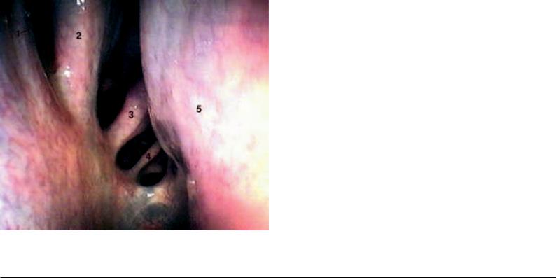
44.3.Endoscopy of the nasal cavity (p. 45): Ventral and middle nasal meatuses, medial part of the nasal cavity, nasal septum, external surfaces of the dorsal and ventral nasal conchae (see to the left of the figure).
Parts of the middle nasal concha and parts of the ethmoidal labyrinth are accessible for inspection endoscopically. A flexible endoscope facilitates an almost complete examination of the ventral and middle nasal meatuses and of the nasal conchae. If necessary, it can also be introduced into the dorsal nasal meatus. The ethmoidal labyrinth can be better and more safely examined with a fiberscope.
Endoscopy of the middle nasal meatus is of importance because of the presence of the nasomaxillary aperture, which occasionally can be identified as a slit in the caudal part of the meatus. Mostly the aperture is covered by the dorsal nasal concha, under the caudal part of which exudates can be observed should the occasion arise.
Introduction of the nasogastric tube (p. 45): In the horse, the most ventral and most spacious of the three nasal meatuses is used for the introduction of instruments, especially the nasogastric tube (stomach tube),. The ventral nasal meatus leads via the nasopharyngeal meatus (choana) directly into the pharynx. In doing this, at the beginning, the tube passes a nasal mucosal fold, the basal fold, which contains a well developed venous plexus (plexus cavernosus conchae), which extends caudally upon the ventral nasal concha.
With the introduction of the tube this fold can be injured and there results more or less considerable hemorrhage from the venous plexus (epistaxis). Improper handling may even fracture a nasal concha, because these are very delicate and fragile. This happens especially if the instruments introduced are not pushed down upon
Fig. 44.3. Endoscopy of the nasal cavity. Endoscopic view in the right caudal nasal region. 1. dorsal nasal concha; 2. middle nasal concha; 3., 4. ethmoidal conchae, 5. nasal septum. (Photo, Prof. Dr. Grabner)
the floor of the nasal vestibule. Apart from that, it is also possible to end up involuntarily in the nasal diverticulum.
Trephination of the nasal cavity: The site of trephination depends on the location of the pathological process. The caudal boundary is a line between both medial angles of the eye. In the middle of the head, the nasal cavity can be approached immediately beside the median line, maximally 4 cm lateral.
44.4.Inspection of the nasal mucosa (p. 45) is of clinical importance. Externally, the pink mucosa of the horse is very well visible in the rostral part of the nasal cavity.
Ethmoidal hematomas are slowly spreading angiomatous masses, which develop from the mucosa of the ethmoidal conchae in the caudal part of the nasal cavity (see Fig. 44.3.). They occur in horses of middle age or older and show a preference to thoroughbreds. The most frequent signs are mostly unilateral hemorrhages of the nose. With increasing size of the hematoma, the nasal cavity, commonly the ventral nasal meatus, becomes partially or entirely occluded. With progressive expansion, the hematomas may intrude into the maxillary and frontal sinuses and occasionally result in a distortion of the facial bones.
44.5.Resection of the nasal conchae: In foals up to 2 years of age a mucoid degeneration of the nasal conchae may occur. Owing to the long roots of the teeth, the caudal maxillary sinus at this time is still very flat. Since with the mucoid degeneration of the ventral nasal concha an empyema of the maxillary sinus is to be expected, initially the latter is trephined in the region of the maxillary septum (= septum sinuum maxillarium). Here, there is commonly found the cupola whose wall consists of a softer, less firm, bone. The degenerated and expanded dorsal nasal concha fills large parts of the nasal and maxillary cavities.
44.6.The site for trephination of the dorsal nasal concha is at the caudal part of the nasal bone, paramedian at the level of the medial angle of the eye (Fig. 34.3.).
44.7.Malformed nasal septum (see p. 51.a'–a'''): The nasal septum may be thickened or bent by a congenital abnormality or fracture. Horses with such a nasal septum demonstrate usually a noise in the upper respiratory way.
44.8.Olfactory components, for example, of the vaginal secretions or urine of mares in heat, are received by the vomeronasal organ. In stallions, this olfactory stimulus leads to sexual excitement and a behavioral response, which is designated Flehmen. In this response, the lips are curled (contraction of the levator labii superioris muscle on each side) and the head is fully extended.
44.9.The oral cavity of the horse is very long and narrow. The narrowness of the mouth opening and the great depth of the oral cavity make clinical examination difficult. Nevertheless, the organs and structures are accessible to an examination of the oral cavity for an average-sized hand.
Endoscopy of the oral cavity offers the possibility of an inspection of the oral mucous membrane, the hard and soft palate, the cheek teeth, the gums and the root of the tongue. It is the one possibility for inspection of the oropharynx and especially the palatine tonsil in the horse.
44.10.The palatine plexus (p. 45.q) is a venous cushion of the palatine mucous membrane. These veins can fill greatly and swell the palatine mucous membrane up to the mouth, which can be observed especially in foals. Laymen call this appearance “frog” (veterinary: “lampas” or “lampers”).
44.11.Vascularization of the tongue—intralingual injection: The body of the tongue is well vascularized. The effective concentration of a medicament within the blood may be reached rapidly by an intralingual injection. Intralingual injection is an extraordinary injection, done if other intravenous injection is impossible because of venous collapse.
For intravenous lingual injection, the branches of the sublingual and lingual veins are used. These veins are visible on the ventral surface of the apex of the tongue and run about 1 cm from the lingual border. As experience shows, the rate of successful intravenous lingual injection in the horse is 82%.
159
46.1.Lymphoid hyperplasia of the pharynx: In its mucosal lamina propria and in its submucosa the pharynx contains collagenic and elastic fibers mixed with lymphatic tissue and mucous glands. The average degree of pharyngeal lymphoid hyperplasia in the foal or yearling is higher than in the adult horse. Even if this finding is usually considered normal for the juvenile horse, the dorsal displacement of the soft palate occurs more easily and more often in those horses that have a pharyngeal lymphoid hyperplasia of third or fourth degree. Higher degrees of pharyngeal lymphoid hyperplasia diminish with increasing age of the horse.
In an endoscopic examination, paresis of the pharynx can be observed as a collapse of the walls of the nasopharynx, which restricts the respiratory way. These signs may occur in horses with a mycosis of the guttural pouch and dysphagia.
46.2.The intrapharyngeal ostium (Fig. 46.4.) is an opening with a “sphincter muscle” for the laryngeal crown and guarantees a direct respiratory way (Fig. 48.2.): nasal cavity-respiratory pharynxlaryngeal crown-trachea. A dislocation of the laryngeal crown into the oropharynx during high oxygen need (extreme stress, races) results in a sudden breakdown of the horse owing to an anoxemia. This phenomenon, called “choking up”, has its cause in a cramp of the sternohyoid and sternothyroid muscles with backward displacement of the larynx. Also retraction of the tongue can dislodge the laryngeal crown in such a way from the intrapharyngeal ostium (“swallowing of the tongue”).
46.3.The pharyngeal ostium of the auditory tube: This opening is the entrance into the guttural pouch in the nasopharynx and is in a transverse plane through the lateral angle of the eye.
The guttural pouch valve is a prominent structure of the nasopharynx. It borders the pharyngeal openings of the Eustachian tubes and is about 3 cm in length. It is pressed against the pharyngeal wall of each side and exhibits an oblique, sinuous course of its ventral free border. It is reinforced by a continuation of the medial cartilage that supports the auditory tube. The slit-like pharyngeal ostium of the auditory tube lies lateral to the guttural pouch valve, is closed normally, and is opened if the horse swallows. In this way, the pressure on both sides of the eardrum becomes equalized.
46.4.Soft palate (Fig. 46.4): It is the caudal continuation of the hard palate and is remarkably long in the horse. In the resting state, it hangs down in front of the epiglottis and is in contact by its free border with the base of the epiglottis. This topographical relation explains the inability of the horse to exert an oral respiration in respiratory problems and, on the other hand, the course of the ingesta through the nasopharynx and the nasal cavity in case of vomitus (in the horse, to be sure, very rare).
Fig. 46.4. Dorsal displacement of the soft palate. With the position of the instrument in the nasal pharynx (ventral in the picture), the endoscopic picture shows a view of the soft palate (in the picture, below) and the intrapharyngeal ostium (in the middle of the picture), as well as the continuing esophageal vestibule. The mucosa-covered corniculate process of the arytenoid cartilage projects from ventral into the intrapharyngeal ostium and accordingly into the laryngeal pharynx. As a result of the extension of the soft palate, that does not however apply to the epiglottis, which cannot be seen. The intrapharyngeal ostium is closed rostrally by the free border of the soft palate (below in the picture), which caudally passes over bilaterally into the palatopharyngeal arch. The reddening is the result of inflam-
160 mation. (Photo, Prof. Dr. Grabner)
Dorsal displacement of the soft palate is diagnosed by the nonvisibility of the epiglottis in endoscopic examination. Normally the epiglottis lies on the soft palate and projects into the nasopharynx. If these conditions are inverse, the soft palate obstructs the aditus laryngis during inspiration and disturbs expiration. It occurs more often in horses that are not yet adult and seems to have a relation to a high-degree of lymphoid hyperplasia of the pharynx. Young horses with a smaller, flabby epiglottis are more inclined to a displacement of the soft palate than adult ones. Tissues of the pharynx and larynx seem to “mature” with increasing age with respect to size, rigidity and coordination.
The cleft soft palate (palatoschisis totalis) is a rare cleft formation in foals and may reach a variable extent; sometimes it involves the total soft palate. In such foals, ingested material is discharged from the nostrils (one or both) and usually they die of pneumonia.
A lateral buccotomy (severance of the cheek from lateral) is a method for treating palatoschisis totalis. Others are access by way of the oral cavity, mandibular symphysiotom or laryngopharyngotomy.
Mandibular symphysiotomy (Fig. 38.8.): For the purpose of a successful implementation of larger surgical operations (e.g., cleft soft palate, palatoschisis totalis), the mandibular symphysis (intermandibular synchondrosis) can be split.
46.5. Guttural pouch: In the horse the auditory tube, which runs between the pharynx and the middle ear, has a characteristic caudoventral diverticulum, the guttural pouch. The opening between tube and guttural pouch is a long slit in the caudoventral tubal wall. Since both the guttural pouch and the auditory tube open into the nasopharynx, they are often involved in respiratory diseases. Since the guttural pouch has a secretory mucous membrane, essentially catarrhal inflammations are found here. The guttural pouch may be subject to a mycotic infection, which erodes its dorsal wall by a diphtheritic inflammation and is responsible for several signs that point to a damage of important blood vessels and nerves. The most spectacular case would be a fatal epistaxis owing to rupture of the internal carotid artery.
Fig. 46.5.1. Endoscopic picture of the right guttural pouch, rostral view. The endoscope was introduced into the guttural pouch through the slit-like opening of the pharyngeal ostium of the auditory tube (see text-figure, p. 46). With its perpendicular orientation, the broad, bright stylohyoideum separates the lateral recess (left) from the medial recess (right). The medial recess contains the perpendicular neurovascular fold, whose distinct (bluish) internal carotid artery reaches the lower border of the picture and whose lighter glossopharyngeal nerve (central in the picture) shears off toward the stylohyoideum. At the upper border of the picture the bean-shaped cranial cervical ganglion of the sympathetic trunk lies to the right in the neurovascular fold. (Photo, Dr. Grabner, Berlin)
Cranial nerves VII, IX, X, XI and XII can be irritated by diseases of the guttural pouch. Damage to cranial nerve IX may cause loss of taste and sensibility at the root of the tongue, partial anesthesia of the pharyngeal mucous membrane and paralysis of the pharyngeal muscles. The horse is then unable to swallow. The ramus sinus carotici, which is responsible for the regulation of blood pressure and heart frequency, may also be encountered with damage to the glossopharyngeal nerve. In this situation, the corresponding regulation by afferent impulses of the pressoreceptors of the carotid sinus and of the chemoreceptors of the glomus caroticum to the medulla oblongata and to the nucleus of origin of the vagus nerve will be disturbed. Such a dysregulation continues on the corresponding efferents to the sinus node and AV-node of the heart.
Damage to cranial nerve X results additionally in partial loss of the sensory and motor functions of the pharynx. With this there exists the danger of an incurable swallow-pneumonia. A possible loss of the cranial and caudal laryngeal nerves causes an absence of the sensory and motor functions of the larynx, with signs of laryngeal paralysis. Injury to cranial nerve XI is clinically not so grave (perhaps twisted neck in case of loss of innervation to the trapezius and sternocleidomastoid [sternocephalicus and cleidomastoid] muscles). If the XIIth cranial nerve is paralyzed, the lingual muscles are involved. The tongue droops from the mouth and cannot be retracted into the oral cavity. A late sequela is an atrophy of the tongue, which occurs also unilaterally if the nerve is damaged unilaterally. Damage to the cranial cervical ganglion of the sympathetic is reflected in a narrowing of the palpebral fissure (loss of tonus of smooth musculature of the eyelids) and by miosis (narrowing of the pupil due to denervation of the dilatator muscle of the pupil).
Paralysis of the facial nerve occurs only rarely in connection with diseases of the guttural pouch. Of course, paralysis of the facial nerve and the spread (of the disease process) onto the temporal bone may damage the inner ear and result in vestibular signs. [394]
[395] [396] [397] [398] [399]
Tympanic guttural pouch of foals: The accumulation of air (tympany) in the guttural pouch of 2–3 weeks old foals is probably caused by a functional disturbance, produced less by an organic defect of the guttural pouch (mucosal fold on the floor of the slitlike opening of the guttural pouch).
VIBORG’s triangle: A triangle located at the transition from the head to the neck, which facilitates external access to the inflamed guttural pouch (Fig. 46.5.2). Its borders are dorsally the tendon of the sternomandibular muscle (sternocephalicus muscle), rostrally the cervical border of the mandible and ventrally the linguofacial vein. Here the skin and cutaneous muscle are first incised, then the parotid gland separated bluntly from the mandible. The blunt dissection process is continued on the medial aspect of the occipitomandibular part of the digastric muscle in a direction toward the ear to reach the guttural pouch. Furthermore, if abscessed, the medial retropharyngeal lymph nodes are incised from Viborg’s triangle. Normally, they are not palpable.
46.6. Anesthesia of the hypoglossal nerve: For anesthesia of the tongue, the lingual nerve and hypoglossal nerve are important. Both nerves are anesthetized from one location. The anesthesia is per-
formed bilaterally. By palpation, the rostral end of the lingual process of the hyoid bone is established. From here one moves 2–3 fingerbreadths rostrally and introduces the needle in a median and perpendicular direction into the body of the tongue.
46.7. Retropharyngeal lymphocenter: This includes the medial (p. 47.27) and lateral retropharyngeal lymph nodes. The former lie on the dorsolateral pharyngeal wall, medial to the mandibular gland and the digastric muscle. They are normally not palpable but, if abscessed, can be incised from VIBORG’s triangle,. The less numerous and smaller lateral retropharyngeal lymph nodes lie ventral to the wing of the atlas on the internal carotid artery and the lateral side of the guttural pouch (p. 47.22).
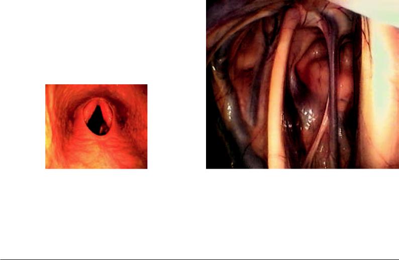
48.1. The larynx of the horse is of special importance for the veterinarian. The purchase-examination should include a thorough palpation and inspection of the larynx. With the so-called “laryngeal roarer”, in 90% of the cases there is in the forefront an unilateral (left-sided) paralysis of the dorsal cricoarytenoid muscle caused by damage to the corresponding recurrent laryngeal nerve. In this case, there occurs a lowering of the arytenoid cartilage (Fig. 48.3. above, right) and—connected to this—a slackening of the vocal fold on the same side. There originates here also the respiratory sound which is characteristic for this disease. In observing the laryngeal lumen with a laryngoscope in such cases, the asymmetry of the rima glottidis is striking.
Treatment is directed to a surgical dilatation of the rima glottidis, and this is obtained by a ventriculectomy and an additional laryngoplasty.
In the case of a ventriculectomy the mucosal lining of the laryngeal ventricle is removed and the caudal thyroid notch furnishes surgical access for this. By the scar-formation that follows, the outer wall of the laryngeal ventricle fuses with the inner wall, including the vocal fold, which is pulled laterally by contraction of the scar tissue with enlargement of the rima glottidis.
Laryngoplasty is directed toward compensating for the loss of contraction of the dorsal cricoarytenoid muscle. Corresponding to the action of the paralyzed muscle, a stout alloplastic band is
|
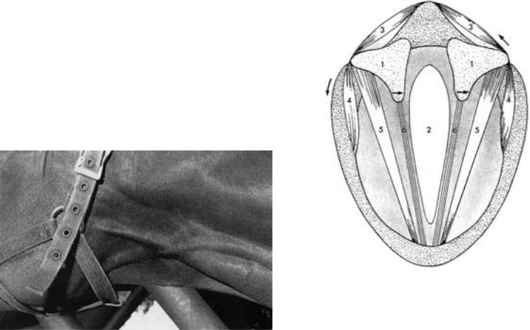
Fig. 48.1. Schematic cross-section through the larynx. 1. Position of the cricoary- |
|
|
tenoid articulation under the arytenoid cartilage, 2. glottic cleft (rima glottidis), 3. |
|
|
cricoarytenoideus dorsalis muscle, 4. cricoarytenoideus lateralis muscle, 5. |
|
|
vocalis muscle, 6. vocal ligament muscle in the vocal fold, the arrows on the right |
|
|
side give the direction of pull by the cricoarytenoideus dorsalis muscle and the |
|
|
widening of the glottic cleft, and on the left side the direction of pull of the |
|
Fig. 46.5.2. Ventral region of the head-neck border demonstrating Viborg’s trian- |
cricoarytenoideus lateralis muscle and the narrowing of the glottic cleft. (After |
|
gle (surgical access to the guttural pouch). (Photo, Prof. Dr. McCarthy, Sydney) |
Dyce/Sack/Wensing [1991]) |
|
|
|
161 |
stretched from the origin of the muscle dorsomedial on the cricoid cartilage to the attachment of the muscle on the muscular process of the arytenoid cartilage. In doing this, the muscular process is drawn caudomedially as in a natural muscular contraction. This results in a craniolateral displacement of the vocal process of the arytenoid cartilage, by which the vocal fold is put under tension for enlargement of the rima glottidis. [400] [401] [402] [403]
48.2.Epiglottic entrapment: This is understood as a “clasping” of the epiglottis. It is brought about by mucosal folds, the aryepiglottic folds, which normally lie below the epiglottis. As thick mucosal membranes they border the laryngeal inlet laterally. Ventrally they are attached to the free border of the epiglottis, in its mucosal covering. Dorsally they course to the lateral and distal surfaces of the arytenoid cartilages and unite below the entrance to the esophagus. The entrapment of the epiglottis happens by dorsal displacement of parts of the aryepiglottic folds. In doing this, these cover the typical vascular pattern, which normally lies upon the epiglottis (Fig. 48.3.).
Epiglottis: Normally the epiglottis of the horse that is not yet adult is smaller and more flabby than in the adult. It lies on the caudal free end of the soft palate. Although the size and the rigidity of the epiglottis are not the only factors that are involved in dorsal displacement of the soft palate (Fig. 46.4.), they appear yet to play a role here. With increasing age the size and rigidity of the epiglottis usually improve.
48.3.Endoscopy of the larynx (laryngoscopy): The larynx of the horse can be inspected by a rhinolaryngoscope, which is introduced through the ventral nasal meatus. In doing this, at the same time, an evaluation of the mucous membrane of the nasal cavity, ethmoturbinalia and pharynx, including the entrance to the auditory tube (EUSTACHIAN tube) is possible.
48.4.Laryngeal ventricle: In the horse it is an outpouching of the laryngeal mucous membrane, which has a depth up to 2.5 cm. It extends between the vocalis muscle and ventricularis muscle in a dorsocaudal direction along the medial surface of the thyroid cartilage. Its blind end lies deeply at the level of the muscular process of the arytenoid cartilage. The laryngeal ventricle of the horse plays a role in laryngeal hemiplegia (left-sided paralysis of the recurrent laryngeal nerve).
48.5.Endotracheal intubation: With endotracheal intubation the angle between the oral cavity and the axis of the aditus laryngis must be considered and compensated by lifting the head. In selection of a tube caliber for endotracheal intubation it is to be kept in mind that the glottic cleft is the narrowest place within the laryngeal cavity (Fig. 48.6.).
48.6.Palpation of the larynx: The larynx is easily accessible for palpation from the ventral side. First, one palpates the arch of the cricoid cartilage, the laminae of the thyroid cartilage (lateral and rostral to the arch), the lamina of the cricoid cartiage and on each side the muscular process of the lateral surface of the arytenoid car-
tilage. Moreover, it is to be kept in mind that the ventral surface of the thyroid cartilage is caudally deeply incised (caudal thyroid notch). In the case of the “roarer”, the muscular process is distinctly palpable, because the dorsal cricoarytenoid muscle that attaches here is atrophied.
Accessibility of the larynx: In dorsal recumbency, the larynx of the horse is reached from ventral between the palpable thyroid cartilage and the arch of the cricoid cartilage. The caudal thyroid notch, which is filled by the cricothyroid ligament, is located in the median plane between the two laminae of the thyroid cartilage. In the “roarer operation” or ventriculotomy, one incises between the two sternohyoid muscles in the median plane and cuts through the cricothyroid ligament. Here is the single surgical access. It provides ventral access to the lateral laryngeal ventricle (hemiplegia), to the arytenoid cartilages (chondritis), to the soft palate (displacement), to the oropharyngeal mucosa (entrapment), to the laryngopharyngeal mucosa (follicular pharyngitis-lymphoid hyperplasia) and to subepiglottic cysts. The lateral ventricle is invaginated and cut away at its borders to the laryngeal cavity.
Ear
50.1. Ear fistula: By an ear fistula one refers to a fistulous tract near the rostral border of the external ear from which a permanent or intermittent secretion occurs. The cause is an erratic tooth or several small malformed teeth. Their removal is indicated in sport and breeding horses for hygienic and cosmetic reasons.
50.2Head tilt occurs in disease of the vestibular apparatus. It is characterized by a tilting of the head-axis, which begins between the ears and runs rostrally between the nostrils. The result is a lower position of the ear, eye, and nostril of one side.
Bending attitude of the head: A bending attitude of head and neck can be observed with a brain lesion. In this, the described axis in the head region remains straight. The head and neck are flexed to one side (pleurothotonus). The bending attitude of the head is not to be confused with head tilt.
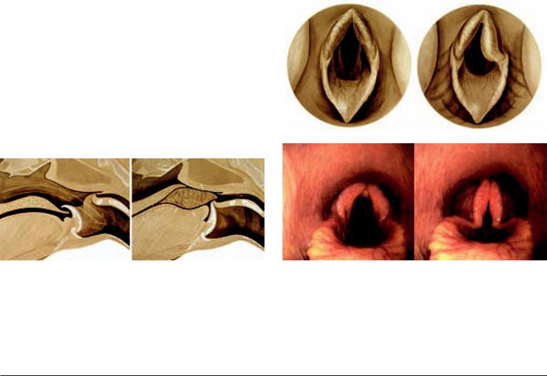
Fig. 48.2. Position of the epiglottis during breathing (left) and swallowing (right). Breathing position (left) and swallowing position (right) of the epiglottis. In breathing, the epiglottis projects through the intrapharyngeal ostium into the nasal pharynx as a part of the laryngeal “crown.” In this case, the air stream arrives from the nasal pharynx through the larynx into the trachea (large arrow). In swallowing the epiglottis, sliding out from the intrapharyngeal ostium, by its retroflexion closes the entrance to the larynx. By elevating the dorsum of the tongue, the bolus passes (small arrow) across the laryngeal entrance into the esophagus. A false passage into the nasal pharynx is prevented by elevation of the soft palate. (Courtesy
162 of Institut f. Veterinär-Anatomie, Berlin)
Fig. 48.3. Illustration (above) and endoscopic picture (below) of the entrance to the larynx. With an opened glottic cleft (left) the vocal folds appear symmetrical and are “framed” by a symmetrical entrance to the larynx. In “roarers” (above right), the glottic cleft is asymmetrical, which is concealed by the collapsed mucosa-covered corniculate process of the arytenoid cartilage (the healthy right vocal fold is visible). The endoscopic pictures (below) reveal the opened (left) and closed (right) glottic cleft and moreover the typical vascular pattern on the surface of the epiglottis, which is concealed in the case of epiglottic entrapment. (Courtesy of Institut f. Veterinär-Anatomie, Berlin)

50.3. Site for taking the pulse—facial artery (Fig. 38.8.): The facial artery is an important structure for taking the pulse. It lies in the vascular notch (incisura vasorum facialium). The latter is a vascular notch at the ventral border of the mandible at the boundary between the body and ramus of the mandible (rostral border of the masseter muscle). As the name implies, here several structures coming from the intermandibular space reflect onto the face. In particular, we are dealing with the facial artery, facial vein and the excretory duct of the parotid gland. The pulse can be palpated if the fingertips press the facial artery medially against the mandible as a hard base.
Central Nervous System
52.1. Function of Cranial Nerves (see pp. 108–111)
Olfactory nerves (I): The olfactory sense of the horse is evaluated only with difficultly. It can be tested, if the horse can smell the food, e.g., the hay, or the hand of the investigator.
Optic nerve (II): The visual sense of the horse can be examined in two ways: 1) With the aid of the menace (threatening) reflex and 2) by leading in the direction of large obstacles. Foals can see already a few hours after parturition; but do not yet know the menace reflex, which they need first to learn in a few days. If a sudden movement is made with the hand in a direction toward the eye, the palpebral fissure is closed.
Oculomotor nerve (III): The function of the oculomotor nerve consists of, among others, the regulation of the size of the pupil (pupillary reflex). The parasympathetic fibers of the oculomotor nerve are responsible for the narrowing of the pupil (miosis; pupillary sphincter muscle), the sympathetic fibers of the ophthalmic nerve for the expansion (mydriasis; pupillary dilatator muscle). The diameter of the pupil permits localization and prognosis of brain injury.
Oculomotor nerve (III), trochlear nerve IV) and abducens nerve (VI): These three cranial nerves coordinate the movement of the eyes. For the examination of eye movements, the head of the patient is evaluated first at rest and then in movement.
Strabismus: abnormal position of the ocular bulb: Injury to the third, fourth and sixth cranial nerves may have strabismus as a consequence. Injury to the oculomotor nerve yields a ventrolateral strabismus (with paralysis of the levator palpebrae superioris muscle); that of the fourth cranial nerve, a rotational strabismus (the dorsal part of the eyeball is shifted laterally), and that of the abducens nerve in a medial strabismus with an inability to retract the ocular bulb. In contrast to the small animals a ventral strabismus in the horse is physiological if the head is elevated.
Trigeminal nerve (V): The sensory part of the trigeminal nerve (ophthalmic nerve, maxillary nerve, and part of the mandibular nerve) is evaluated by control of the corneal and palpebral reflexes and sensibility in the head region. The latter is performed best on the nasal mucosa. Injury to the motor part of the trigeminal nerve (mandibular nerve) results in a paralysis of the lower jaw, masticatory disturbances, saliva flow, the tongue’s hanging from the mouth as well as an atrophy of the muscles, which are innervated by it (masseter, pterygoid, and temporalis muscles, and rostral part of the digastric muscle).
Abducent nerve (VI) and facial nerve (VII) (see pp. 110/111). Vestibulocochlear nerve (VIII): Nystagmus (involuntary move-
ment of the eye) with a peripheral lesion of the vestibular system is rare in the horse, so that its occurrence usually speaks for a central injury.
Glossopharyngeal nerve (IX), vagus nerve (X), and accessory nerve (XI): They provide sensory and motor innervation to the larynx and pharynx (IX, X) as well as parts of the muscles of the neck and shoulder. Besides brain diseases also peripheral injuries may be the cause for disturbances of pharyngeal and laryngeal function (e.g., inflammation of the guttural pouch).
Hypoglossal nerve (XII): It supplies the motor innervation to all extrinsic and instrinsic muscles of the tongue. Injury results in a deviation of the tongue to the normal side. A distinct atrophy may develop. In diffuse, severe cerebral damage a protrusion of the tongue is possible (injury to the upper motor neurons, which control the function of the hypoglossal nerve).
52.2. Brain stem and spinal cord can, following a protozoan or herpes viral infection, be altered by degeneration or by inflammation.
Consequences, among others, are lameness, ataxia, and muscle atrophy. [404] [405]
Fig. 48.6. “Glass-head” with the hyoid apparatus emphasized in yellow (cf., textfigure, p. 34) and the laryngeal muscles marked in red (cf., p. 51). (Courtesy of Institut f. Veterinär-Anatomie, Berlin)
Fig. 52.1. Canial nerves. Assignment of nerve fiber qualities to their areas of innervation.
163
52.3.Cerebellar ataxia occurs especially in foals. Cerebellar functions in the coordination of movement are disturbed and lead to the loss of neuromuscular reflexes. [404] [405]
52.4.Cerebrospinal fluid: A clear, colorless plasma ultrafiltrate of the brain and spinal cord. It is formed by the choroid plexuses of the ependymal lining of the cerebral ventricle, and the blood vessels of the pia mater as well as the encephalic arachnoidea. The main place of absorption is an arachnoid villus in the cerebral vein or venous sinus. Other sites of absorption are the veins and lymph vessels around the roots of the spinal nerves. The direction of flow of the cerebrospinal fluid is from the ventricles across the mesencephalic aqueduct to the central canal. The cerebrospinal fluid may be used for diagnostic purposes. The site for puncture to obtain cerebrospinal fluid (cerebellomedullary cistern, cisterna magna) lies on the cross-point of the dorsal midline of the head with a transverse line through both cranial borders of the atlas.
54.1.A spinal ataxia occurs especially in young horses. Owing to a spondylolisthesis (a gliding of vertebrae), compressions occur in the cervical spinal cord with interruption of motor and sensory pathways. [406] [378] [407]
54.2.Epidural hemorrhages are traumatically caused and lead to a loss of function of conduction pathways in the spinal cord. [378]
54.3.For epidural anesthesia the interarcuate space between the sacrum and the first caudal vertebra or between the first two caudal vertebrae is chosen. [408] [378] [409]
54.4.Cerebrospinal fluid is obtained for diagnostic examination from the subarachnoid space. To obtain larger amounts of cerebrospinal fluid, with the head lowered (bent on the neck), the atlantooccipital space is entered 6.5 cm caudal to the occipital protuberance, into the cerebellomedullary cistern. To obtain smaller amounts it is also possible to enter the interarcuate lumbosacral space, which lies median on a line between the caudal ends of the two tubera coxae. A funnel-like access to the interarcuate lumbosacral space lies between the spinous processes of the last lumbar vertebra and the first sacral vertebra, which converge at their bases. To obtain cerebrospinal fluid the needle must be introduced through the dura mater into a partial enlargement of the subarachnoid space that lies ventral to the end of the spinal cord. [383] [410]
More references related to the head: [411] [412] [413] [414]
Neck and Back |
BIANCA PATAN |
56.1. Because in young horses, the radiographically demonstrable epiphyseal lines of fusion of the cervical vertebral column may be confused with fracture lines, it is important for the clinician to know the time of closure of the epiphyseal lines. At the atlas an epiphyseal line is demonstrable only in dorsoventral view. It is closed at 12 months. [415] On the other hand, on cervical vertebrae vC2–7, there can be demonstrated on the vertebral body in lateral view, one cranial (closure at 2–5 years) and one caudal (closure at
4–6 years) epiphyseal line each. The axis has moreover an apophy-
seal line of fusion to the dens, which closes at the age of 7–12 months. [416] [417] Further epiphyseal lines of fusion of the atlas and axis are demonstrable only by computed tomography. [415]
On the basis of changes in form of the cervical vertebrae, occasionally there occurs a narrowing (stenosis) of the vertebral canal and, in consequence of this, compression of the spinal cord (cervical vertebral malformation/malarticulation, cervical stenotic myelopathy, wobbler-syndrome). [418] [419] The resulting neurological signs are expressed as a rule in a disturbance in coordination of movement (spinal ataxia). Among others, it is typically a staggering or stumbling gait, exaggerated or slackened movements of the limbs (dysmetria) and disturbed deep sensibility. [419] Depending on the effect of the attitude of the neck on compression of the spinal cord, two forms are distinguished. Dynamic (functional) stenosis appears only with flexion (commonly in the region of vC2–6) or extension of the cervical vertebral column (predominantly vC6–vT1); whereas, static (absolute) stenosis is independent of the position in which the neck is held. [420] [419] A further differentiation is possible according to causative factors. Type I is based on embryologically conditioned changes (in part, osteochondritic) of the cervical vertebral column, for which reason, as a rule, foals or young horses of the age of 6 months to 2 years are affected. [420] [419] In this case, dynamic as well as static stenosis can appear. [420] These are caused, among others, by an enlargement of the caudal epiphysis or by a bowing in the region of the caudal epiphysis in direction toward the spinal cord (intravertebral stenosis). [421] [420] Also, deformation of the articular processes (especially of cervical vertebrae vC3–5) is possible, which may result in an instability of the cervical vertebral column and by this in subluxations (partial luxations) of the cervical vertebrae. [421] [419] In addition, a caudal elongation of the dorsal vertebral arch may develop, so that the latter projects caudally over the cranial epiphysis of the next vertebral body. The consequence is a dynamic (intervertebral) stenosis, in which case vC2–5 are especially affected. [420] In the case of Type II, in which no indications of developmental changes in the area of the cervical vertebrae are present, we are presumably dealing with the late consequence of traumata. Degenerative changes of the vertebral joints (especially vC5–7) are typical. In this case, on the basis of osseous proliferations or on account of the proliferation of articular or periarticular soft tissues, there is brought about a pressure damage of the spinal cord. [420]
[419] Usually it appears for the first time at an age of 1–3 years, but it has been observed also in older horses (5–10 years). [419] The usual radiographic examination of the cervical vertebral column results often only in a suspect diagnosis. Precise information concerning the extent and location of spinal cord compression is possible with the aid of myelography (radiographic examination of the cervical vertebral column after injection of radiographic contrast medium into the subarachnoid space). [420] Treatment of foals with Type I changes in the early stage can be conservative (restrictive feeding and limitation of movement). [420] [418] In advanced stages as well as in the case of Type II changes, surgical treatment is possible in selected cases. [420] In doing this both in dynamic and static stenosis a vertebral arthrodesis (surgical stiffening of the intervertebral joint) in the extension attitude of the neck can be performed. This results in a stabilization of the cervical vertebral column. In static stenosis, also a dorsal laminectomy (removal of part
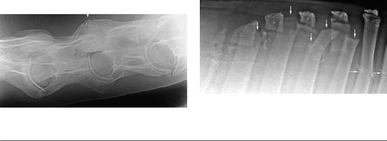
Fig. 56.1. Older vertical fracture in the region of the left cranial articular process of the 6th cervical vertebra (arrow), right = caudal, 3 years old trotter mare. (Courtesy
164 of Klinik für Orthopädie bei Huf- u. Klauentieren, Veterinärmed. Univ. Wien)
Fig. 56.2.1. Multiple fractures of the thoracic vertebral spinous processes in the region of the withers (arrow) with displacement of several fracture-fragments; asterisk: cartilaginous caps of the spinous processes with isolated ossification centers, right = caudal, 3 years old warmblood-gelding. (Courtesy of Klinik für Orthopädie bei Huf- u. Klauentieren, Veterinärmed. Univ. Wien)