

Budras_Анатомия лошади
.pdfMale Reproductive Organs
UQL|XbVxUmAVqC+HnIZ42uaraA==|1288009198
(left lateral view)
9
g
18
|
|
|
|
|
|
|
|
13 |
|
l |
||
|
|
|
|
|
f |
|
|
|
|
|
||
|
|
|
|
|
|
11 |
|
|
|
|
||
|
|
d |
|
|
|
|
10 |
|
|
k |
||
|
|
|
|
|
|
|
|
|
||||
|
|
|
e |
|
|
|
||||||
|
|
|
|
|
|
|
|
|
||||
|
|
|
|
|
|
|
|
|
|
j |
||
|
|
b |
|
|
|
h |
|
|||||
|
|
|
|
|
|
|
||||||
|
|
|
|
|
|
|
|
|
|
|||
|
a |
|
|
|
|
|
s |
i |
|
n |
|
|
|
|
|
|
|
|
|
|
|
||||
|
|
|
|
|
|
|
|
|
|
|||
|
|
|
|
|
|
|
|
|
|
|
|
|
|
|
c |
|
|
|
|
|
|
|
|
||
Prepuce: |
|
|
|
|
|
|
|
|
|
|
|
|
|
Preputial fold |
q |
o |
|
|
|
|
|
|
|||
|
|
|
|
|
|
|
|
|
||||
|
|
|
|
|
|
|
|
|
|
|||
1 |
External layer |
|
|
|
|
|
|
|
|
t |
|
|
2 |
Internal layer |
|
|
r |
|
|
|
|
|
|
|
|
3 |
Preputial ring |
|
|
|
|
|
|
|
|
|
|
|
4 |
Preputial orifice |
|
|
|
|
|
|
|
|
|
|
|
|
5 |
Internal lamina |
|
|
|
|
|
|
|
|
||
|
|
6 External lamina |
|
|
|
|
|
|
|
|
||
a |
Right transversus abdominis |
g |
Ureter |
m |
Root of penis |
b |
Right internal abd. oblique |
h |
Ext. pudendal vessels |
n |
Body of penis |
c |
Right rectus abdominis |
i |
Accssory ext. pudendal vein |
o |
Corpus cavernosum |
d |
Proximal mesorchium (Vascular fold) |
j |
Middle artery of penis |
p |
Urethra and corpus |
e |
Vaginal ring |
k |
Suspensory lig. of penis |
q |
Free part of penis |
f |
Deferent duct |
l |
Urethralis |
r |
Depth of preputial cavity |
|
7 Bulbospongiosus and |
m |
bulb of penis |
|
8Ischiocavernosus and crus of penis
9 Retractor penis
(See p. 19, 72, 75, 83)
sSupf. inguinal lymph nodes
tMedial surface of right testis
uSpermatic cord (mesorchium)
vMesofuniculus, mesorchium at v'
wLig. of tail of epididymis
xProper lig. of testis
Left Testis and Epididymis |
Accessory Reproductive Glands |
||||
(lateral view) |
|
|
|
|
(dorsal view) |
|
g |
|
|
|
f |
|
|
|
|
||
|
|
|
|
|
|
|
|
||||
10 Ampulla of |
duct |
||||
u |
11 |
Seminal vesicle |
|
|
|
Prostate: |
|
|
|
|
|
|
|
|
v |
12 |
Right lobe |
|
|
13 |
Left lobe |
|
|
|
|
|
|
||
|
|
Epididymis: |
|
|
|
14 |
Head |
|
l |
|
|
|
|
|
|
16 |
Tail |
|
|
|
w |
Testicular bursa |
|
|
|
17 |
|
|
|
|
x |
Bulbourethral gland and bulboglandularis |
|
|
|
18 |
|
|
|
|
19 |
Left testis |
8 |
7 |
|
|
|
|
|
|
20 |
Testicular artery |
|
|
|
|
|
|
9 |
85
8. Perineum, Pelvic Diaphragm, and Tail
a) PERINEUM and PERINEAL REGION are terms that refer to the area surrounding anus and vulva in the female, and anus and the root of the penis in the male. Specifically, the perineal region is on the surface of the body, and therefore has no depth.
The perineum lies deep to the perineal region; it is the body wall that closes the pelvic cavity caudally. Since this particular part of the body wall by necessity includes the anal canal and urogenital tract, it is considerably more complicated than ordinary body wall, like that of the flank, for example. The boundaries of the perineum are the floor of the bony pelvis ventrally, the sacrosciatic ligaments laterally, and the sacrum and first few caudal vertebrae dorsally. In caudal view, therefore, the perineum is triangular.
The perineal region is only skin deep. It comprises the narrow median strip between the rounded semimembranosi, the anus and vulva, and extends from the root of the tail to the ventral commissure of the vulva, which in older mares falls below the level of the pelvic floor. (The region may be divided into anal and urogenital parts by a line connecting the ischial tubers which can only be discerned by deep palpation.) In the male the perineal region extends to the base of the scrotum. The prominence of the two semimembranosi and their proximity to the midline precludes formation of an ischiorectal fossa. For the same reason the caudal border of the sacrosciatic ligament cannot be palpated as is the case in dog and cattle.
b) The PELVIC DIAPHRAGM is the muscular basis of the perineum. It is formed by the levator ani and coccygeus (see pp. 19.i; e, and 83.o; n) of which the latter slightly overlaps the lateral surface of the former. The levator ani arises from the ischial spine and adjacent medial surface of the sacrosciatic ligament. Most of its fibers end on the anus where they mingle with those of the external anal sphincter.
The coccygeus has a similar origin but passes more dorsally to insert on the first few caudal vertebrae. The two muscles are sandwiched between layers of fascia that are an integral part of the pelvic diaphragm. The space between the two coccygeus muscles dorsal to the anus is closed by the external anal sphincter (see p. 83.p) and the smooth internal anal sphincter and rectococcygeus muscles. The latter is a dorsal gathering of the outer longitudinal muscle layer of the rectum that passes caudally beyond the level of the anus to the undersurface of the tail vertebrae (see Fig. on this page).
The hiatus between the pelvic diaphragm and the caudal margin of the pelvic floor is closed by the urogenital diaphragm that is penetrated by the urogenital canal accompanied in the male by the bulbospongiosus (see p. 83.9) and in the mare by the corresponding constrictor vestibuli and constrictor vulvae muscles (see p. 83.s; t).
Another muscle in the area is the paired retractor penis (clitoridis)
(see p. 85.9) which arises from the second caudal vertebra and descends, deep to the levator ani, on each side of the rectum. It decussates ventral to the rectum and from here descends to the ventral surface of the penis, but does not reach the clitoris in the mare.
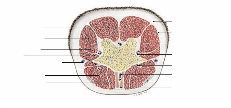
Transverse Section of the Root of the Tail
The perineal body is a musculofibrous node of tissue between anus and vestibule and consequently is a median structure. It comprises the muscular connection between external anal sphincter and constrictor vestibuli, the internal anal sphincter, the subanal decussation of the retractor clitoridis, and a fibrous plate (perineal septum) that passes craniodorsally from the vestibule to the rectum.
The birth process in the mare is occasionally accompanied by perineal lacerations; in severe cases a foot of the foal can push through the dorsal wall of the vestibule and the ventral wall of the rectum, tearing apart the tissues of the perineal body.
In the male the urethra can be palpated as it turns around the ischial arch. It is accessible to the surgeon at this point when urinary calculi have to be removed.
The pudendal and caudal rectal nerves (see p. 81.j; k) supply the perineum with motor and sensory innervation. They arise from spinal nerves S2–S4 where their roots communicate. The nerves furnish supf. perineal nerves to the skin of the perineal region as far as the base of udder and scrotum.
The pudendal nerve at first lies on the deep surface of the sacrosciatic ligament but soon buries into it. At the lesser sciatic foramen it communicates with the caudal cutaneous femoral nerve and releases the deep perineal nerve (see p. 81.i; l). The latter reinforces the supf. perineal nerves and then passes deeply to the muscles of the perineum. The continuation of the pudendal nerve crosses the internal pudendal artery and, near the median plane, turns around the ischial arch to innervate penis and clitoris.
The caudal rectal nerve runs parallel to the pudendal nerve and at first supplies the coccygeus and levator ani. Further branches go to the anal sphincter and via the supf. perineal nerve to the skin.
c) The TAIL of the horse contains about 20 vertebrae of which the last few are thin rods without discernable processes. The vertebrae are surrounded by several muscles most of which arise from the sacrum (Mm. sacrocaudales); the muscles are enclosed by fascia on the outside of which is the skin. There are four sacrocaudales muscles: dorsalis medialis (see p. 83.k), dorsalis lateralis (see p. 83.l), ventralis lateralis (see p. 83.m), and ventralis medialis (see also the
Fig. on this page). The much smaller intertransverse muscles connect neighboring transverse processes. The smooth rectococcygeus, as already mentioned, continues the longitudinal muscle layer of the rectal wall to the first few caudal vertebrae. The retractor penis (clitoridis) passes ventrally from the second caudal vertebra. Blood vessels and nerves closely accompany the caudal vertebrae. The largest are the median caudal vessels (from the caudal gluteal) which lie ventral to the vertebrae associated, at the root of the tail, with the rectococcygeus (see Fig. on this page). Taking the pulse from the artery is not as convenient as in cattle in which the sacrocaudal muscles are not as well developed.
|
Skin |
Sacrocaudalis dorsalis medialis |
|
|
Fascia |
Sacrocaudalis dorsalis lateralis |
|
|
Filamentous caudal end of dura mater |
Dorsal caudal (nerve) plexus |
|
Intertransversarii |
Dorsolateral caudal vessels |
Cutaneous branch of internal pudendal vein |
vCy4 |
|
|
Ventral caudal (nerve) plexus |
Ventrolateral caudal vessels |
|
|
Sacrocaudalis ventralis lateralis |
Median caudal vessels |
Sacrocaudalis ventralis medialis |
Tendon of coccygeus |
|
Rectococcygeus |
86
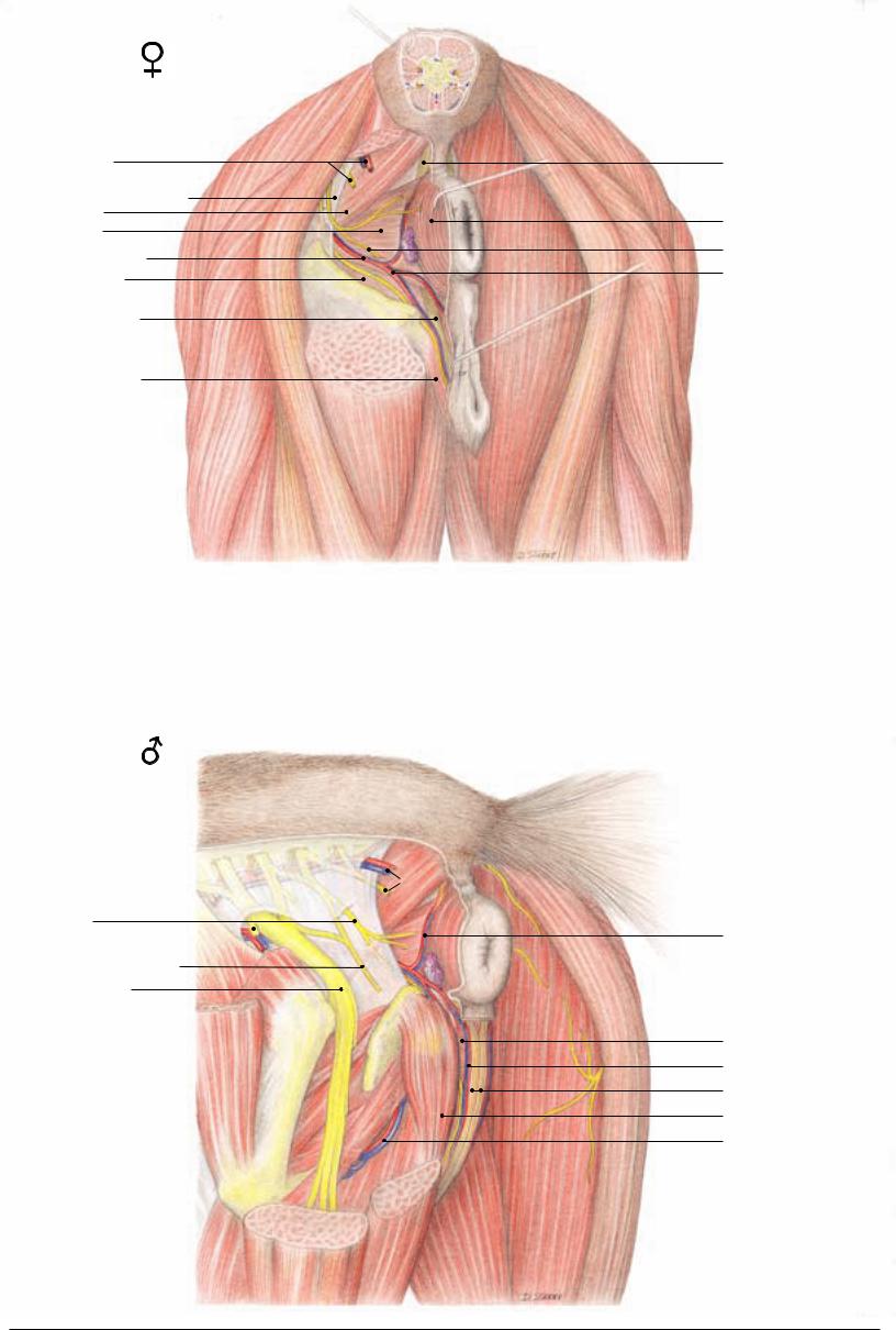
Regio perinealis
(caudal view)
d
1 Caudal rectal n., Caudal gluteal a./v.
Broad sacrotuberous ligament 2 Coccygeus m.
3 Levator ani m.
Internal pudendal a./v.
a
4 Obturator internus
5 Retractor clitoridis m.
6 Constrictor vulvae m.
e
f
g
a |
Ischiadic tuber |
|
d |
Biceps femoris m. |
b |
Greater trochanter |
|
e |
Semitendinosus m. |
c |
Third trochanter |
|
f |
Semimembranosus m. |
|
|
|
g |
Gracilis m. |
|
|
|
|
|
|
|
|
|
|
(lateral view)
2
1
10 Pudendal n. Cranial gluteal a./v. 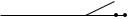
11 Caudal cutaneous femoral n.
Ischiadic (sciatic) n.
3
8
m
a
b
i
h
h
k
j
l
c
f
g
e
hGemellus mm.
iInternal obturator m.
jExternal obturator m.
kQuadratus femoris m.
 1
1
 11
11
e
f
7Rectococcygeus m.
8External anal sphincter
9Deep perineal nerve Ventral perineal a./v.
lAdductor magnus m.
mAnorectal ln.
Caudal rectal a./v.
12 Bulbospongiosus m.
a./v. of the bulb of the penis 13 Retractor penis m.
Ischiocavernosus m. Dorsal a./v. of the penis
87

Chapter 10: Selected Body Systems in Tabular Form
1. Muscles
Muscle |
Origin |
Insertion |
Innervation |
Function |
Comments |
Medial shoulder and arm muscles (p. 7)
Teres major |
Caudal border of |
Teres tuberosity of |
Axillary n. |
(7.1) |
scapula and sub- |
humerus |
|
|
scapularis |
|
|
Subscapularis |
Subscapular fossa of |
(7.3) |
scapula |
Coracobrachialis |
Coracoid process of |
(7.19) |
scapula |
Articularis humeri |
Proximal to medial rim |
|
of glenoid cavity |
Biceps brachii |
Supraglenoid tubercle |
(7.25) |
|
Brachialis |
Proximocaudal surface |
(7.20) |
of humerus |
Tensor fasciae |
Caudal border of |
antebrachii |
scapula; insertion ten- |
(7.21) |
don of latissimus dorsi |
Lesser tuberosity of |
Subscapular and |
humerus |
axillary nn. |
Proximomedial surface |
Musculocutaneous n. |
of humerus |
|
Neck of humerus |
Axillary n. |
Radial tuberosity; me- |
Musculocutaneous n. |
dial collateral lig.; via |
|
lacertus fibr. on tendon |
|
of extensor carpi rad. |
|
Proximomedial surface |
Musculocutaneous n.; |
of radius |
lateral parts of muscle |
|
by radial n. in half the |
|
horses |
Deep fascia of forearm; |
Radial n. |
olecranon |
|
Flexes shoulder joint
Predominantly an extensor of shoulder joint
Extensor of shoulder joint; adductor of limb
Tenses shoulder joint capsule
Extensor of shoulder joint; flexor of elbow joint; via lacertus fibr. stabilizer of carpus (in stay-apparatus)
Flexor of elbow joint
Tenses forearm fascia; extends elbow joint
Wide, flat muscle
Multipennate; functions as (contractile) medial collateral ligament of shoulder joint
Has a synovial bursa or a tendon sheath associated with tendon of origin
Lies on medial surface of shoulder joint capsule
Intertubercular bursa under tendon of origin; has int. tendon; consists of two parts assumed to be principally postural and locomotor
Long fibers for considerable shortening during contraction; in half the horses additional innervation by radial n.
Lateral shoulder and arm muscles (p. 7)
Deltoideus |
|
|
Axillary n. |
• Clavicular part |
Clavicular inscription |
Crest of humerus |
Advances limb |
(M. cleidobrachialis) |
|
|
|
(7.22) |
|
|
|
• Scapular part |
Aponeurotically from |
Deltoid tuberosity of |
Flexes shoulder joint |
(7.6) |
scapular spine; fleshy |
humerus |
|
|
from caudal border of |
|
|
|
scapula |
|
|
Teres minor |
Distal half of caudal |
Proximal to deltoid |
Axillary n. |
(7.13) |
border of scapula |
tuberosity of humerus |
|
Supraspinatus |
Supraspinous fossa, |
Greater and lesser |
Suprascapular n. |
(7.5) |
scapular cartilage and |
tubercles of humerus |
|
|
spine |
|
|
Infraspinatus |
Infraspinous fossa, |
Fleshy on greater tuber- |
Suprascapular n. |
(7.10) |
scapular cartilage and |
cle of humerus; strong |
|
|
spine |
tendon to lat. surface of |
|
|
|
geater tubercle distal to |
|
|
|
lat. insertion of |
|
|
|
supraspinatus |
|
Triceps brachii |
|
Olecranon tuber |
Radial n. |
|
|
(underlain by small |
|
|
|
bursa) |
|
• Long head |
Caudal border of |
|
|
(7.15) |
scapula |
|
|
• Lateral head |
Deltoid tuberosity and |
|
|
(7.16) |
vicinity |
|
|
• Medial head |
Medial surface of |
|
|
(7.17) |
humerus, middle third |
|
|
Anconeus |
Border of olecranon |
Lat. surface of |
Radial n. |
(7.24) |
fossa |
olecranon |
|
Flexes shoulder joint |
Covered by deltoideus |
Extends and stabilizes |
Biceps tendon of origin |
shoulder joint |
passes between the two |
|
insertion tendons of |
|
supraspinatus |
Extends (and flexes) shoulder joint; functions as (contractile) lat. collateral ligament
Extends elbow joint, its long head also flexes shoulder joint
Extends elbow joint; raises joint capsule to prevent its being pinched during extension of joint
Multipennate; its strong tendon underlain by (infraspinatus) bursa
Medial head is relatively the weakest
Not easily separated from lat. head of triceps
88

Muscle |
Origin |
Insertion |
Innervation |
Function |
Comments |
Caudomedial forearm muscles (p. 7) |
|
|
|
|
|
Supf. digital flexor |
Medial epicondyle of |
Distal collat. tubercles |
Ulnar n. |
Flexes digit and carpus; |
Receives access. lig. from |
(7.32) |
humerus |
of prox. phalanx; prox. |
|
extends elbow joint |
radius; forms sleeve |
|
|
collat. tubercles of |
|
|
around deep flexor ten- |
|
|
middle phalanx |
|
|
don at fetlock joint |
Deep digital flexor |
|
Flexor surface of distal |
|
Flexes digit and carpus; |
Receives access. lig. from |
(7.35) |
|
phalanx |
|
extends elbow joint |
carpus; passes through |
• Humeral head |
Medial epicondyle of |
|
Median and ulnar nn. |
|
supf. flexor sleeve; bursa |
|
humerus |
|
|
|
betw. tendon and navicu- |
• Ulnar head |
Medial on olecranon |
|
Ulnar n. |
|
lar bone |
• Radial head |
Middle of caudal |
|
Median n. |
|
|
|
surface of radius |
|
|
|
|
Flexor carpi ulnaris |
|
Access. carpal bone |
Ulnar n. |
Flexes carpus |
Its humeral head under- |
(7.41) |
|
|
|
|
lain by a bursa |
• Humeral head |
Medial epicondyle of |
|
|
|
|
|
humerus |
|
|
|
|
• Ulnar head |
Medial on olecranon |
|
|
|
|
Flexor carpi radialis |
Medial epicondyle of |
Prox. end of Mc2 |
Median n. |
Flexes carpus |
At its origin underlain by |
(7.28) |
humerus |
|
|
|
a bursa that communi- |
|
|
|
|
|
cates with that of flexor |
|
|
|
|
|
carpi ulnaris |
Metacarpus |
|
|
|
|
|
Interosseus (medius) |
Proximocaudal on |
Prox. sesamoid bones |
Deep branch of ulnar n. Counteracts overexten- |
Contains little muscular |
|
|
Mc3, and palmar |
|
|
sion of fetlock joint |
tissue; sends extensor |
|
carpal lig |
|
|
|
branches around prox. |
|
|
|
|
|
phalanx to common digi- |
|
|
|
|
|
tal extensor |
Craniolateral forearm muscles: |
|
|
|
|
|
Most of which arise from the lateral epicondyle of the humerus (p. 7) |
|
|
|||
Common digital |
Radial head (Phillip’s |
Proximodorsal on prox. Radial n. |
Carpal and digital |
In the digit the tendon |
|
extensor |
muscle) (7.34) |
phalanx (together with |
|
extensor |
receives the extensor |
(7.33) |
|
tendon of lat. dig. ex- |
|
|
branches of the in- |
|
|
tensor) |
|
|
terosseus |
|
Humeral head (7.33) |
|
|
|
|
|
Lat. epicondyle of |
Extensor process of dis- |
|
|
|
|
humerus |
tal phalanx (and of |
|
|
|
|
Ulnar head (Thierness’ |
middle phalanx) |
|
|
|
|
muscle) |
|
|
|
|
Lateral digital extensor |
Prox. end of radius and |
Proximodorsal on |
Radial n. |
Extensor of fetlock |
Insertion tendon com- |
(7.36) |
ulna |
prox. phalanx |
|
joint (not of digit) |
bines with Phillip’s |
|
|
|
|
|
muscle and is underlain |
|
|
|
|
|
by a bursa |
Extensor carpi radialis |
Lat. supracondylar |
Proximodorsal on Mc3 |
Radial n. |
Extends carpus |
Tendon receives the lac- |
(7.31) |
crest and radial fossa |
|
|
|
ertus fibr. and passes un- |
|
|
|
|
|
der the insertion tendon |
|
|
|
|
|
of the extensor obliquus |
Ulnaris lateralis |
Lat. epicondyle of |
Its short tendon on ac- |
Radial n. |
Flexes (!) carpus |
Only the long tendon has |
(M.extensor carpi |
humerus |
cess. carpal bone; its |
|
|
a tendon sheath |
ulnaris) |
|
long tendon prox. on |
|
|
|
(7.38) |
|
Mc4 |
|
|
|
Extensor carpi obliquus |
Craniolateral, middle |
Prox. on Mc2 |
Radial n. |
Extends carpus |
Covered by the digital |
(M.abductor pollicis |
of radius |
|
|
|
extensors; its tendon has |
longus) |
|
|
|
|
a tendon sheath and is |
(7.42) |
|
|
|
|
underlain by a bursa |
Muscles of the hip joint (p. 19)
Tensor fasciae latae |
Coxal tuber |
Together with the fascia Cran. gluteal n. |
(19.20; |
|
lata on patella, lat. |
73.i) |
|
patellar lig., and cran. |
|
|
border of tibia; with |
|
|
supf. gluteal muscle on |
|
|
third trochanter |
Flexes hip joint; protracts hindlimb; extends stifle; tenses fascia lata
Forms cran. border of thigh; its caudal portion blends with supf. gluteal muscle
89

Muscle |
Origin |
Insertion |
Innervation |
Function |
Comments |
Muscles of the group |
|
|
|
|
|
Supf. gluteal muscle |
Coxal tuber |
Third trochanter and |
Cranial and caudal |
Flexes hip joint; pro- |
Blends with tensor fasci- |
(19.11) |
(gluteal fascia) |
fascia lata |
gluteal nn. |
tracts and abducts |
ae latae |
|
|
|
|
hindlimb |
|
Middle gluteal muscle |
Longissimus lumbo- |
Greater trochanter |
Cranial glutal n. |
Extends hip joint; |
|
(19.3) |
rum; gluteal surface of |
|
|
abducts hindlimb |
|
|
ilium; sacrum; sacroili- |
|
|
|
|
|
ac and sacrosciatic ligg. |
|
|
|
|
Accessory gluteal |
Gluteal surface of ilum |
Just distal to greater |
Cranial gluteal n. |
|
Considered deep portion |
muscle |
|
trochanter |
|
|
of middle gluteal muscle; |
(19.9) |
|
|
|
|
tendon underlain by |
|
|
|
|
|
trochanteric bursa |
Deep gluteal muscle |
Ischial spine |
Greater trochanter |
Cranial gluteal n. |
Abducts hindlimb |
Deep to caudal portion |
(19.6) |
|
|
|
|
of middle gluteal muscle |
Caudal thigh muscles: Double innervation by caudal gluteal and tibial nerves (p. 19)
Biceps femoris |
Vertebral head: Spinous |
(19.22) |
and transv. processes of |
|
last three sacral verte- |
|
brae; Sacrosciatic lig. |
|
and tail fascia; Pelvic |
|
head: ischial tuber |
Patella; lat. and middle |
Caudal gluteal and sci- |
Extends hip and stifle |
patellar ligg.; cran. |
ataic nn. |
joints; with its caudal |
border of tibia; crural |
|
division flexes stifle; |
fascia; and via its tarsal |
|
abducts hindlimb; ex- |
tendon on calcaneus |
|
tends hock joint |
Semitendinosus |
Vertebral head: last |
(19.1) |
sacral and first two |
|
caudal vertebrae; tail |
|
fascia and sacrosciatic |
|
ligament; Pelvic head: |
|
ventral aspect of ischial |
|
tuber |
Semimembranosus |
Vertebral head: first |
(19.23) |
caudal vertebra; sacro- |
|
sciatic ligament; Pelvic |
|
head: ventromedial |
|
aspect of ischial tuber |
Cranial border of tibia; |
Caudal gluteal and |
Limb supporting |
Bursa between ischial |
crural fascia; via tarsal |
sciatic nn. |
weight: extends hip, |
tuber and vertebral head |
tendon on calcaneus |
|
stifle, and hock joints; |
of muscle |
|
|
Limb not supporting |
|
|
|
weight: flexes stifle, |
|
|
|
retracts and adducts |
|
|
|
limb |
|
Medial condyles of |
Caudal gluteal and |
Limb supporting |
Blends with adductor |
femur and tibia |
sciatic nn. |
weight: extends hip and |
|
|
|
stifle joints; Limb not |
|
|
|
supporting weight: re- |
|
|
|
tracts, adducts, and ro- |
|
|
|
tates limb inward |
|
Deep muscles of the hip joint (pp. 19 and 87)
Gemelli (87.h) |
Dorsal border of |
Trochanteric fossa of |
Muscular brr. of |
Rotate thigh outward |
|
|
ischium |
femur |
sciatic n. |
|
|
Internal obturator |
Internal surface of |
Trochanteric fossa of |
Muscular brr. of |
Rotates thigh outward |
As tendon crosses lesser |
(19.7; |
ischium and pubis from |
femur |
sciatic n. |
|
sciatic notch it is sur- |
87.i) |
border of obturator |
|
|
|
rounded by a tendon |
|
foramen to pelvic sym- |
|
|
|
sheath |
|
physis |
|
|
|
|
Quadratus femoris |
Ventral surface of |
Caudal surface of |
Muscular brr. of |
Assists in extending hip |
Covered medially by |
(87. k) |
ischium |
femur near third |
sciatic n. |
joint |
adductor |
|
|
trochanter |
|
|
|
External obturator |
Ventral surface of |
Trochanteric fossa of |
Obturator n. |
Rotates thigh outward; |
Obturator n. passes |
(87.j) |
pelvis from border of |
femur |
|
adducts limb |
through a gap in this |
|
obturator foramen |
|
|
|
muscle |
Medial muscles of the thigh: adductors (p. 19)
Gracilis |
Pelvic symphysis via |
Crural fascia, medial |
Obturator n. |
Adducts limb (also |
The tendons of origin of |
(19.21; |
symphysial tendon |
patellar ligament, and |
|
extends stifle) |
right and left muscles |
73.8; |
|
cranial border of tibia |
|
|
form the symphysial ten- |
87.g) |
|
|
|
|
don; the muscle forms |
|
|
|
|
|
the caudal border of the |
|
|
|
|
|
femoral triangle |
Adductor |
Ventral surface of |
Caudal surface and |
Obturator n. |
Adducts and retracts |
|
(19.19) |
pelvis; symphysial |
medial epicondyle of |
|
limb |
|
|
tendon |
femur |
|
|
|
Pectineus |
Pubis and iliopubic |
Medial surface of |
Obturator and femoral |
Adducts limb; flexes |
The tendons of origin of |
(19.14; |
eminence of the other |
femur |
nn. |
hip joint |
right and left muscles |
73.6; |
side |
|
|
|
form the prepubic ten- |
77.5) |
|
|
|
|
don |
90

Muscle |
Origin |
Insertion |
Innervation |
Function |
Comments |
Extensors of the stifle (p. 19) |
|
|
|
|
|
Sartorius |
Int. iliac fascia and |
Medial aspect of stifle |
Femoral n. |
Flexes hip joint; pro- |
Forms cranial border of |
(19.10; |
insertion tendon of |
|
|
tracts and adducts limb |
femoral triangle |
73.18) |
psoas minor |
|
|
|
|
Quadriceps femoris |
|
Via the intermediate |
Femoral n. |
Flexes hip joint (rectus |
Tonus stabilizes patella; |
(19.15; |
|
patellar ligament on the |
|
fem.); extends and sta- |
prox. and distal infra- |
73.j) |
|
tibial tuberosity |
|
bilizes stifle |
patellar bursae lie deep |
• Rectus femoris |
Shaft of ilium cranial to |
|
|
|
to insertion tendon |
(19.o) |
acetabulum |
|
|
|
(intermediate patellar |
• Vastus lateralis |
Proximolateral on |
|
|
|
lig.) |
(19.p) |
femur |
|
|
|
|
• Vastus medialis |
Proximomedial on |
|
|
|
|
(19.n) |
femur |
|
|
|
|
• Vastus intermedius |
Proximodorsal on |
|
|
|
|
|
femur |
|
|
|
|
Special flexor of the stifle: caudal to the stifle (p. 19)
Popliteus |
Lat. condyle of femur |
Caudomedial border |
Tibial n. |
(19.27) |
|
of tibia |
|
Extensors of the hock, flexors of the digit: on caudal surface of leg (p. 19)
Gastrocnemius |
With medial and |
As part of common |
Tibial n. |
(19.26) |
lateral heads from cor- |
calcanean tendon on |
|
|
responding supracondy- |
calcanean tuber |
|
|
lar tuberosities of the |
|
|
|
femur |
|
|
Soleus |
Prox. end of fibula |
Joins common |
Tibial n. |
(19.28) |
|
calcanean tendon |
|
Supf. digital flexor |
Supracondylar fossa |
Plantar on distal end of |
Tibial n. |
(19.31) |
of femur |
proximal phalanx and |
|
|
|
prox. collateral tuber- |
|
|
|
cles of middle phalanx |
|
Deep digital |
|
Plantar on distal |
Tibial n. |
flexor |
|
phalanx |
|
• Lat. digital flexor |
Caudal surface of tibia |
|
|
(19.34) |
with tibialis caudalis |
|
|
• Tibialis caudalis |
Caudal surface of tibia |
|
|
(19.33) |
with lat. dig. Flexor |
|
|
• Med. digital flexor |
Lat. tibial condyle |
|
|
(19.29) |
|
|
|
Flexes stifle |
Tendon of origin is sur- |
|
rounded by a pouch of |
|
the stifle joint |
Extends hock; flexes |
|
stifle |
|
Rudimentary muscle; |
Forms m. triceps surae |
could extend the hock |
with both heads of gas- |
|
trocnemius |
Being mainly tendinous, the prox. part acts in the reciprocal mechanism, the distal part supports fetlock and pastern joints
Extends hock; flexes digit
Large subtendinous bursa over calcanean tuber; forms sleeve for deep dig. flexor at fetlock joint
Their tendons combine to pass over the sustentaculum tali
Joins common deep flexor tendon in metatarsus
Fexors of hock and extensors of digit: Craniolateral on leg (p. 19)
Tibialis cranialis |
Lat. condyle and |
(19.36) |
tuberosity of tibia |
Peroneus tertius |
Lat. condyle of femur |
(19.37) |
|
Long digital extensor |
Lat. condyle of femur |
(19.40) |
|
Lateral digital extensor |
Lat. collat. lig of stifle |
(19.41) |
and nearby tibia and |
|
fibula |
Dors. br.: on T3 and |
Peroneal n. |
prox. end of Mt3; Med. |
|
br.: on T1+2 |
|
By four branches on all |
Peroneal n. |
tarsal bones (except |
|
T1+2) and on Mt3 |
|
Extensor process of dis- |
Peroneal n. |
tal phalanx; secondarily |
|
on proximal and mid- |
|
dle phalanges |
|
Joins long extensor |
Peroneal n. |
tendon |
|
Extensor digitalis |
From the lat. brr. Of |
Joins tendon of long |
Peroneal n. |
brevis |
peroneus tertius inser- |
extensor |
|
|
tion tendon |
|
|
Metatarsus
Interosseus muscle |
Mt3, calcaneus, and T4 Prox. sesamoid bones |
Tibial n. |
(suspensory ligament) |
|
|
Flexes hock joint |
The medial branch of its |
|
insertion tendon is |
|
known also as the |
|
“cunean tendon” |
Entirely tendinous; |
Originates together with |
constituent of reci- |
long dig. extensor from |
procal mechanism |
the lat. condyle of the |
|
femur |
Extends digit; flexes |
Held to dorsal surface of |
hock |
hock by three retinacula; |
|
in digit receives extensor |
|
brr. from interosseus |
Extends digit; flexes |
|
hock |
|
Rudimentary: could |
|
extend digit |
|
Counteracts overexten- |
In the foal, the inter- |
sion of fetlock joint |
osseous is fairly muscu- |
|
lar. In the adult the |
|
interosseous is entirely |
|
tendinous. Its extensor |
|
branches pass onto the |
|
dorsal aspect of the |
|
proximal phalanx and |
|
join the long digital ex- |
|
tensor tendon. |
91

Muscle |
Origin |
Insertion |
Innervation |
Function |
Comments |
Facial Musculature (p. 37) |
|
|
|
|
|
Cutaneous muscle of |
Thin bundles between |
|
Facial n. |
Moves the skin; retracts |
|
the face |
larynx and mouth |
|
|
angle of the mouth |
|
(37.a) |
|
|
|
|
|
M. sphincter colli profundus: Muscles of the external ear (p. 37) |
|
|
|||
Supf. cervicoauricularis |
Ext. occipital protuber- |
Ext. surface of auricle |
Caudal auricular n. |
Raises auricle |
Has double innervation |
(37.29) |
ance and nuchal lig. |
|
(from facial), and great |
|
|
|
|
|
auricular n. |
|
|
Cervicoscutularis |
Nuchal crest |
Caudomedial on scuti- |
Facial and caudal |
Elevates auricle |
Well developed |
|
|
form cartilage |
auricular nn. |
|
|
Cervicoauricularis |
Nuchal lig. and caudal |
Lateral border of |
Facial and caudal |
Moves auricle laterally |
Partially covered by supf. |
profundus and |
surface of occipital |
auricle |
auricular nn. |
|
cervicoauricularis |
medius |
bone |
|
|
|
|
(37.l) |
|
|
|
|
|
Interscutularis |
Connects right and |
Also attaches on ext. |
From facial n.: |
Stabilizes scutiform |
|
(37.o) |
left scutiform cartilages |
sagittal crest and tem- |
auriculopalpebral n. |
cartilage |
|
|
|
poral line |
and rostral auricular |
|
|
|
|
|
brr. |
|
|
Frontoscutularis |
|
|
|
Stabilizes scutiform |
|
(37.m) |
|
|
|
cartilage |
|
• Frontal part |
Temporal line |
Rostal and lateral on |
From facial n.: |
|
Both parts well isolated |
• Temporal part |
Zygomatic arch |
scutiform cartilage |
auriculopalpebral n. |
|
|
|
|
|
and rostral auricular |
|
|
|
|
|
brr. |
|
|
Supf. scutuloauricularis |
Scutiform cartilage |
Rostromedial on |
From facial n.: |
Straightens and moves |
May be divided into |
|
|
auricle |
auriculopalpebral n. |
auricle medially |
three parts |
|
|
|
and rostal auricular |
|
|
|
|
|
brr. |
|
|
Zygomaticoauricularis |
Zygomatic arch |
Ventromedial on |
From facial n.: |
Moves auricle medially |
|
(37.n) |
|
auricle |
auriculopalpebral n. |
|
|
|
|
|
and rostral auricular |
|
|
|
|
|
brr. |
|
|
Parotidoauricularis |
Fascia covering |
Ventrolateral on the |
Cervical br. of |
Depressor of auricle |
Lies on parotid gland |
(37.28) |
parotid gland |
base of the auricle |
facial n. |
|
|
Styloauricularis |
Ext. acoustic meatus |
Ventromedial on the |
Caudal auricular n. |
Not known |
Lies on ext. acoustic |
|
|
base of the auricle |
(from facial) |
|
meatus |
Muscles of the lips and cheek (p. 37)
Orbicularis oris |
Forms a closed circle |
|
Dorsal and ventral buc- |
Closes the mouth |
No attachment on bone |
(37.1) |
around the mouth |
|
colabial brr. of facial n. |
opening |
|
Buccinator |
Border of upper jaw |
Border of lower jaw |
Dorsal and ventral |
Forms muscular basis |
May be divided into |
(37.7) |
and coronoid process |
|
buccolabial brr. of |
of cheek; returns food |
buccal and molar parts |
|
of mandible |
|
facial n. |
to the central cavity of |
|
|
|
|
|
the mouth |
|
Zygomaticus |
Facial crest |
Angle of mouth |
Auriculopalpebral br. |
Retracts angle of mouth |
|
(37.5) |
|
|
of facial n. |
|
|
Caninus |
Rostral end of facial |
Lateral border of |
Dorsal and ventral |
Elevates upper lip and |
Passes between the two |
(37.3) |
crest |
nostril |
buccolabial brr. of |
widens nostril |
parts of the levator |
|
|
|
facial n. |
|
nasolabialis |
Levator labii |
Lacrimal bone |
By common tendon |
Dorsal and ventral |
Elevates upper lip; |
Covers infraorbital |
superioris |
|
with its fellow between |
buccolabial brr. of |
“flehmen” reaction |
foramen; passes deep to |
(37.6) |
|
the nostrils in the upper |
facial n. |
|
levator nasolabialis |
|
|
lip |
|
|
|
Depressor labii |
Mandibular ramus |
Blends with orbicularis |
Dorsal and ventral |
Depresses and retracts |
Its tendon covers the |
inferioris |
|
oris in lower lip |
buccolabial brr. of |
lower lip |
mental foramen |
(37.2) |
|
|
facial n. |
|
|
92

Muscle |
Origin |
Insertion |
Innervation |
Function |
Comments |
Muscles of the eye lids and nose (p. 37)
Orbicularis oculi |
Forms a closed circle around palpebral fissure |
Auriculopalpebral n. |
Closes palpebral |
Better developed in |
|
(37.23 and |
|
|
(from facial n.) |
fissure |
upper lid |
43.c) |
|
|
|
|
|
Levator anguli oculi |
Base of zygomatic |
Medial end of upper |
Auriculopalpebral n. |
Elevates medial part of |
A small muscle |
(37.22) |
process of frontal bone |
lid |
(from facial n.) |
upper lid |
|
Levator nasolabialis |
Frontal and nasal |
Supf. part: blends with |
Auriculopalpebral n. |
Elevates upper lip; |
Caninus passes between |
(37.4) |
bones |
orbicularis oris; deep |
(from facial n.) |
enlarges nostril |
the two parts of the |
|
|
part: into upper lid |
|
|
muscle |
Malaris |
Dorsal to facial crest |
Lower lid |
Auriculopalpebral n. |
Pulls lower lid |
A small muscle |
(37.9) |
|
|
(from facial n.) |
ventrally |
|
Muscles of mastication (pp. 37, 39 and 45)
Supf. Muscles of the intermandibular space
Digastricus |
Paracondylar process |
Ventral border of |
(39.17) |
|
mandible; its occipito- |
|
|
mandibular part to |
|
|
angle of mandible |
Caudal belly: digastric |
Opens mouth; elevates |
The two bellies are seper- |
br. of facial n.; Rostral |
hyoid apparatus |
ated by an intermediate |
belly: mylohyoid n. of |
|
tendon which penetrates |
mandibular n. |
|
the tendon of the stylo- |
|
|
hyoideus |
Mylohyoideus |
Lingual surface of |
Basihyoid and lingual |
Mylohyoid n. (from |
Elevates floor of |
Consists of rostral and |
(39.15; |
mandible (mylohyoid |
process; and median |
mandibular n.) |
mouth; presses tongue |
caudal parts |
45.k) |
line) |
raphe |
|
against hard palate |
|
Lateral Muscles of |
|
|
|
|
|
Mastication |
|
|
|
|
|
Temporalis |
Temporal fossa and |
Coronoid process of |
Masticatory n. (from |
Mastication (elevates |
|
(39.11) |
medial surface of |
mandible |
mandibular n.) |
mandible and presses it |
|
|
zygomatic arch |
|
|
against maxilla) |
|
Masseter |
|
Large area caudolateral |
Masticatory n. |
Mastication (elevates |
Penneate muscle |
(39.9) |
|
on mandible |
(from mandibular n.) |
mandible and presses it |
|
• Supf. part |
Facial crest |
|
|
against maxilla |
|
• Deep part |
Zygomatic arch |
|
|
|
|
Medial Muscles of |
|
|
|
|
|
Mastication |
|
|
|
|
|
Ptergoideus |
Pterygoid process of |
Convave medial |
Pterygoid nn. (from |
Synergist to masseter; |
(39.12) |
basisphenoid and |
surface of mandible; |
mandibular n.) |
in unilateral contrac- |
• —medialis |
vicinity |
condyle of mandible |
|
tion: moves lower jaw |
• —lateralis |
|
and vicinity |
|
laterally |
The mandibular n. lies between the two muscles; the lateral muscle is fleshy
Muscles of the eye (p. 41 and Anatomy of the Dog)
Muscles of the pharynx (p. 47)
Stylopharyngeus |
Medial on dorsal third |
Dorsolateral |
Glossopharyngeal n. |
Dilates pharynx |
The only muscle that |
(47.10) |
of stylohyoid |
pharyngeal wall |
(IX) |
|
dilates the pharynx |
Muscles of the Soft |
|
|
|
|
|
Palate |
|
|
|
|
|
Tensor veli palatini |
Muscular process of |
Around hamulus of |
Mandibular n. (V3) |
Tenses soft palate |
|
(47.7) |
petrous temporal bone, |
pterygoid bone into the |
|
|
|
|
pterygoid bone, lateral |
aponeurosis of the soft |
|
|
|
|
surface of auditory tube |
palat |
|
|
|
Levator veli palatini |
Muscular process of |
In the soft palate |
Pharyngeal plexus (IX |
Elevates soft palate |
|
(47.8) |
petrous temporal bone, |
|
and X) |
|
|
|
lateral surface of audi- |
|
|
|
|
|
tory tube |
|
|
|
|
93

Muscle |
Origin |
Insertion |
Innervation |
Function |
Comments |
Rostral Pharyngeal |
|
|
|
|
|
Constrictors |
|
|
|
|
|
Pterygopharyngeus |
Pterygoid bone |
Pharyngeal raphe |
Pharyngeal plexus |
Constricts and |
Difficult to separate from |
(47.9) |
|
|
(IX and X) |
protracts pharynx |
palatopharyngeus; |
|
|
|
|
|
crosses lat. surface of |
|
|
|
|
|
levator veli palatini |
Palatopharyngeus |
Via aponeurosis of soft |
(47.11) |
palate from palatine |
|
and pterygoid bones |
Middle Pharyngeal |
|
Constrictor |
|
Hypopharyngeus |
Medial surface of ven- |
(47.12) |
tral end of stylohyoid; |
|
thyrohyoid and thyroid |
|
lamina |
Caudal Pharyngeal |
|
Constrictors |
|
Thyropharyngeus |
Thyroid cartilage |
(47.13) |
|
Cricopharyngeus |
Arch of cricoid |
(47.14) |
cartilage |
Intrinsic muscles of the larynx (p. 49)
Cricothyroideus |
Ventrolateral on cricoid |
(49.12) |
cartilage |
Cricoarytenoideus |
Ipsilateral half of |
dorsalis |
cricoid lamina |
(49.7) |
|
Pharyngeal raphe and |
Pharyngeal plexus |
Constricts and |
Lies medial to levator |
dorsal border of thy- |
(IX and X) |
protracts pharynx |
veli palatini |
roid cartilage |
|
|
|
Pharyngeal raphe |
Pharyngeal plexus |
Constricts pharynx |
May be divided into |
|
(IX and X) |
|
ceratoand chondro- |
|
|
|
pharyngei |
Pharyngeal raphe |
Pharyngeal plexus |
Constricts pharynx |
Blends with crico- |
|
(IX and X) |
|
pharyngeus |
Pharyngeal raphe |
Pharyngeal plexus |
Constricts pharynx |
Blends caudally with the |
|
(IX and X) |
|
longitudinal musculature |
|
|
|
of the esophagus |
Caudal border of |
Cranial laryngeal n. |
Narrows glottic cleft; |
|
thyroid lamina |
(from vagus) |
tenses vocal folds |
|
Muscluar process of |
Recurrent laryngeal n. |
Enlarges glottic cleft |
|
arytenoid cartilage |
(from vagus) |
|
|
Cricoarytenoideus |
Rostrolateral on cricoid |
Muscular process of |
Recurrent laryngeal n. |
Narrows glottic cleft |
|
lateralis |
cartilage |
arytenoid cartilage |
(from vagus) |
|
|
(49.6) |
|
|
|
|
|
Arytenoideus |
Muscular processes of |
Median raphe dorsal to |
Recurrent laryngeal n. |
Narrows glottic cleft |
Unpaired muscle |
transversus |
right and left arytenoid |
arytenoid cartilages |
(from vagus) |
|
|
(49.5) |
cartilages |
|
|
|
|
Thyroarytenoideus |
|
|
Recurrent laryngeal n. |
Narrows glottic cleft |
|
|
|
|
(from vagus) |
|
|
• Ventricularis |
Ventromedial on |
Muscular process of |
|
|
|
(49.9) |
thyroid cartilage, and |
arytenoid cartilage; |
|
|
|
|
on cricothyroid liga- |
some fibers blend with |
|
|
|
|
ment |
arytenoideus transv. |
|
|
|
• Vocalis |
Ventromedial on |
Muscular and vocal |
|
|
|
(49.11) |
thyroid cartilage |
processes of arytenoid |
|
|
|
|
|
cartilage |
|
|
|
Thyroarytenoideus |
Muscular process of |
Dorsal border of |
Recurrent laryngeal n. |
Constricts glottic cleft |
Inconstant and present |
accessorius |
arytenoid cartilage |
thyroid lamina |
(from vagus) |
|
only in the horse; may be |
|
|
|
|
|
represented by a liga- |
|
|
|
|
|
ment |
Tensor ventriculi |
Cuneiform process of |
Fans to lateral surface |
Recurrent laryngeal n. |
Tenses lateral laryngeal |
Thin muscle, present |
laryngis |
epiglottic cartilage |
of laryngeal ventricle |
(from vagus) |
ventricle |
only in the horse |
Muscles of the tongue and hyoid apparatus (fan into the tongue or attach on the basihyoid)
(p. 45 and 51)
M. lingualis proprius |
Intrinsic tongue muscle |
Hypoglossal n. (XII) |
Simultaneous contrac- |
|
|
|
tion of transv. and per- |
|
|
|
pendicular bundles |
|
|
|
stiffens the tongue |
Extrinsic Tongue Muscles
Consists of longitudinal, transverse, and perpendicular bundles
Styloglossus |
Lateral surface of |
Ends near the tip of the Hypoglossal n. (XII) |
Pulls tongue caudodor- |
(51.10) |
stylohyoid |
tongue |
sally; in unilateral con- |
|
|
|
traction, to the side |
Right and left muscles fuse near the tip of the tongue
94