

Budras_Анатомия лошади
.pdf[164] [165] [166] Possible conservative therapy consists of an orthopedic horse shoeing including hoof trimming, systemic and local medication as well as an adequate exercise regime. [167] [168] [169] Recently also the application of shock-wave-treatment (extracorporeal shock wave therapy) is recommended. [170] Beyond that, also different surgical procedures are described. To promote perfusion of the digit, perivascular sympathectomy (section of sympathetic nerve fibers) in the region of the medial or lateral palmar digital artery and vein, in combination with a fasciotomy, has been proposed. [99] As a further possibility, desmotomy of the ligaments between the long pastern bone and the navicular bone is described. [171] [172] With a positive result from anesthesia of the palmar digital nerves (TPA 2), neurectomy of the lateral and medial palmar digital nerves at the pastern can be performed as a final measure. [121] [168]
14.1. Synovial structures on the Thoracic Limb
Besides the diagnostic nerve anesthesias, also often intrasynovial anesthesias of joints or certain synovial sheaths of synovial bursae are performed in examination for lameness. For this reason the most often used sites of injection of these structures are important for diagnosis of lameness. Also, for the implementation of an endoscopic examination (arthroscopy, tendovaginoscopy or bursoscopy) knowledge of the anatomy of the concerned structure is a basic precondition. Besides that, the synovial structures are of a special clinical importance, because the lesions in the region of these structures are always connected to a risk of its infection and by this to the necessity of an immediate adequate therapy. A solid knowledge of the exact localization and extension of the synovial spaces is for this reason also essential for the evaluation of lesions.
14.2. The shoulder joint is covered by relatively strong muscles. For this reason, the danger of an infection caused by a wound is more rare. But also non-infectious inflammation of the shoulder joint is relatively rarely observed. Occasional causes of lameness are lesions of the articular cartilage or of the subchondral bone, related to a disorder of endochondral ossification (osteochondrosis). [173] [108] In radiographs a flattening or indentation of the bony contour or of isolation of a cartilage-bone-chip is visible. Such alterations can be observed both in the region of the glenoid cavity of the scapula and the humeral head. Usually the caudal half of the joint is involved. [173] [108] Moreover, cyst-like defects may occur in the subchondral bone. These are usually localized at the transition between the middle and caudal thirds of the glenoid cavity of the scapula. [108] Therapy can be conservative [108] or surgical
(arthroscopical curettage of the altered cartilage or removal of the osteochondral fragment [173]).
Especialy in Shetland ponies, there occasionally appears also a dysplasia (defective development) of the shoulder joint, which is accompanied by subluxation and a secondary degenerative arthri-
tis. [174] [175] As collateral ligaments are absent in the shoulder joint, the insertional tendons of the subscapularis, infraspinatus and supraspinatus muscles are responsible for the collateral stability of the joint. [176] Collateral instability of the shoulder joint can be caused by a lesion of the nerves of the brachial plexus and the accompanying loss of function of the muscles, which act as a replacement for the collateral ligaments.
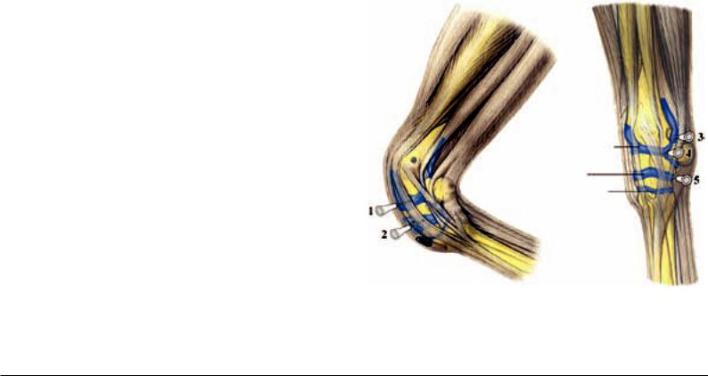
Injection of the shoulder joint for intraarticular anesthesia or local therapy is performed cranial to the tendon of the infraspinatus muscle, which is easily palpable. To do this, the needle is introduced at the depression between the cranial and caudal parts of the greater tubercle of the humerus and advanced about 6–8 cm in a caudomedial and slightly distal direction (in a direction toward the elbow of the opposite side; Fig. 14.2., 1). [114] [177] If a second access is necessary to flush the joint, the shoulder joint can be injected additionally about 10 cm caudal and 4 cm distal to the site just described. The needle is directed 20° dorsally and to a depth of 8 cm. [177] Anesthesia of the shoulder joint is obtained with 25–30 cc of a 2% local anesthesia solution [114], in which case the injection of a larger volume (more than 10 cc) may, owing to diffusion, lead to a paresis of the adjacent suprascapular nerve. [177]
For arthroscopy of the shoulder joint, access is as a rule also cranial to the tendon of the infraspinatus and directly proximal to the depression between the cranial and caudal parts of the greater tubercle of the humerus. [178] [179] The needle is introduced at an angle of about 25° in a caudodistal direction. [179] The second access for the instruments will be chosen depending on the location of the lesion. Usually a caudolateral portal for the instruments
(about 6 cm caudal and 4 cm distal to the arthroscopic portal) is utilized. [178] [179] Sometimes also a cranial access of the instruments directly distal to the arthroscopic portal is possible. An alternate access is located about 1 cm caudal to the tendon of the infraspinatus (between the infraspinatus muscle and the teres minor muscle) muscle. Access for the instruments is, in this case, about 2–4 cm caudal to the arthroscopic portal. [179]
14.3. Pathologic changes in the region of the elbow joint are likewise relatively seldom the cause for lameness. Occasionally, cystlike subchondral defects occur, which are usually located medially in the proximal epiphysis of the radius. [180] [181] Osteochondral changes or traumata are discussed as the cause. [182] Besides conservative therapy, a surgical procedure in the form of an extra-artic- ular curettage of the subchondral lesion is described. [180]
In contrast to the shoulder joint, the lateral part of the elbow joint is covered only by little soft tissue. For this reason, in the event of lacerations of the elbow joint region, the joint is relatively easily
Shoulder joint
Subtendinous bursa of the infraspinatus muscle Intertubercular bursa
Fig. 14.2. Injection of the shoulder joint (1) and the intertubercular bursa (2). (Courtesy of Institut f. Veterinär-Anatomie, Berlin)
Subcutaneous bursa over the olecranon
Subtendinous bursa of the
triceps brachii muscle
Elbow (cubital) joint
Subtendinous bursa of the extensor carpi ulnaris (ulnaris lateralis) muscle
Subcutaneous bursa
over the lateral prominence of the head of the radius
Fig. 14.3. Injection of the elbow joint: 1) cranial access, 2) proximolateral access.
(Courtesy of Institut f. Veterinär-Anatomie, Berlin)
125
opened and can become infected. In such a case, a possible communication of the joint with the 5 cm long subtendinous bursa of the ulnaris lateralis muscle [183] as well as a shift of the skin over the articular region during movement of the limb must be taken into consideration, so that even a supposed distant wound may have a communication with the elbow joint. To examine for possible articular infection, the joint is penetrated and the synovia is examined cytologically.
Injection of the elbow joint is traditionally performed cranial to the lateral collateral ligament, which is found between the lateral epicondyle of the humerus and the tuberosity of the radius for the attachment of the ligament (both bony points are readily palpable).
[177] [184] The needle is introduced about 2.5 cm cranial to the collateral ligament and about 3.5 cm proximal to the ligament’s insertion on the radius. The skin is penetrated horizontally and the needle directed slightly caudally and advanced to a depth of 5–6 cm (Fig. 14.3. 1). [114] [177] Alternatively, the joint can also be penetrated by way of the proximolateral part of the caudal recess in the region of the olecranon fossa. To do this, the needle is introduced into the palpable depression between the olecranon and the lateral supracondylar crest of the humerus at an angle of about 45°. The needle is advanced mediodistally and slightly cranially about 4–7 cm (Fig. 14.3, 2). [114] [183] For intra-articular anesthesia of the elbow joint, about 20–25 cc of a 2% local anesthesia solution is injected. [114] The anesthesia of the elbow joint that was performed in former times by way of the subtendinous bursa of the ulnaris lateralis muscle is very uncertain owing to the fact that the two synovial structures communicate only in about one-third of horses. [183]
For arthroscopy of the elbow joint, access can be caudomedial, between the flexor carpi radialis muscle and the flexor carpi ulnaris muscle, where a large part of the caudal articular surfaces of the humerus, radius and ulna can be demonstrated. [185] A second access for the instrumentation is possible caudal and slightly proximal to the arthroscopic portal. To avoid injury to the ulnar nerve as well as the collateral ulnar artery and vein, access should not be proximal to the articular cavity of the humeroradial joint. [185] Alternatively, access to the caudal region of the elbow joint is also possible laterally by way of the large synovial outpouching in the olecranon fossa. [179] To demonstrate the cranial articular surface of the humeral condyles, arthroscopic access can be gained directly cranial to the lateral collateral ligament. In this case, access for instrumentation is located in the region of the belly of the extensor carpi radialis muscle or of the common digital extensor muscle.
[185]
14.4. The carpal joint is composed of three horizontally arranged joints. The proximal joint (antebrachiocarpal joint or radiocarpal joint) is located between the radius and the proximal row of carpal bones and permits flexion of about 95°. [186] The middle joint
(mediocarpal joint, in clinical usage also intercarpal joint) is between the proximal and distal row of carpal bones and can be flexed about 45°. [186] It communicates always with the distal joint (carpometacarpal joint), in which case the synovial spaces—among others—communicate between the third and fourth carpal bones. A communication of the intercarpal joint with the antebrachiocarpal joint is rare. [187] The carpometacarpal joint has palmarodistal outpouchings, which are present laterally and medially between the corresponding splint bone and the cannon bone. They extend about
2.5 cm in a distal direction. [187]
Besides the chip and slab fractures in the region of the carpus, degenerative changes of the carpal joints are the most frequent problems. They are usually associated with an increased filling of the joint, which can be most easily palpated on the flexed limb medially and laterally beside the tendon of the extensor carpi radialis muscle. [31]
In the carpal joint region, also a great number of synovial sheaths and bursae are found (Fig. 14.13.). These can also be excessively filled due to inflammatory processes and have to be considered in the differential diagnosis. Also, with injuries in the carpal region, which are accompanied by effusion of synovia, an opening of the carpal joint, of the synovial sheaths or of the synovial bursa must be considered in the differential diagnosis.
For injection of the antebrachiocarpal joint and the intercarpal joint, the named joints are flexed, in which case they open dorsally.
Medial to the insertion tendon of the extensor carpi radialis muscle
or between this tendon and the tendon of the common digital exten-
126
sor muscle, both joints are palpable as a depression. The needle is inserted in this depression perpendicular to the skin and advanced into the particular joint (Fig. 14.4., 1/2). Care must be taken for the tendons and their tendon sheaths. [114] [184] An additional access to both joints is possible by way of the corresponding palmarolateral recess. [114] [188] The corresponding articular outpouching of the antebrachiocarpal joint extends proximal to the accessory carpal bone and lies directly cranial to the carpal synovial sheath, which in case of increased filling of these synovial structures can easily lead to mistakes. [114] Injection of the joint is performed between the readily palpable tendon of the lateral digital extensor muscle and the tendon of the ulnaris lateralis muscle, directly proximal to the accessory carpal bone (section 14.4., 3). [114] Alternatively, the injection can also be performed farther distal, about the level of the middle of the accessory carpal bone. [188] At this site, a slight depression is palpable between the distal radius and the ulnar carpal bone into which the needle is introduced perpendicular to the skin (section 14.4., 4.). [114] [188] In doing this, care must also be taken for the surrounding tendons and tendinous sheaths. The palmarolateral recess of the intercarpal joint is located about 2–2.5 cm farther distal between the palmar border of the ulnar carpal bone and the fourth carpal bone (section 14.4., 5.).
[114] [188] For anesthesia of each corresponding joint about 5–10 cc of a 2% local anesthesia solution is injected, [114] The carpometacarpal joint does not have to be injected individually, because it will be desensitized by anesthesia of the intercarpal joint owing to the communication of the synovial spaces. In doing this, consideration must be given to the fact that it is possible for a diffusion of anesthesia solution from the palmarodistal outpouching of the carpometacarpal joint to the origin of the suspensory ligament as well as to the palmar metacarpal nerves. This can lead to misinterpretations. [114] Moreover, diffusion processes have been described between the intercarpal joint and the antebrachiocarpal joint. [189]
For arthroscopy of the antebrachiocarpal and of the intercarpal joint, access is also medial to the terminal tendon of the extensor carpi radialis muscle or between that tendon and the tendon of the common digital extensor muscle. [179] [190] The palmar part of the joints is accessible via the respective palmarolateral articular recess, in which case, after filling of the joint, the instruments are introduced in the center. [179] Besides that, also an access to the antebrachiocarpal joint from caudomedial is described. [191]
Antebrachiocarpal joint
Mediocarpal (clinical: intercarpal) joint Carpometacarpal joint
Fig. 14.4. Injection of the carpal joint: dorsal access into the antebrachiocarpal joint (1) or into the mediocarpal- (middle carpal-) joint (2), proximal (3) or distal (4) access into the palmarolateral recess of the antebrachiocarpal joint, (5) access into the palmarolateral recess of the mediocarpal- (middle carpal-) joint. (Courtesy of Institut f. Veterinär-Anatomie, Berlin)
14.5. The fetlock joint is a compound hinge joint, which permits a slight side-to-side movement only in extreme flexion. [176] In the weight-bearing limb, the joint is in hyperextension, in which the dorsal extension angle is about 140°. [152] To prevent excessive hyperextension of the joint, the fetlock joint is supported palmarly by different ligamentous and tendinous structures (so-called suspensory apparatus). [176]
The collateral ligaments of the fetlock joint consist each of two parts, which are well demonstrated sonographically. The somewhat more dorsally located superficial part is relatively thin and runs in a vertical direction. The deeper part is on the other hand obliquely distopalmarly oriented and attaches not only to the proximal phalanx, but also to the corresponding sesamoid bone. [192] Lesions of the collateral ligaments, which can arise from avulsion fractures of the proximal phalanx, are usually due to trauma and lead to an instability or luxation of the fetlock joint. [193] The joint capsule forms a dorsal and a palmar articular recess, in which case the dorsal recess extends proximally about 2 cm and the palmar recess about 4–5 cm proximally. With increased filling, the latter is palpable or even visible (so-called “windpuffs”) between the insertional branches of the suspensory ligament and the cannon bone (McIII). It must not be confused with the proximal part of the digital synovial sheath of the flexor tendons, which is palmar to the suspensory ligament. [176]
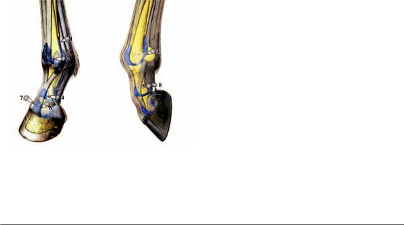
Diagnostic or therapeutic injection of the fetlock joint can be performed in the region of both articular outpouchings, in which case access to the palmar recess is the easier. The traditional proximal injection site lies between the cannon bone (McIII) and the suspensory ligament at the level of the “button” of the splint bone and proximal to the collateral ligament of the sesamoid bone, in which case the needle is advanced in a slightly distal direction (Fig. 14.5., 1).
[188] [184] [194] Owing to the fact that at this site there are prominent villi of the joint capsule, which often cause an obstruction of the needle and, besides that, the danger of hemorrhage into the joint is relatively high, it would be better to tap the joint somewhat farther distally. [195] To do this, the fetlock joint is slightly flexed.
The needle is inserted perpendicularly through the skin dorsal to the neurovascular bundle in the palpable depression between McIII and the lateral sesamoid bone and advanced through the collateral ligament of the sesamoid bone (Fig. 14.5., 3). [195] [188] Injection into
Fig. 14.5. Injection of the digital articulations: (1) proximal access into the palmar recess of the fetlock joint, (2) injection of the dorsal fetlock joint recess, (3) distal access into the palmar recess of the fetlock joint with the limb flexed, (4) injection of the dorsal pastern joint recess, (5) palmar access into the pastern joint, (6/7) injection of the dorsal coffin joint recess, (8) injection of the palmaroproximal coffin joint recess. a: subtendinous bursa of the common digital extensor muscle, b: subtendinous bursa of the lateral digital extensor muscle, c: bursa under the extensor branch of the suspensory ligament (interosseous muscle). (Courtesy of Institut f. Veterinär-Anatomie, Berlin)
the dorsal joint sac can be lateral or medial to the extensor tendon at the level of the articular cleft. The needle is advanced horizontally under the extensor tendon (Fig. 14.5., 2). [188] For anesthesia of the fetlock joint, 10 cc of 2% local anesthesia solution is injected.
[114]
For arthroscopy of the fetlock joint in the region of the dorsal articular outpouching, access is also medial and lateral to the tendon of the common digital extensor muscle, in which case, lateral access is directly through the lateral digital extensor tendon. For demonstration of the palmar parts of the joint, there is besides medial or lateral access possible in the center of the palmaroproximal articular recess. The instrument portal is installed either distal or proximal to the arthroscopic portal according to where the lesion is found. [190]
In athletic horses, inflammatory changes of the fetlock joint are relatively often associated with lameness. A possible cause is osteochondrotic lesions in the region of the articular cartilage or of the subchondral bone. These can be demonstrated radiographically as irregularities of contour or fragmentation and are usually located dorsally on the sagittal ridge of the cannon bone. [51] [52] Occasionally there also occur intra-articular fragments dorsoproximally or palmaroproximally on the proximal phalanx (chip-/Birkeland fractures, section 4.13.). [67] [61] [51] If lameness is present, usually surgical therapy (arthroscopic curettage of the altered articular cartilage or removal of fragments) is indicated. [61] [51] [52]
Subchondral cystoid defects are a further possible cause of lameness. These are mostly found on the weight-bearing surface of the medial condyle of the cannon bone. [196] Therapy can be arthroscopic or by extra-articular curettage (eventually with drilling of the subchondral bone [196] or by an additional transplantation of bone substance from the coxal tuber). [197]
A special form of arthritis of the fetlock joint is chronic proliferative synovitis (villonodular synovitis) which is characterized by a great increase in the synovial pads (localized duplicatures of the synovial membrane medial and lateral in the region of the dorsoproximal fixation of the joint capsule to the bone). Repeated traumata and irritations of the tissue as a result of hyperextension of the fetlock joint at high speed are considered to be the cause. A striking feature is chronic filling of the joint as well as typical radiographic changes in the form of an indentation in the region of the dorsodistal cannon bone (directly proximal to the sagittal ridge). Therapy can be performed conservatively or surgically (arthroscopic removal of altered tissue). [198] [199]
14.6. The pastern joint is a saddle joint, which—besides movement in the sagittal plane (to flex and extend)—also allows a slight passive side-to-side and torsion-movement (about 4°). [200] [176] The joint capsule forms a proximal palmar and dorsal recess. [176]
Injection into the dorsal pouch of the pastern joint is performed lateral or medial to the extensor tendon about 1 cm distal to the attachment of the collateral ligament at the well palpable distal tubercles of the proximal phalanx. The needle is directed horizontally or slightly distally under the extensor tendon (Fig. 14.5., 4).
[188] Alternatively, injection into the palmar recess of the pastern joint can be realized from lateral (Fig. 14.5., 5). [201] [188] To do this, dorsal to the neurovascular bundle, the depression between the palmar border of the proximal phalanx and the lateral branch of the superficial flexor tendon (proximal to its insertion at the flexor tuberosity) is palpated. At this depression proximal to the distal tubercles of the proximal phalanx, the needle is introduced and advanced into the joint pouch in a slightly distal direction. [201] Additional access is possible at the palmaroproximal border of the middle phalanx (directly proximal to the proximal tubercle of the middle phalanx); the needle is directed as nearly horizontally as possible. [188] For anesthesia of the pastern joint, 5–10 cc of 2% anesthesia solution is injected. [114]
Arthroscopy of the pastern joint is necessary only in rare cases. For this, access is in the region of the dorsal pouch of the joint, medially and laterally beside the tendon of the common digital extensor muscle. [179]
Arthrotic changes of the pastern joint (high ringbone) are usually observed bilaterally. They are relatively frequently the cause of lameness in athletic horses. [202] [203] Increased deposition of bone dorsally on the proximal middle phalanx and distally on the proximal phalanx are typical and radiographically visible (Fig.
4.13., b). In advanced cases, these may result in bridge-like forma-
tions and ankylosis (stiffening of the joint) and then they are usual-
127
ly also palpable and even visible. [204] Treatment depends on the degree of the disease. For advanced stages an arthrodesis (surgical stiffening of the joint by different procedures of osteosynthesis) is possible, [202] [205] [203] [206] in which case arthrodesis of the pastern joint of the pelvic limb is described as having a better chance of success than the thoracic limb. [205] [202]
14.7.The coffin joint is a compound saddle joint, which besides flexion and extension, has also a certain side-to-side movement and torsion movement (thoracic limb up to 15°, pelvic limb to 18°) that permit a compensation for irregularities of the ground. [207] [200] The joint capsule has a palmar and a dorsal pouch, [176] in which case the dorsal pouch, which is located under the tendon of the common digital extensor muscle, extends 1–2 cm proximal to the hoof capsule. With increased filling of the coffin joint it is palpable as a fluctuating swelling directly above the coronet. [111]
Especially in case of injuries in the dorsal region of the coronet, there may be an opening of the dorsal joint pouch and by this an infection of the coffin joint. Beyond that, also foreign bodies (nail) may enter via the solear surface of the hoof and lead to an opening of the joint in the palmarodistal recess. Here usually the deep flexor tendon and the distal sesamoid impar ligament (distal sesamoid– distal phalanx ligament) will be penetrated. [208]
Injection of the coffin joint for local treatment or intra-articular anesthesia is usually performed in the region of the dorsal jointpouch. Here the skin is penetrated about 1.5 cm proximal to the coronet and 1.5–2 cm from the midline of the limb. The needle is advanced in a palmarodistal and axial direction into the articular pouch. (Fig. 14.5, 6.). [188] [112] Alternatively the joint may also be injected in the midline with the needle directed slightly distally
(Fig. 14.5.7.). [114] [188] Further access is by way of the palmaroproximal recess, in which the needle is introduced in the depression between the proximal border of the ungular cartilage and the palmarodistal border of the middle phalanx in a dorsodistal and axial direction (Fig. 1.5., 8). [209] 188] For anesthesia, maximally 6 cc of 2% local anesthesia solution is injected. [114] Because of the direct proximity to the navicular bursa and navicular bone, including ligaments of the navicular bone, a diffusion of the anesthetic solution to these structures is possible. [210] [189] [211] For this reason, anesthesia of the coffin joint is not entirely specific. [211] Also, diffusion of the adjacent palmar digital nerves is discussed, [112] which would explain the observed desensitization of the entire sole following anesthesia of the coffin joint. [212] [213]
Arthroscopy of the coffin joint is also possible in the region of the dorsal joint pouch. Access is 2–3 cm proximal to the coronet, directly beside the extensor tendon (about 2 cm lateral or medial to the midline of the limb). [214]
Because of the enormous biomechanical stress on the coffin joint
(especially upon uneven ground or turning) the joint is relatively frequently subject to pathological changes. [207] In addition to inflammation of the coffin joint, also arthrotic changes (low ringbone) that result in lameness are not rare. Radiographically demonstrable articular or periarticular increases in bone substance are typical. In advanced cases, proximal to the coronet, these form a prominence which is palpable or even visible. [204]
Occasionally there are also cystoid defects in the coffin bone, which usually communicate with the coffin joint and often are associated with a lameness. [215] Such lesions are usually found centrally in the proximal coffin bone and can be well demonstrated radiographiclly. [215] [216] Besides conservative therapy, a surgical procedure in the form of a curettage of the lesion (arthroscopic or extra-articular via a defect of the hoof wall) is described. [182]
14.8.The intertubercular bursa (bicipital bursa) is located between the intermediate tubercle of the humerus and the tendon of the biceps brachii muscle that rides on it. Inflammation of the synovial bursa may cause a typical shoulder lameness. Injection is performed from lateral, in which case the needle is introduced about 4 cm proximal to the distal border of the deltoid tuberosity of the humerus, between the biceps brachii muscle and the humerus in a proximomedial and slightly cranial direction. It is then advanced along the cranial margin of the bone (Fig. 14.2., 2). Alternatively, the cranial part of the greater tubercle of the humerus can serve as a landmark. Here, the injection site is about 3–5 cm distal and 6–7 cm caudal to this readily palpable bony point. For anesthesia of the bursa, 10–20 cc of 2% local anesthesia solution is injected. [114]
For endoscopy of the intertubercular bursa, the access is 2–3 cm
128
proximal to the deltoid tuberosity. A second access is possible 2–3 cm proximal to the cranial part of the greater tubercle of the humerus near the tendon of origin of the biceps brachii, in which case the instrument is introduced and directed distomedially. [217]
14.9.A subcutaneous olecranon bursa frequently arises over the olecranon tuber, often as a result of contusions (e.g., by insufficient bedding or the pressure of a horseshoe while lying down). With inflammation, it usually becomes visibly enlarged (so-called “capped elbow”). [218]
14.10.Also, the precarpal subcutaneous bursa can be increasingly filled due to inflammation (hygroma), which as a rule can be traced to trauma in the region of the dorsal carpus. In a differential diagnosis, this has to be distinguished from an increased filling of the extensor tendon sheaths or a rather more rare synovial hernia of the intercarpal or antebrachiocarpal joint. [31]
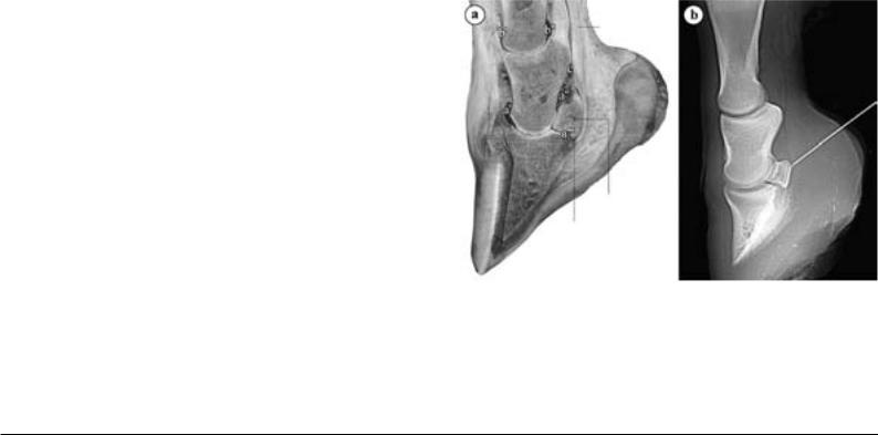
14.11.The navicular bursa (podotrochlear bursa) is located between the navicular bone and the deep flexor tendon that rides over it (Fig. 14.11., a). It belongs to the complex of podotrochlea (see podotrochleosis, section 12.7). For injection of the navicular bursa, the projection of the navicular bone onto the side wall of the hoof serves as a landmark. This is located 1 cm distal to the coronet in the middle between the dorsal and palmar borders of the hoof capsule. [219] The needle is introduced on the palmar midline about 1 cm proximal to the coronet and advanced in the direction of the navicular bone until bone-contact is made (usually at a depth of 4–5 cm; Fig. 14.11., b). [219] This is best performed under radiographic control to avoid an erroneous injection of the adjacent coffin joint. [114] Alternatively, the needle may be introduced at the deepest place between the heels and advanced dorsodistally in the direction of the navicular bone. For anesthesia, 3–5 cc of 2% anesthesia solution is injected into the bursa. Owing to diffusion, certainly after 20 minutes a desensitization of the coffin joint is also possible. [189] [220]
For endoscopy of the navicular bursa, access is proximal to the ungular cartilage between the border of the deep flexor tendon and the neurovascular bundle, in which case, entry is dorsal to the deep flexor tendon in a distoaxial direction. In this way, the synovial membrane of the synovial bursa, the insertion of the navicular ligaments, the palmar or plantar surface of the navicular bone as well as the dorsal surface of the deep flexor tendon can be demonstrated.
[221]
Deep flexor tendon
Distal sesamoid (navicular) bone
Podotrochlear (navicular) bursa
Fig. 14.11. a) Longitudinal section through the distal limb to demonstrate the podotrochlea. Observe the close relationship of the podotrochlear bursa to the palmar recess of the coffin joint and to the distal outpouching of the digital synovial sheath, b) Radiograph used to monitor the position of the needle in injection of the podotrochlear bursa.
a: coffin joint (a': dorsal recess, a”: palmaroproximal recess, a''': palmarodistal recess), b: pastern joint (b': dorsal recess, b'': palmar recess), c: digital synovial sheath. (Courtesy of Dr. Patan)
14.12.An increased filling of the synovial sheath of the common digital or lateral digital extensor muscle in the carpal region occurs occasionally in newborn foals or foals a few days old, in which case especially immature foals are involved. The cause is as a rule a rupture of the corresponding extensor tendon. With the typical swelling, a knuckling over (non-physiological flexure of the fetlock joint) is usually observed when the limb is advanced. [154]
14.13.The carpal flexor sheath surrounds the superficial and deep flexor tendons in the carpal canal and, with an increased filling, is visible proximal and distal to the flexor retinaculum. [114] Injection of the synovial sheath is possible caudolaterally, about 5 cm proximal to the accessory carpal bone between the tendons of the lateral digital extensor muscle and the ulnaris lateralis muscle (Fig.
14.13., 1). [114] [112] Because the palmarolateral recess of the antebrachiocarpal joint is in the immediate neighborhood at this site, there is always the danger of an unintentional articular injection. [114] Alternatively, the synovial sheath can be injected directly distal to the flexor retinaculum, in which case the needle is introduced between the lateral splint bone and the deep flexor tendon in a slightly proximal direction (Fig. 14.13., 2). [114] [112] For anesthesia of the carpal synovial sheath, 10–15 cc of 2% local anesthesia solution is necessary. [114] Owing to diffusion, also here a desensitization of the palmar or palmar metacarpal nerves is possible, which can lead to misinterpretation. [112]
Endoscopy of the carpal flexor sheath is performed on the slightly flexed carpal joint in the lateral forearm region. The arthroscopic portal is located about 3 cm proximal to the distal epiphysis of the radius and about 2.5 cm caudal to the radius between the tendons of the lateral digital extensor and ulnaris lateralis muscles. [222]
Access for the instrumentation is distal to this point. Among other things, the radial head of the deep digital flexor muscle, the proximal check ligament (accessory ligament) of the superficial digital flexor muscle, the distal radial epiphysis, the tendons of the superficial and deep digital flexor muscles, the accessory carpal bone as well as the adjacent articular pouches of the antebrachiocarpal and intercarpal joints can be demonstrated. [223] [222]
The other tendon sheaths located in the carpal region, which have to be considered in differential diagnosis in case of injuries in the carpal region, are shown in Fig. 14.13.
14.14.The (about 14–20 cm long) fetlock flexor tendon sheath
(digital synovial sheath) surrounds mainly the deep digital flexor tendon and only partially the superficial digital flexor tendon. [224]
[134] It begins proximally at the level of the button of the splint bone and reaches distally to the middle of the pastern bone. [224]
At the latter site, it borders directly on the navicular bursa and the coffin joint (Fig. 14.11., a). [134] [225] With increased filling, the synovial sheath is visible proximal to the palmar anular ligament between the suspensory ligament and the deep flexor tendon or distal below and at the sides of the four-cornered plate formed by the proximal digital anular ligaments. [224] Because an expansion of the synovial sheath is not possible in the region of the palmar anular ligament, an indentation can be observed here in lateral view (see anular-ligament-syndrome, section 10.4.). Proximal to the fetlock joint, the superficial flexor tendon forms within the synovial sheath a ring-like sleeve around the deep flexor tendon (manica flexoria), which is very well demonstrated sonographically. [225] Especially with filling of the tendon sheath, proximal to the manica flexoria, the lateral and medial attachment of the deep flexor tendon to the wall of the tendon sheath (so-called mesotendon) is easily recognized sonographically. [225] In the fetlock region the deep flexor tendon is dorsally connected with the wall of the tendon sheath (vinculum tendinis), which likewise with increased filling can be well demonstrated sonographically. [226]
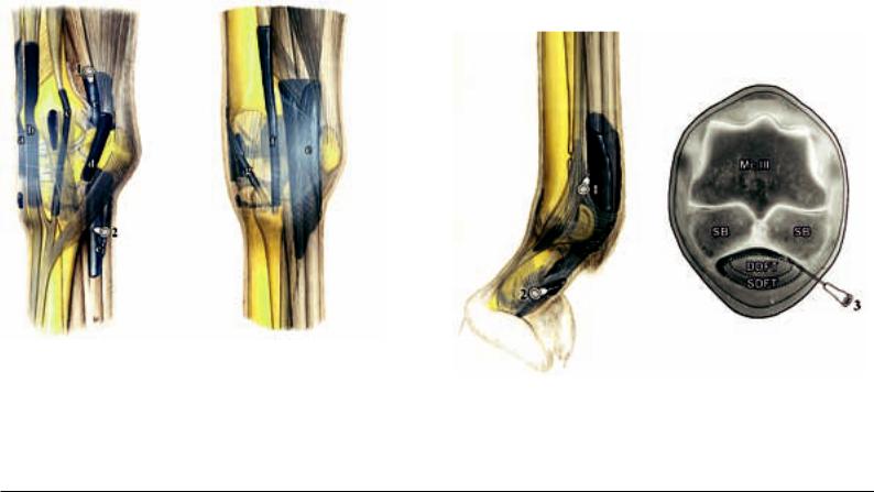
Injection of the fetlock flexor tendon sheath (digital synovial sheath) is possible in the proximolateral pouch about 2 cm proximal to the sesamoid bones between the suspensory ligament and the deep flexor tendon or between the deep and superficial flexor tendons (Fig. 14.14., 1). [112] Because at this site a distinct hypertrophy of the villi of the synovial membrane can be present, obtaining synovial fluid is sometimes difficult. [114] An alternate site of injection is in the midline of the pastern in the region of the distal blind sac, which is bounded by the four-cornered plate of the proximal digital anular ligaments and the distal digital anular ligament (Fig. 14.14., 2). [114] With a slight filling of the tendon sheath, this injection is also difficult, for which reason a further access was described. Here, with the fetlock joint slightly flexed, the needle is inserted at the level of the middle of the sesamoid bone, palmar to the neurovascular bundle between the sesamoid bone and the flexor tendons nearly perpendicularly through the skin and advanced through the palmar anular ligament in the direction of the intersesamoid region to a depth of 1.5–2 cm (Fig. 14.14., 3). [227] For anesthesia of the digital synovial sheath, 10–15 cc of 2% anesthesia solution is injected. [114] Because of diffusion, here also a desensitization of the medial and lateral palmar digital nerves is possible, which may lead to misinterpretations. [112]
(lateral view) |
(medial view) |
Fig. 14.13. Tendon sheaths and synovial bursae in the carpal region.
a: tendon sheath of the extensor carpi radialis muscle, b: tendon sheath of the common digital extensor muscle, c: tendon sheath of the lateral digital extensor muscle, d: tendon sheath of the extensor carpi ulnaris (ulnaris lat.) muscle, e: carpal flexor synovial sheath with proximal (1) and distal (2) sites of injection, f: tendon sheath of the flexor carpi radialis muscle, g: tendon sheath of the abductor pollicis longus (oblique carpal ext.) muscle. (Courtesy of Institut f. VeterinärAnatomie, Berlin)
Fig. 14.14. Injection of the digital synovial sheath: 1) proximal access, 2) access to the distal, blind, end, 3) injection at the middle of the proximal sesamoid bones. McIII Third metacarpal (cannon) bone, DDFT: deep digital flexor tendon, SB: sesamoid bone, SDFT: superficial digital flexor tendon. (Courtesy of Institut f.
Veterinär-Anatomie, Berlin)
129
For endoscopy of the digital synovial sheath, access is at the proximal border of the palmar anular ligament; [228] a second access is possible directly distal to the palmar anular ligament. [229] Here entrance into the synovial sheath is at each border of the superficial digital flexor tendon about 1–2 cm palmarly from the neurovascular bundle. [229] [225] In this manner, both the region between the palmar anular ligament and superficial flexor tendon as well as the region between superficial and deep flexor tendons can be inspected. [225]
Pelvic Limb |
BIANCA PATAN |
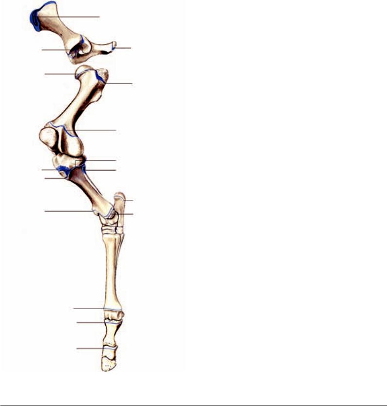
16.1. For the fusion of the epiphyses and apophyses of the pelvic limb, basically, the same holds as for the thoracic limb. Closure of the epiphyses of the pelvic limb is given in Fig. 16.1. Epiphyseal fractures are most often located in the region of the proximal and distal growth plates of the femur and tibia, [4] [5] as well as distally on the cannon bone (Mt III). [4] One of the most frequent angular limb deformities of the pelvic limb is tarsus valgus, which how-
UQL|XbVxUmAVqC+HnIZ42uaraA==|1288052437
approx. 12 months
24–36 months
9–12 months
30–36 months
17–24 months
approx. 6 months
approx. 12 months
8–12 months
approx. 10 months
approx. 10–12 months
18–30 months
24–30 months
24–30 months
from 1–2 months
approx. 16–24 months
approx. 3 months
Fig. 16.1. Apophyseal and epiphyseal lines of fusion of the pelvic limb of the horse (closure of the growth plates after Butler et al., 2000). (Courtesy of Institut f. Vet-
130 erinär-Anatomie, Berlin)
ever is observed much more rarely than carpus valgus (cause and therapy, see carpus valgus, section 4.1.). Tarsus varus deformities are on the other hand rather rare. A special form is the so-called
“windswept foal”, in which on the one pelvic limb a tarsus valgus and on the other a tarsus varus is present. Another relatively frequent deviation of the axis of the pelvic limb is the varus position at the fetlock joint, where usually the distal epiphyseal growth plate of
Mt III, more rarely the proximal one of the proximal phalangeal bone is involved. Valgus deformities of the fetlock joint are rather rare. With surgical correction of limb deformities of the fetlock joint it is to be noted that in this region the epiphyses close much earlier than the distal epiphysis of the radius or of the tibia. [8]
16.2.With respect to pelvic fractures, two different forms can be differentiated according to the location of the fracture lines. Fractures at the pelvic border (blow-up fractures of the tuber coxae, ischiadic or sacral tubers) are usually accompanied by a slight to moderate lameness of the pelvic limb and heal usually within 3–4 months. On the other hand, pelvic ring fractures (e.g., symphysial fractures) result in a severe lameness or the horse is unable to rise. Here the pelvis fractures usually at several sites because of its configuration as a relatively rigid ring. Such fractures are always associated with the danger of rupture of blood vessels, which may result in a greater blood loss. If the acetabulum is not involved in the fracture, healing can also take place with conservative treatment. With acetabular fractures, a severe arthrosis of the hip joint (coxarthrosis) must be expected and with this a permanent lameness. Fractures of the pelvic symphysis in large horses are untreatable. [230]
Also, spontaneous stress fractures of the wing of the ilium occur occasionally in young thoroughbreds in training (especially in hur- dle-racehorses). If the dosal surface is involved, then the fracture can occasionally be demonstrated sonographically; otherwise, to make the diagnosis, scintigraphy is the procedure of choice. [231]
16.3.For the femur, occasionally fractures of the third trochanter are observed, fewer of the greater trochanter, in which the fracture fragments are usually displaced owing to the pull of the attaching musculature. Sometimes such fractures can be demonstrated sonographically; besides that, scintigraphic examination is valuable in pointing to the diagnosis. [231]
Especially in foals there occasionally occur diaphysial fractures, which frequently exhibit several fracture lines. The success rate of the surgical therapy by suitable osteosynthetic procedures depends on the age or the weight of the foals. [232]
Moreover, sometimes avulsion fractures occur in the region of the origin of the peroneus tertius muscle and long digital extensor muscle at the lateral femoral condyle. [233] [234] This chiefly involves young horses in which the bone tissue is still weakly developed. On the other hand, in older animals rather a rupture of the peroneus tertius muscle in its middle is observed. [233] The therapy is usually surgical, in which, depending on its size, the fracture fragment is removed arthroscopically or fixed by internal fixation.
[233] [234]
16.4.The medial ridge of the femoral trochlea is distinctly larger than the lateral and has a nose-like bulge upon which the patella can hook.
16.5.Fractures of the patella usually have a traumatic origin and are especially often observed in show-jumpers in which the horse has collided with an obstacle in the stifle region. [235] Sagittal fractures of the medial part of the patella occur most often. This involves the patellar cartilage and with this the attachment of the medial patellar ligament, in which an involvement of the femoropatellar joint is usually also present. [235] [236] Beyond that, also transverse or crush fractures or fractures of the basis patellae may occur. [236] Especially fractures of the apex are described in connection with desmotomy of the medial patellar ligament (see proximal fixation of the patella, section 24.1). [231] Conservative therapy is possible in case of a fracture without involvement of the joint or with smaller, non-dislocated fragments.
[236] On the other hand a surgical approach is indicated where fragments up to the size of one quarter of the patella can be arthroscopically or arthrotomically removed. [235] [236] For larger frac- ture-fragments, internal fixation is necessary. [237]
16.6.Besides traumatic fractures of the tibia, especially in two year old thoroughbreds and trotters in training, incomplete stress or fatigue fractures (fissures) also occur. [16] [238] In young thoroughbreds often the caudolateral corticalis of the middle or distal diaphysis is involved, [15] [16] but also such fractures are not rarely observed proximolaterally or proximocaudally. [15] [238] On the other hand, stress fractures in trotters are almost exclusively in the middle region of the diaphysis. [238] Because the fissure lines often are not radiographically demonstrable, investigation by scintigraphy is the method of choice to find the diagnosis. [231] [15]
Occasionally, also traumatic intra-articular fractures are observed in the region of the lateral malleolus of the tibia. Therapy is surgical, in which case the fracture-fragment is usually arthroscopically removed. [239]
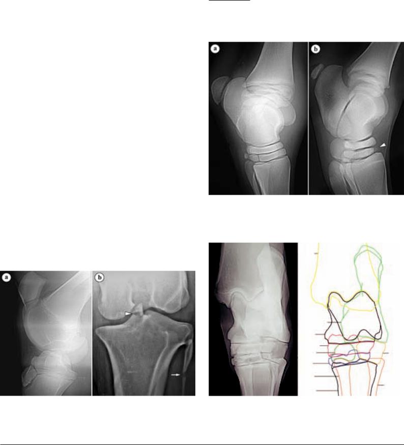
16.7.The intercondylar eminence of the tibia has two prominences that are well demonstated on radiographs. The medial tubercle is the more prominent and more frequently involved in fractures than the lateral (Fig. 16.7., b). Such fractures are sometimes seen in association with lesions of the cranial cruciate ligament, which attaches cranial to the intercondylar eminence. Therapy depends on the size of the fracture-fragment (arthroscopic removal or internal fixation). [240] [241]
16.8.Owing to the exposed position of the tibial tuberosity, trauma in this region (e.g., by collision with an obstacle during jumping) may easily result in a fracture. The fracture-fragment is often displaced proximocranially by the pull of the inserting middle patellar ligament (teminal tendon of the quadriceps femoris muscle).
[243] The femoropatellar joint is not usually involved. In young horses (less than 3 years), the growth plate of the tibial tuberosity, which is not yet closed, must be considered in the differential diagnosis (Fig. 16.7., a). In fractures without joint involvement, therapy can be conservative; otherwise, a surgical approach is indicated (internal fixation or removal of smaller fracture-fragments). [243]
16.9.The fibula often has several ossification centers, between which a radiographically demonstrable split (radiolucent area) may persist (Fig. 16.7., b). Such translucent lines must not be confused with a rather rare fracture of the fibula. [244] In most horses the distal fibula is reduced. Its distal epiphysis corresponds to the lateral malleolus of the tibia, which thus has its own ossification center.
[3] In some pony breeds, a complete fibula (fibula completa) is developed in an isolated case. Such a malformation occurs often combined with flexural limb deformities or angular limb deformities (especially tarsus valgus). [17]
16.10.With respect to incomplete ossification of the tarsal bones principally the same is valid as for that of the carpal bones, in which on the pelvic limb the central tarsal bone and the third tarsal bone
are especially involved (Fig. 16.10). [27] Stress on the insufficiently ossified tarsal bones may result in a collapse and consequently in a wedge-like deformity in the dorsala region. Sometimes there also appears fragmentation of the bones concerned. [23] [27] Such form changes of the tarsal bones are connected usually to limb deformities (tarsus valgus or cow-hocked). [245] [23]
16.11. Fractures of the tarsal bones are sometimes hard to demonstrate owing to the complex radiography (Fig. 16.11.). Most often slab fractures are present (but distinctly more rarely than on the carpus), which are usually located dorsolaterally on the third tasal bone and central tarsal bone. [246] [247] Thoroughbreds and trotters are mainly involved, occasionally also quarterhorses. [248] A wedge-shaped conformation of the bone is considered a predisposing factor. In young thoroughbreds, this is relatively frequently observed in the dorsolateral region of the third tarsal bone. [249]
aRemarks concerning the designation of direction on the pelvic limb: Corresponding to the relations on the forelimb, in the region of the distal limb (up to and including the tarsus) the terms “dorsal” or “plantar” (toward the sole of the foot) are used in place of the designations “cranial” or, respectively, “caudal.”
Fig. 16.10. a) Physiologic development of the tarsal bones of a 13 days old warmblood foal, b) Incomplete ossification of tarsal bone III (arrowhead) of a 14 days old warmblood foal. (Radiographs: Klinik fır Orthopädie bei Huf- u. Klauentieren, Veterinärmed. Univ. Wien)
Fig. 16.7. a) Growth plate lines of fusion in the stifle joint region of a 6.5 months old warmblood foal, b) Fracture of the intercondylar eminence of the tibia (arrowhead), fibula with radiolucent line (arrow) between separate ossification centers, 13 years Shetland pony. (Radiographs: Klinik für Orthopädie bei Huf- u. Klauentieren, Veterinärmed. Univ. Wien)
Tibia
Calcaneus |
|
Talus |
|
T centr. |
|
T I+II |
T IV |
T III |
|
Mt II |
|
Mt III |
Mt IV |
|
Fig. 16.11. Tarsal joint of an adult horse (dorsoplantar radiograph) with schematic representation of the individual bones.
Mt II – IV: metatarsal bones II – IV, T I+II, III – IV: tarsal bones I+II, III – IV, T centr: central tarsal bone. (Courtesy of Klinik für Orthopädie bei Huf- u. Klauentieren,
Veterinärmed. Univ. Wien)
131

[250] Because in cases of conservative therapy, secondary degenerative changes are to be expected in adjacent joints (tarsometa- tarsal-, distal or proximal intertarsal joints), especially in racehorses a surgical therapy (internal fixation) is recommended. [246] [247]
16.12.Fractures of the talus and calcaneus are altogether rather rare and mostly brought about by trauma. [251] To be sure, in thoroughreds and trotters, incomplete sagittal fractures of the talus occasionally occur. These usually start from the proximal articular surface and are located between the ridges. [252]
Occasionally there occur rounded, accessory bone pieces plantarly at the proximal end of the trochlea of the talus (trigonal bone of the tarsus) or distally at the medial ridge of the talus. These are to be regarded as rare anatomical variations. [253]
16.13.The first and second tarsal bones are ordinarily fused at the time of birth. [3] In some horses this fusion is incomplete, or even two independent bones are developed. [253]
18.1.An injury of the obturator nerve (e.g., as a result of a dystocia or pelvic fractures) may lead to a loss of function of the adductors of the pelvic limb. In case of unilateral damage the horses stand with the pelvic limbs widely spread; with bilateral damage the horses lie immovably with straddled pelvic limbs. [107]
18.2.The gluteal muscles can be utilized for intramuscular injection. The injection site is midway between the coxal tuber and the root of the tail and the needle is introduced in a craniodistal direction. [110]
A painful condition of the musculature (especially of the accessory gluteal muscle) in the region between the coxal tuber and the greater trochanter of the femur is designated as the “gluteal syndrome”. This is especially observed in trotters; athletic horses are also occasionally involved. The cause is not exactly known. Presumably the basis is a partial rupture of the muscle in the area of its origin on the wing of the ilium. [254]
18.3.Severe trauma in the hip, thigh or gluteal region as well as a deeply intramuscularly located abscess may lead to a paralysis of the ischiadic nerve. The result is a loss of sensory and motor functions in the area of supply of the tibial and peroneal nerves, which have their origin from the ischiadic nerve. The affected limb can only be elevated and flexed to a slight degree. Besides this, a collapse of the hock joint is typical as well as a knuckling over of the fetlock joint (non-physiological flexure). [107]
18.4.If only the tibial nerve is injured, it may result in a loss of function of the extensors of the hock and flexors of the digits. As a consequence, the involved limb can be lifted, but weight bearing by the hock joint is not possible. Besides the collapse in the hock joint, a knuckling over of the fetlock joint is usually also observed. [107]
Anesthesia of the tibial nerve, which may be a part of the lameness examination, will be described with the nerve blocks of the pelvic limb (section 22.1., c).
18.5.An occasionally occurring abnormality of the gait of the pelvic limb is designated “fibrotic/ossifying myopathy.” It is characterized by fibrotic changes in the region of the flexor muscles of the stifle joint and is particularly observed in quarterhorses. [255]
[256] The semitendinosus muscle is predominantly involved; more rarely, also the semimembranosus, gracilis or biceps femoris muscles. [255] [257] [258] Besides degenerative lesions, [259] trauma and infections (e.g., after intramuscular injections), [258] a cause that is also considered is an increased tension on the insertion of the semitendinosus muscle in case of an intensive stress on the pelvic limb (e.g., by sliding stops in western riding). [255] A typical clinical sign is a shortened forward phase in the stride, [256] in which the forward movement is abruptly broken off and the hoof is brought back to the ground after a short backward movement. [260] [231] Usually also a thickening in the area of insertion of the muscle involved is palpable. [256] [261] Sonographic investigation gives additional indications of altered areas within the musculature. [256] [261] As therapy, three different surgical procedures are described: myotomy or myectomy of the semitendinosus muscle
(section or partial removal of the muscle at the level of the stifle joint and removal of intramuscular adhesions), [256] [261] tenoto-
my (section of its tibial insertion tendon) [257] [261] as well as myotenotomy (section of muscle and tendon of insertion). [258]
The thigh musculature (semimembranosus or semitendinosus) like the rump musculature can be used for intramuscular injection. The injection site is midway between the ischiadic tuber and the back of the stifle. [110] A possible complication is the development of a “fibrotic myopathy.” [258]
18.6.In the region of the calcanean tuber, the tendon of the superficial digital flexor muscle is broadened to form the calcanean cap. [134] It is kept in position by tendinous bands, which are attached laterally and medially at the calcanean tuber. [262] [134] Damage to these bands may result in a luxation of the superficial digital flexor tendon, in which case usually a lateral displacement is observed.
[262] Besides conservative therapy, also a surgical fixation of the calcanean cap is described. [262] [263]
18.7.Paralysis of the peroneal neve (e.g., by an unfavorable position in anesthesia, trauma or tissue-irritating intramuscular injections) may lead to a loss of function of the flexors of the hock and extensors of the digit. A nearly complete loss of the angulation of the hock joint with maximal flexion of the digital joints is typical. With advance of the affected limb, the hoof is dragged on the ground. If the horse has to bear weight on the limb, then it stands on the dorsal region of the fetlock. [107]
For anesthesia of the peroneal nerve, see anesthesia of the pelvic limb [Fig. 22.1.1.).
18.8.The peroneus tertius muscle is discussed together with the passive stay-apparatus of the pelvic limb (section 24.2).
18.9.The medial tendon of insertion of the tibialis cranialis muscle is also designated the spavin tendon. It attaches medioplantarly on the fused first and second tarsal bones, occasionally on the medial splint bone (Mt II) and is underlain by a synovial bursa. [264] Sometimes inflammatory changes of the tendon or synovial bursa are observed in connection with spavin.
18.10. Myotenectomy (section/partial removal of the tendon and muscle belly) of the lateral digital extensor muscle is a possible option for surgical therapy of stringhalt. [265] This disease is an abnormality of the gait of the pelvic limb, which is triggered by different neuromuscular disturbances (among others, by a herbal intoxication, a trauma or a painful process in the region of the distal limb). [265] [266] [267] Affected horses typically demonstrate an exaggerated action of the pelvic limb with each stride, in which the hock joint and stifle joint are suddenly moved convulsively and the limb raised high.
20.1. A thrombotic occlusion in the region of the termination of the aorta may lead to the clinical picture of intermittent limping. [268] The thrombus is usually located in the aorta or in the external iliac artery or internal iliac artery, sometimes also the femoral artery is involved. [269] [268] The cause is not exactly known. Besides hemodynamic factors and disorders of blood clotting, also parasitic causes are mentioned. [268] A typical clinical sign is a stressinduced lameness of the pelvic limb, [269] [268] which presumably has its origin in hypoxemia of the affected limb. To confirm the diagnosis, a rectal sonographic examination can be performed.
Additional information may be obtained from Doppler sonographya of the femoral artery. [271] Besides medicinal therapy, also surgical removal of the thrombus by way of the femoral artery
(access in the region of the femoral triangle) is described. [270]
aDoppler sonography yields information concerning blood-flow velocity and changes in the hemodynamic. The basic physical phenomenon is the change in the frequency of sound waves in their reflection from locally changing (moving) interfaces (Doppler effect) in which case the erythrocytes in the bloodstream act as moving reflectors.
132

22.1. Anesthesia of Nerves of the Pelvic Limb
Basically, diagnostic nerve anesthesia of the distal limb (up to and including high four-point anesthesia) corresponds to what was given on the thoracic limb (Fig. 22.1.1.).
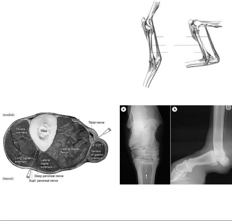
a)High 4-point anesthesia is performed about 3 cm distal to the tarsometatarsal joint. Similar to the thoracic limb, it may be misinterpreted by an unintentional injection of the tarsal synovial sheath or the distoplantar outpouching of the tarsometatarsal joint. [114]
b)In addition to the medial and lateral plantar metatarsal nerves, which both are derivatives of the deep branch of the lateral plantar nerveb and thus from the tibial nerve, there are on the pelvic limb also dorsal metatarsal nerves II and III, which originate from the deep peroneal nerve (Fig. 22.1.1.). [113] To eliminate cortical pain in the dorsal region of Mt III, the dorsal metatarsal nerves are also anesthetized by a subcutaneous deposit of 2–3 cc of anesthesia solution (high 6-point anesthesia). [114] [113] With this procedure, injection of dorsal common digital vein II or the great metatarsal artery III (a. metatarsea dorsalis III) should be avoided.
c)With the limb lifted up, the tibial nerve is palpable as a firm cord, about 6 mm thick, between the common calcanean tendon and the lateral digital flexor muscle. [114] Anesthesia of the tibial nerve is performed from the medial side, about 10 cm proximal to the calcanean tuber. Subfascially, fan-like, 10–20 cc of anesthesia solution is injected (Figs. 22.1.1. and 22.1.2.). [114] [113] The plantar part of the tarsus, the distal Achilles tendon (common calcanean tendon)
bAccording to the Nomina Anatomica Veterinaria (1994), synonymous designation for the N. digitalis plantaris com. III
as well as the structures which were already considered in the 4- point block are desensitized. [113]
d) Anesthesia of the tibial nerve is usually combined with anesthesia of the fibular nerve (peroneal nerve). With this technique, the distal tibia, the entire hock joint, the tarsal synovial sheath as well as the total distal limb are desensitized. [114] Anesthesia of the peroneal nerve is performed from the lateral side, about 10 cm proximal to the tarsus, where its superficial branch is well palpable between the long digital extensor muscle and the lateral digital extensor muscle. [114] [113] At this site, the needle is introduced directed toward the tibia and the deep peroneal nerve deeply with 10–15 cc anesthesia solution fan-like injected. [114] Since the nerve takes its course near the cranial tibial artery and vein, injection of the vessels demonstrates roughly the proper site for the needle. In withdrawing the needle, the superficial peroneal nerve is also anesthetized by a subcutaneous injection of 5–10 cc anesthesia solution (Fig. 22.1.1. and 22.1.2.). [114]
22.2. Dorsal metatarsal artery III, the great metatarsal artery, is the main artery of the metatarsus of the pelvic limb. To take the pulse, this vessel can be palpated proximolaterally on the cannon bone between Mt III and the lateral splint bone (Mt IV). [272] [273] The distal continuation of the vessel runs between Mt III and Mt IV in a gradually oblique distoplantar direction, [272] which has to be considered if the distal splint bone is surgically removed. [274]
Lateral view
Tibial nerve anesthesia
Fibular (peroneal) nerve
anesthesia
Lateral dorsal
metatarsal nerve
HPA, position of high plantar
nerve block Lateral plantar nerve
Communicating branch
MPA, position of deep 4-point anesthesia
Abaxial sesamoid block
Lateral plantar digital nerve
TPA 2
TPA 1
Plantar view |
Dorsal view |
Caudal cutaneous |
Tibial nerve |
|
|
sural nerve, tibial n. |
Supf. fibular (peroneal) n. |
|
|
|
Deep fibular (peroneal) n. |
|
Saphenous n. |
Medial plantar nerve |
|
Lateral plantar nerve |
|
Communicating branch
MPA, position of deep 4-point anesthesia
Dorsal branch, plantar digital n., anesthesia
TPA 2
TPA 1
HPA, position of high plantar (4-point, 6-point) nerve block
Medial plantar metatarsal nerve
Lateral plantar metatarsal nerve
Medial dorsal metatarsal nerve Lateral dorsal metatarsal nerve
Dorsal branch, med/lat plantar dig. N.
Lateral plantar digital nerve
Medial plantar digital nerve
(lateral) |
(medial) |
(lateral) |
Fig. 22.1.1. Relevant nerves of the pelvic limb and their sites for anesthesia for lameness diagnosis. In the right part of the figure the tibial nerve including its distal continuation is yellow and the terminal branches of the common fibular (peroneal) nerve are dark green. HPA: high plantar nerve block, MPA: low 4-point anesthesia, TPA 1
and 2: deep (distal and proximal) plantar digital nerve blocks. (Courtesy of Institut f. Veterinär-Anatomie, Berlin)
133
22.3. The medial saphenous vein (or its cranial branch) is located medially on the crus. Especially in foals, it can be used as an alternative to the external jugular vein for venepuncture or installation of a catheter. [110]
24.1. The alternate passive fixation of the pelvic limb in the stifle joint, which enables the horse to stand without fatigue is determined predominantly by the action of the quadriceps femoris muscle. [275] [134] By that, the stifle joint is so far extended by contraction of this muscle, that the patella, drawn proximally, glides on the nose-like bulge of the medial ridge of the trochlea and can be anchored there with the help of the medial parapatellar fibrocartilage and the middle and medial patellar ligaments. [134] The dislodging of the fixed patella is brought about again by the contraction of the quadriceps femoris muscle, whereby the patella is turned simultaneously by the pull of the biceps femoris muscle. [275] [134] Disorders of the gliding movement of the patella on the medial ridge of the trochlea are designated proximal patellar fixation. If the freeing of the patella is only delayed (habitual patellar fixation), with flexion of the stifle joint there is a typical jerky movement of the affected limb. [275] The uneven movement of the patella is sometimes audible as a snapping sound. [276] With stationary patellar fixation, flexion of the stifle joint is impossible. The limb is kept stiff and extended since, owing to the reciprocal apparatus, the hock joint also cannot be flexed. [275] [276] Proximal patellar fixation usually occurs bilaterally. A decrease in tonus of the quadriceps femoris muscle is regarded as a main cause. For this reason especially young horses or horses in poor condition are affected. Beyond that, a steep attitude of the pelvic limbs is predisposing. [277] [275] [276] Initially, therapy can be conservative, whereby the exclusion of predisposing factors as well as a monitored training for muscle build-up have a special importance. [231] [277] [276] As a last measure, surgical therapy is also possible in the form of a desmotomy of the medial patellar ligament 1–2 cm proximal to its attachment to the tibia. [231] [275] Certainly, as a sequel often fragmentation of the distal patella or contour changes of the cranial border of the tibia are observed. [231] These, however, as a rule, have no clinical relevance. [275] Recently also a percutaneous splitting of the proximal third of the medial patellar ligament is described. [278] [276]
Particularly in newborn Shetland pony foals a lateral subluxation or luxation of the patella is occasionally observed. To differentiate a traumatically induced patellar dislocation from this inherited change, the term “patella-ectopy” is also employed. [279] [280] Usually this condition has its basis in an inherited dysplasia (lack of development) of the trochlea of the femur, in which mainly the lateral ridge of the trochlea is affected. [281] [282] [283] Patellar dislocation can vary according to the extent of the defective deve-
Fig. 22.1.2. Cross-section of the left hind limb of a horse (about a handsbreadth proximal to the tuber calcanei) to demonstrate the sites of injection of the tibial nerve and deep and superficial fibular (peroneal) nerves. 1: cranial tibial a./v., 2: cranial branch of the medial saphenous v. and saphenous a./n., 3: caudal branch of the saphenous a./medial saphenous v., MDF: medial digital flexor muscle, PT: fibularis (peroneus) tertius muscle, SDFT: superficial digital flexor tendon (plantar tendon), TC: tibialis caudalis muscle. (Courtesy of Dr. Patan)
lopment. In severe cases affected foals demonstrate a typical dogsitting posture because an extension of the stifle joint is impossible. [281] Therapy consists of a surgical deepening of the trochlear groove with an associated repositioning of the patella. [281] [283]
This can be combined with a tightening of the medial joint capsule including the fascia as well as a section of the lateral femoropatellar ligament and parts of the aponeurosis of the biceps femoris muscle. [281]
24.2. The reciprocal apparatus in the crural region allows only an equal movement of stifle and hock joints. It is formed cranially by the tendinous peroneus tertius muscle and caudally by the common calcanean tendon (especially by the plantar tendon of the superficial digital flexor muscle (Fig. 24.2.). [284] Occasionally the peroneus tertius muscle suffers a traumatically induced rupture, which is often located in the middle crural region. [233] [284] A typical clinical sign is loss of the reciprocal apparatus and with this the possibility of extension of the hock joint with the stifle joint flexed. In this condition, the common calcanean tendon is an entirely relaxed and undulating structure. [284] To demonstrate the lesion of the peroneus tertius muscle as well as to monitor the healing process, sonographic examination is the first choice. [285]
Stifle joint
Common calcaneal tendon
Tibia
Fibularis (peroneus)
tertius muscle
Hock (tarsal) joint
Fig. 24.2. Semidiagrammatic representation of the reciprocal apparatus of the pelvic limb of the horse. (Courtesy of Institut f. Veterinär-Anatomie, Berlin)
Fig. 24.3. a) Dorsoplantar radiograph of the tarsal joint with desmopathy of the insertion of the suspensory ligament (interosseous muscle) at its origin (arrow), 11 years warmblood mare, b) high degree of hyperextension of the fetlock joint and distal displacement of the proximal sesamoid bones because of a functional loss of the proximal portion of the suspensory apparatus (damage to the suspensory ligament ), 24 years old warmblood gelding. (Courtesy of Klinik für Orthopädie bei Huf- u. Klauentieren, Veterinärmed. Univ. Berlin)
134