

1.2Portal Vein and Its Tributaries
C. Goerg
The portal venous system is comprised of four regions: intrahepatic branches of the portal vein; portal vein at the porta hepatis; splenic vein; and splanchnic veins. Blood from the splanchnic region (including the spleen) normally flows in hepatopetal fashion.
Anatomy


Size |
Size and shape. |
The diameter of the portal |
|
vein displays a |
somewhat wider range be- |
||
● Portal vein at the hilum: 1.0–1.5 cm |
|||
tween individuals (see Fig.1.93). In fasting pa- |
|||
● Splenic vein at the hilum: 0.5–1.0 cm |
|||
tients, a diameter of up to 15 mm is considered |
|||
|
|||
Flow |
normal. Usually, the portal vein has an oval |
||
● Mean flow velocity of the portal vein: |
shape in the transverse plane. The mean portal |
||
15–20 cm/s
flow velocity has been reported as 15–20 cm/s, with a wide range of values here as well.
Under normal conditions the diameter of the splenic vein at the hilum should be less than 7 mm ( 1.6e,f). The mesenteric vein is compressible and exhibits respiratory caliber variations of more than 15%.
Topography 


Position
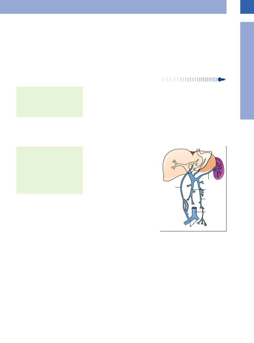
●Splenic vein posterior to the pancreas
●Mesenteric vein parallel to the superior mesenteric artery
●Portal vein posterior to the bile duct and hepatic artery
●Intrahepatic bifurcation of the portal vein into left and right branch
Lead Structure
● Posterior to the pancreatic head
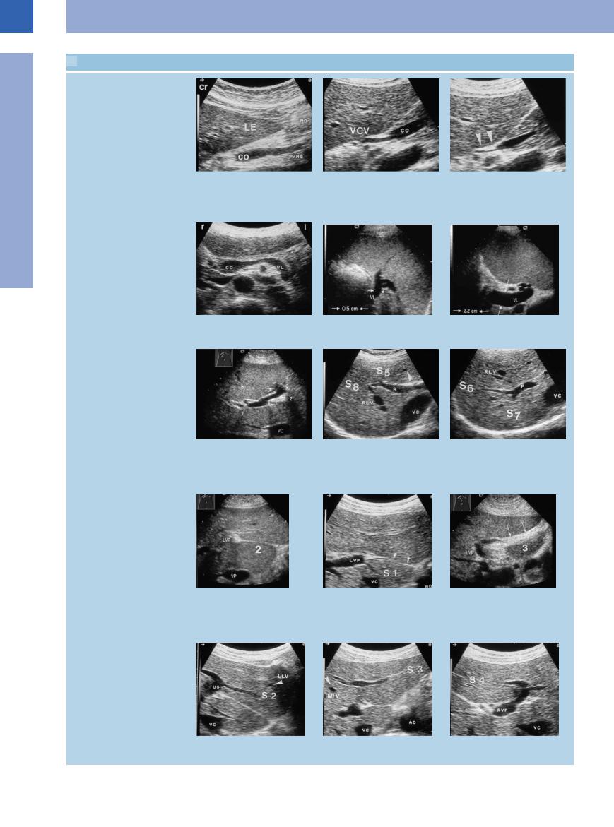
The main portal tract receives the blood flow of the left gastric vein cranially, in the region of the hilum ( 1.6a–c). The splenic vein is regarded as the leading structure for portrayal of
the pancreas and can be followed into the splenic hilum ( 1.6 d–f). The superior mesenteric vein parallels the superior mesenteric artery and can be detected, together with its branches, in the inferior parts of the mesentery (Fig.1.83).
Within the liver the portal trunk divides like a “staghorn” into the left and right portal branches ( 1.6 g). The main right portal branch gives off the anterior branch, which supplies segments V and VIII, while its posterior branch supplies segments VI and VII (
 1.6h,i). The left portal branch divides into the so-called horizontal and umbilical parts, supplying segments I, II, III, and IV (
 1.6j–o). Anomalies of the intrahepatic branches of the portal vein are rare.
Fig. 1.83 Portocaval collaterals accessible to ultrasound in |
portal hypertension.
1
Portal Vein and Its Tributaries
39
1
Vessels
 1.6 Topographic Anatomy of the Portal Vein and Its Tributaries
Portal vein and its tributaries
a Inflow of the superior mesenteric vein |
b and c Junction of the left gastric vein |
c Expiration (arrows). |
(VMS) into the venous confluence. LE = |
(VCV) with the confluence of the superior |
|
liver; CO = venous confluence; MA = |
mesenteric and splenic veins (co); respi- |
|
stomach. |
ratory variation in the lumen diameter. |
|
|
b Inspiration. |
|
d Transverse epigastric view with splenic |
e Splenic vein (VL, arrows) with normal |
f Splenic vein (VL) in portal hypertension. |
vein (VL). CO = venous confluence. |
lumen diameter. |
|
Intrahepatic right and left branches of the portal veins with liver segments
g Subcostal view of the liver with left and |
h Right anterior branch of the portal vein |
i Posterior branch of the main right |
right branches of the portal vein. VC = |
(A) with branches supplying the anterior |
portal vein trunk (P) supplying the |
vena cava; 1, 2, 3 = segments I–III. |
segments V (S 5) (inferior) and VIII (S 8) |
posterior segments VI (S 6) (inferior) and |
|
(superior). RLV = right hepatic vein. |
VII (S 7) (superior) of the right hepatic |
|
|
lobe. RLV = right hepatic vein; VC = vena |
|
|
cava. |
j–l The left portal vein (LVP) supplies |
k Segment I. Anterior venous ligament |
l The ligamentum teres stretches be- |
segments I–IV (S 1–S 3). |
(arrows). AO = aorta; VC = vena cava. |
tween the left branch of the portal vein |
j The venous ligament runs from the left |
|
(3) and the umbilicus. LVP = left portal |
branch of the portal vein (2) to the |
|
vein. |
posteroinferior aspect of the liver. LVP = |
|
|
left portal vein; VC = vena cava. |
|
|
m Segment II. US = umbilical segment, |
n Segment III. AO = aorta; VC = vena cava. o Segment IV. RVP = right branch of |
LLV = left hepatic vein. |
portal vein. |
40