

Литература / Европа+рекомендации+по+лечению+ССЗ+у+беременных+2018
.pdfEuropean Heart Journal (2018) 39, 3165–3241 doi:10.1093/eurheartj/ehy340
ESC GUIDELINES
2018 ESC Guidelines for the management of cardiovascular diseases during pregnancy
The Task Force for the Management of Cardiovascular Diseases during Pregnancy of the European Society of Cardiology (ESC)
Endorsed by: the International Society of Gender Medicine (IGM), the German Institute of Gender in Medicine (DGesGM), the European Society of Anaesthesiology (ESA), and the European Society of Gynecology (ESG)
Authors/Task Force Members: Vera Regitz-Zagrosek* (Chairperson) (Germany), Jolien W. Roos-Hesselink* (Co-Chairperson) (The Netherlands), Johann Bauersachs (Germany), Carina Blomstro¨m-Lundqvist (Sweden), Renata Cıfkova (Czech Republic), Michele De Bonis (Italy), Bernard Iung (France), Mark Richard Johnson (UK), Ulrich Kintscher (Germany), Peter Kranke1 (Germany), Irene Marthe Lang (Austria), Joao Morais (Portugal), Petronella G. Pieper (The Netherlands),
Patrizia Presbitero (Italy), Susanna Price (UK), Giuseppe M. C. Rosano (UK/Italy), Ute Seeland (Germany), Tommaso Simoncini2 (Italy), Lorna Swan (UK),
Carole A. Warnes (USA)
* Corresponding authors. Vera Regitz-Zagrosek, Charite´ Universitaetsmedizin Berlin, Institute for Gender in Medicine, CCR, DZHK, partner site Berlin, Hessische Str 3-4, 10115 Berlin, Germany, Tel: þ49 30 450 525 288, Fax: þ49 30 450 7 525 288, E-mail: vera.regitz-zagrosek@charite.de. Jolien W. Roos-Hesselink, Department of Cardiology, Erasmus Medical Center Rotterdam, Dr Molewaterplein 40, 3015CGD, Rotterdam, Netherlands, Tel: þ31 10 7032432, E-mail: j.roos@erasmusmc.nl
ESC Committee for Practice Guidelines (CPG) and National Cardiac Societies document reviewers: listed in the Appendix.
1Representing the European Society of Anaesthesiology (ESA)
2Representing the European Society of Gynecology (ESG)
ESC entities having participated in the development of this document:
Associations: Acute Cardiovascular Care Association (ACCA), European Association of Cardiovascular Imaging (EACVI), European Association of Percutaneous Cardiovascular Interventions (EAPCI), European Heart Rhythm Association (EHRA), Heart Failure Association (HFA).
Councils: Council on Cardiovascular Nursing and Allied Professions, Council on Cardiovascular Primary Care, Council on Hypertension, Council on Valvular Heart Disease.
Working Groups: Aorta and Peripheral Vascular Diseases, Cardiovascular Pharmacotherapy, Cardiovascular Surgery, Grown-up Congenital Heart Disease, Myocardial and Pericardial Diseases, Pulmonary Circulation and Right Ventricular Function, Thrombosis.
The content of these European Society of Cardiology (ESC) Guidelines has been published for personal and educational use only. No commercial use is authorized. No part of the ESC Guidelines may be translated or reproduced in any form without written permission from the ESC. Permission can be obtained upon submission of a written request to Oxford University Press, the publisher of the European Heart Journal and the party authorized to handle such permissions on behalf of the ESC (journals.permissions@oxfordjournals.org).
Disclaimer. The ESC Guidelines represent the views of the ESC and were produced after careful consideration of the scientific and medical knowledge and the evidence available at the time of their dating. The ESC is not responsible in the event of any contradiction, discrepancy and/or ambiguity between the ESC Guidelines and any other official recommendations or guidelines issued by the relevant public health authorities, in particular in relation to good use of health care or therapeutic strategies. Health professionals are encouraged to take the ESC Guidelines fully into account when exercising their clinical judgment as well as in the determination and the implementation of preventive, diagnostic or therapeutic medical strategies. However, the ESC Guidelines do not override in any way whatsoever the individual responsibility of health professionals to make appropriate and accurate decisions in consideration of each patient’s health condition and in consultation with that patient and the patient’s caregiver where appropriate and/or necessary. Nor do the ESC Guidelines exempt health professionals from taking careful and full consideration of the relevant official updated recommendations or guidelines issued by the competent public health authorities in order to manage each patient’s case in light of the scientifically accepted data pursuant to their respective ethical and professional obligations. It is also the health professional’s responsibility to verify the applicable rules and regulations relating to drugs and medical devices at the time of prescription.
VC The European Society of Cardiology and The European Society of Hypertension 2018. All rights reserved. For permissions please email: journals.permissions@oup.com.

3166 |
ESC Guidelines |
Document Reviewers: Christi Deaton (CPG Review Coordinator) (UK), Iain A. Simpson
(CPG Review Coordinator) (UK), Victor Aboyans (France), Stefan Agewall (Norway), Emanuele Barbato (Italy), Pavel Calda2 (Czech Republic), Antonio Coca (Spain), Ioan Mircea Coman (Romania), Julie De Backer (Belgium), Victoria Delgado (The Netherlands), Giovanni Di Salvo (UK), Samantha Fitzsimmons (UK), Donna Fitzsimons (UK), Madalina Garbi (UK), Sofie Gevaert (Belgium), Gerhard Hindricks (Germany), Guillaume Jondeau (France), Jolanda Kluin (The Netherlands), Christos Lionis
(Greece), Theresa A. McDonagh (UK), Pascal Meier (UK/Switzerland), Philip Moons (Belgium), Antonis Pantazis (UK), Massimo Francesco Piepoli (Italy), Bianca Rocca (Italy), Marco Roffi (Switzerland), Stephan Rosenkranz (Germany), Andrea Sarkozy (Belgium), Evgeny Shlyakhto (Russia), Candice K. Silversides (Canada), Karen Sliwa (South Africa), Miguel Sousa-Uva (Portugal), Juan Tamargo (Spain), Sara Thorne (UK), Marc Van de Velde1 (Belgium), Bryan Williams (UK), Jose Luis Zamorano (Spain)
The disclosure forms of all experts involved in the development of these Guidelines are available on the ESC website www.escardio.org/guidelines
Online publish-ahead-of-print 25 August 2018
...................................................................................................................................................................................................
Keywords |
Guidelines • Pregnancy • Cardiovascular disease • Risk assessment • Management • Congenital heart dis- |
|
ease • Valvular heart disease • Hypertension • Heart failure • Arrhythmia • Pulmonary hypertension • |
|
Aortic pathology • Cardiomyopathy • Drug therapy • Pharmacology |
Table of Contents |
|
Table of Contents. ............................................................................................. |
3166 |
List of tables . ....................................................................................................... |
3168 |
Abbreviations and acronyms. ......................................................................... |
3168 |
1. Preamble . ......................................................................................................... |
3169 |
2. Introduction . ................................................................................................... |
3171 |
2.1 Why do we need new Guidelines on the management of |
|
cardiovascular diseases in pregnancy? . ................................................... |
3171 |
2.2 New format of the Guidelines . ......................................................... |
3171 |
2.3 Why these Guidelines are important . ............................................. |
3171 |
2.4 Methods . ................................................................................................... |
3171 |
2.5 What is new? . ......................................................................................... |
3172 |
3. General considerations. ............................................................................... |
3173 |
3.1 Epidemiology . ......................................................................................... |
3173 |
3.2 Physiological adaptations to pregnancy . ......................................... |
3174 |
3.3 Pre-pregnancy counselling . ................................................................. |
3174 |
3.3.1 Risk of maternal cardiovascular complications. . . . . . . . . |
3174 |
3.3.2 Risk of obstetric and offspring complications . . . . . . . . . . |
3174 |
3.3.3 Pregnancy heart team. ............................................................. |
3176 |
3.4 Cardiovascular diagnosis in pregnancy. . . . . . . . . . . . . . . . . . . . . . . |
3176 |
3.4.1 Electrocardiography . ............................................................... |
3176 |
3.4.2 Echocardiography . ................................................................... |
3176 |
3.4.3 Exercise testing. ......................................................................... |
3177 |
.
.
.
.
.
.
.
.
.
.
.
.
.
.
.
.
.
.
.
.
.
.
.
.
.
.
.
.
.
.
.
.
.
.
.
.
.
.
.
.
.
.
.
.
.
.
.
.
.
.
.
.
.
.
.
.
.
.
.
.
.
.
.
.
.
.
.
.
.
.
.
.
.
.
3.4.4 Ionizing radiation exposure . ................................................. |
3177 |
3.4.5 Chest radiography and computed tomography . ............ |
3177 |
3.4.6 Cardiac catheterization . ......................................................... |
3177 |
3.4.7 Magnetic resonance imaging. ................................................. |
3177 |
3.5 Genetic testing and counselling . ....................................................... |
3177 |
3.5.1 Pre-natal diagnosis. ................................................................... |
3178 |
3.6 Foetal assessment . ................................................................................. |
3178 |
3.6.1 Screening for congenital heart disease . . . . . . . . . . . . . . . . |
3178 |
3.6.2 Assessing foetal wellbeing. ..................................................... |
3178 |
3.7 Interventions in the mother during pregnancy . ........................... |
3178 |
3.7.1 Percutaneous therapy . ........................................................... |
3178 |
3.7.2 Cardiac surgery with cardiopulmonary bypass . . . . . . . . |
3178 |
3.8 Timing and mode of delivery: risk for mother and child . . . . . . . |
3179 |
3.8.1 Timing of delivery . ................................................................... |
3179 |
3.8.2 Labour induction . ..................................................................... |
3179 |
3.8.3 Vaginal or caesarean delivery. ............................................... |
3179 |
3.8.4 Delivery in anticoagulated women (not including |
|
mechanical valve; see section 5) . ................................................... |
3179 |
3.8.5 Urgent delivery on therapeutic anticoagulation. . . . . . . . |
3179 |
3.8.6 Haemodynamic monitoring during delivery . . . . . . . . . . . |
3180 |
3.8.7 Anaesthesia/analgesia . ............................................................. |
3180 |
3.8.8 Labour . ......................................................................................... |
3180 |
3.8.9 Perimortem caesarean section . ........................................... |
3180 |

ESC Guidelines |
|
3.8.10 Post-partum care. ................................................................... |
3180 |
3.8.11 Breastfeeding . ......................................................................... |
3180 |
3.9 Infective endocarditis . ........................................................................... |
3180 |
3.9.1 Prophylaxis. ................................................................................. |
3180 |
3.9.2 Diagnosis and risk assessment . ............................................. |
3180 |
3.9.3 Treatment . ................................................................................. |
3180 |
3.10 Methods of contraception and termination of pregnancy, |
|
and in vitro fertilization. ............................................................................... |
3181 |
3.10.1 Methods of contraception . ................................................. |
3181 |
3.10.2 Sterilization . ............................................................................. |
3181 |
3.10.3 Methods of termination of pregnancy . . . . . . . . . . . . . . . |
3181 |
3.10.4 In vitro fertilization . ............................................................... |
3181 |
3.11 Recommendations . ............................................................................. |
3182 |
4. Congenital heart disease and pulmonary hypertension . . . . . . . . . . . |
3182 |
4.1 Introduction . ........................................................................................... |
3182 |
4.2 Pulmonary hypertension and Eisenmenger’s syndrome. . . . . . . |
3183 |
4.2.1 Pulmonary hypertension . ....................................................... |
3183 |
4.2.2 Eisenmenger’s syndrome. ....................................................... |
3183 |
4.2.3 Cyanotic heart disease without pulmonary hypertension . |
|
..................................................................................................................... |
3184 |
4.3 Specific congenital heart defects . ..................................................... |
3184 |
4.3.1 Left ventricular outflow tract obstruction. . . . . . . . . . . . . |
3184 |
4.3.2 Atrial septal defect. ................................................................... |
3184 |
4.3.3 Ventricular septal defect . ....................................................... |
3184 |
4.3.4 Atrioventricular septal defect . ............................................. |
3184 |
4.3.5 Coarctation of the aorta . ....................................................... |
3184 |
4.3.6 Pulmonary valve and right ventricular outflow tract disease . |
|
.................................................................................................................... |
3184 |
4.3.7 Congenital aortic stenosis. ..................................................... |
3185 |
4.3.8 Tetralogy of Fallot . ................................................................... |
3185 |
4.3.9 Ebstein’s anomaly. ..................................................................... |
3185 |
4.3.10 Transposition of the great arteries . . . . . . . . . . . . . . . . . . |
3185 |
4.3.11 Congenitally corrected transposition of the great arteries . |
|
.................................................................................................................... |
3185 |
4.3.12 Fontan circulation . ................................................................. |
3186 |
4.4 Recommendations . ............................................................................... |
3186 |
5. Aortic diseases. ............................................................................................... |
3186 |
5.1 Maternal and offspring risk . ................................................................. |
3187 |
5.2 Specific syndromes . ............................................................................... |
3187 |
5.2.1 Marfan syndrome. ..................................................................... |
3187 |
5.2.2 Bicuspid aortic valve . ............................................................... |
3187 |
5.2.3 Vascular Ehlers–Danlos syndrome . ................................... |
3187 |
5.2.4 Turner syndrome. ..................................................................... |
3187 |
5.2.5 Other autosomal dominant aortopathies . . . . . . . . . . . . . |
3187 |
5.3 Management . ........................................................................................... |
3187 |
5.3.1 Follow-up and medical therapy. ........................................... |
3187 |
5.3.2 Interventions . ............................................................................. |
3188 |
5.3.3 Delivery. ....................................................................................... |
3188 |
5.4 Recommendations . ............................................................................... |
3189 |
6. Valvular heart disease . ................................................................................. |
3190 |
6.1 Stenotic valve lesions . ........................................................................... |
3190 |
.
.
.
.
.
.
.
.
.
.
.
.
.
.
.
.
.
.
.
.
.
.
.
.
.
.
.
.
.
.
.
.
.
.
.
.
.
.
.
.
.
.
.
.
.
.
.
.
.
.
.
.
.
.
.
.
.
.
.
.
.
.
.
.
.
.
.
.
.
.
.
.
.
.
.
.
.
.
.
.
.
.
.
.
.
.
.
.
.
.
.
.
.
.
.
.
.
.
.
.
.
.
.
.
.
.
.
.
.
.
.
.
.
.
.
.
.
.
.
.
.
.
.
.
.
.
.
.
.
.
.
.
.
.
.
.
.
.
.
.
.
.
.
.
.
.
.
.
.
.
.
.
.
.
.
|
3167 |
6.1.1 Mitral stenosis . ........................................................................... |
3190 |
6.1.2 Valvular aortic stenosis . ......................................................... |
3190 |
6.2 Regurgitant lesions . ............................................................................... |
3191 |
6.2.1 Mitral and aortic regurgitation. ............................................. |
3191 |
6.2.2 Tricuspid regurgitation. ........................................................... |
3191 |
6.3 Atrial fibrillation in native heart valve disease. ............................... |
3191 |
6.4 Prosthetic valves . ................................................................................... |
3191 |
6.4.1 Choice of valve prosthesis . ................................................... |
3191 |
6.4.2 Pregnancy risk with bioprostheses. ..................................... |
3192 |
6.5 Mechanical prostheses and anticoagulation . ................................. |
3192 |
6.5.1 Maternal risk . ............................................................................. |
3192 |
6.5.2 Obstetric and offspring risk . ................................................. |
3192 |
6.5.3 Management . ............................................................................. |
3193 |
6.6 Recommendations . ............................................................................... |
3195 |
7. Coronary artery disease. ............................................................................. |
3197 |
7.1 Aetiology . ................................................................................................. |
3197 |
7.2 Presentation and diagnosis. ................................................................. |
3197 |
7.3 Management . ........................................................................................... |
3197 |
7.4 Pharmacotherapy . ................................................................................. |
3197 |
7.5 Intervention. ............................................................................................. |
3197 |
7.5.1 Stent choice and antiplatelet therapy . . . . . . . . . . . . . . . . |
. 3197 |
7.6 Pre-existing CAD . ................................................................................. |
3198 |
7.7 Labour and delivery . ............................................................................. |
3198 |
7.8 Recommendations . ............................................................................... |
3198 |
8. Cardiomyopathies and heart failure . ....................................................... |
3198 |
8.1 Peripartum cardiomyopathy . ............................................................. |
3198 |
8.1.1 Diagnosis . ................................................................................... |
3198 |
8.1.2 Prognosis and counselling . ..................................................... |
3198 |
8.2 Dilated cardiomyopathy . ..................................................................... |
3198 |
8.2.1 Prognosis and counselling . ..................................................... |
3199 |
8.3 Management of heart failure during and after pregnancy . ........ |
3199 |
8.3.1 Acute/subacute heart failure and cardiogenic shock |
|
during or after pregnancy . ............................................................... |
3199 |
8.3.2 Bromocriptine and peripartum cardiomyopathy . . . . . |
. 3201 |
8.3.3 Devices and transplantation. ................................................. |
3201 |
8.3.4 Anticoagulation . ....................................................................... |
3201 |
8.3.5 Delivery and breastfeeding . ................................................... |
3201 |
8.4 Hypertrophic cardiomyopathy. ......................................................... |
3201 |
8.4.1 Management . ............................................................................. |
3201 |
8.4.2 Delivery. ....................................................................................... |
3202 |
8.5 Recommendations . ............................................................................... |
3202 |
9. Arrhythmias . ................................................................................................... |
3203 |
9.1 Introduction . ........................................................................................... |
3203 |
9.2 Maternal risk . ........................................................................................... |
3203 |
9.3 Obstetric and offspring risk . ............................................................... |
3203 |
9.4 Supraventricular tachycardia . ............................................................. |
3203 |
9.5 Atrial fibrillation and atrial flutter . ..................................................... |
3203 |
9.5.1 Anticoagulation . ....................................................................... |
3203 |
9.6 Ventricular tachycardia . ....................................................................... |
3203 |
9.7 Bradyarrhythmias . ................................................................................. |
3204 |

3168
9.7.1 Sinus node dysfunction . ......................................................... |
3204 |
9.7.2 Atrioventricular block . ........................................................... |
3204 |
9.8 Interventions. ........................................................................................... |
3204 |
9.8.1 Electrical cardioversion . ......................................................... |
3204 |
9.8.2 Catheter ablation. ..................................................................... |
3204 |
9.8.3 Implantable cardioverter-defibrillator and pacing . . . . . . |
3204 |
9.9 Recommendations . ............................................................................... |
3206 |
10. Hypertensive disorders . ........................................................................... |
3207 |
10.1 Diagnosis and risk assessment . ....................................................... |
3207 |
10.1.1 Blood pressure measurement. ........................................... |
3207 |
10.1.2 Laboratory tests. ..................................................................... |
3207 |
10.2 Definition and classification of hypertension in pregnancy . . . |
3207 |
10.3 Prevention of hypertension and pre-eclampsia . . . . . . . . . . . . . |
3207 |
10.4 Management of hypertension in pregnancy . . . . . . . . . . . . . . . . . |
3208 |
10.4.1 Background . ............................................................................. |
3208 |
10.4.2 Non-pharmacological management . . . . . . . . . . . . . . . . . |
3208 |
10.4.3 Pharmacological management. ........................................... |
3208 |
10.5 Delivery . ................................................................................................. |
3208 |
10.6 Prognosis after pregnancy . ............................................................... |
3209 |
10.6.1 Blood pressure post-partum . ............................................. |
3209 |
10.6.2 Hypertension and lactation . ............................................... |
3209 |
10.6.3 Risk of recurrence of hypertensive disorders in a |
|
subsequent pregnancy . ..................................................................... |
3209 |
10.6.4 Long-term cardiovascular consequences of |
|
gestational hypertension . ................................................................. |
3209 |
10.6.5 Fertility treatment . ................................................................. |
3209 |
10.7 Recommendations . ............................................................................. |
3209 |
11. Venous thrombo-embolic disease during pregnancy and |
|
the puerperium . ................................................................................................. |
3210 |
11.1 Epidemiology and maternal risk . ..................................................... |
3210 |
11.2 Risk factors for pregnancy-related venous thrombo-embolism |
|
and risk stratification . ................................................................................... |
3210 |
11.3 Prevention of venous thrombo-embolism . . . . . . . . . . . . . . . . . . |
3210 |
11.4 Management of acute venous thrombo-embolism . . . . . . . . . . |
3210 |
11.4.1 Pulmonary embolism . ........................................................... |
3210 |
11.4.2 Acute deep vein thrombosis . ............................................. |
3211 |
11.5 Recommendations . ............................................................................. |
3211 |
11.5.1 Management of delivery . ..................................................... |
3211 |
12. Drugs during pregnancy and breastfeeding . . . . . . . . . . . . . . . . . . . . . |
3212 |
12.1 General principles. ............................................................................... |
3212 |
12.1.1 Pharmacokinetics in pregnancy. . . . . . . . . . . . . . . . . . . . . . |
3212 |
12.1.2 Drug classes in pregnancy. ................................................... |
3213 |
12.2 US Food and Drug Administration classification . . . . . . . . . . . . |
3214 |
12.3 Internet databases . ............................................................................. |
3214 |
12.4 Pharmaceutical industry . ................................................................... |
3214 |
12.5 Recommendations . ............................................................................. |
3214 |
13. Gaps in the evidence. ................................................................................. |
3231 |
14. Key messages. ............................................................................................... |
3231 |
15. ‘What to do’ and ‘what not to do’ messages from the |
|
Guidelines . ........................................................................................................... |
3233 |
.
.
.
.
.
.
.
.
.
.
.
.
.
.
.
.
.
.
.
.
.
.
.
.
.
.
.
.
.
.
.
.
.
.
.
.
.
.
.
.
.
.
.
.
.
.
.
.
.
.
.
.
.
.
.
.
.
.
.
.
.
.
.
.
.
.
.
.
.
.
.
.
.
.
.
.
.
.
.
.
.
.
.
.
.
.
.
.
.
.
.
.
.
.
.
.
.
.
.
.
.
.
.
.
.
.
.
.
.
.
.
.
.
.
.
.
.
.
.
.
.
.
.
.
.
.
.
.
.
.
.
.
.
.
.
.
.
.
.
.
.
.
.
.
.
.
.
.
.
.
.
.
|
ESC Guidelines |
|
16. Appendix . |
..................................................................................................... |
3236 |
References . ......................................................................................................... |
|
3237 |
List of tables |
|
|
Table 1. Classes . .........................................................of recommendation |
3170 |
|
Table 2. Level . .............................................................................of evidence |
3171 |
|
Table 3. Modified World Health Organization classification of |
|
|
maternal cardiovascular . .........................................................................risk |
3175 |
|
Table 4. Predictors . . . . . . . . . . . . . . . .of maternal and neonatal events |
3176 |
|
Table 5. Aortic . ...................................................................................diseases |
3188 |
|
Table 6. Recommended surveillance levels at time of delivery in |
|
|
women with arrhythmias . ............................................................................... |
3205 |
|
Table 7. Drugs . .....................................................................and safety data |
3215 |
|
Abbreviations and acronyms |
|
|
ABPM |
Ambulatory blood pressure monitoring |
|
ACE |
Angiotensin - converting enzyme |
|
ACE-I |
Angiotensin - converting enzyme inhibitor |
|
ACR |
Albumin:creatinine ratio |
|
ACS |
Acute coronary syndromes |
|
AF |
Atrial fibrillation |
|
AHF |
Acute heart failure |
|
AMI |
Acute myocardial infarction |
|
aPTT |
Activated partial thromboplastin time |
|
ARB |
Angiotensin receptor blocker |
|
ARNI |
Angiotensin receptor neprilysin inhibitor |
|
AS |
Aortic stenosis |
|
ASD |
Atrial septal defect |
|
ASI |
Aortic size index |
|
AT |
Atrial tachycardia |
|
AUC |
Area under the curve |
|
AV |
Atrioventricular |
|
BMI |
Body mass index |
|
BP |
Blood pressure |
|
BSA |
Body surface area |
|
CAD |
Coronary artery disease |
|
CARPREG |
CARdiac disease in PREGnancy |
|
CCB |
Calcium channel blocker |
|
CI |
Confidence interval |
|
CO |
Cardiac output |
|
CoA |
Coarctation of the aorta |
|
CPG |
Committee for Practice Guidelines |
|
CT |
Computed tomography |
|
CVD |
Cardiovascular disease |
|
DBP |
Diastolic blood pressure |
|
DCM |
Dilated cardiomyopathy |
|

ESC Guidelines
DES |
Drug-eluting stent |
DVT |
Deep vein/venous thrombosis |
ECG |
Electrocardiogram |
EF |
Ejection fraction |
ESC |
European Society of Cardiology |
FDA |
US Food and Drug Administration |
HCM |
Hypertrophic cardiomyopathy |
HF |
Heart failure |
HFrEF |
Heart failure with reduced ejection fraction |
5-HT1A |
5-hydroxytryptamine (serotonin) |
HTAD |
Heritable thoracic aortic disease |
ICD |
Implantable cardioverter-defibrillator |
ICU |
Intensive care unit |
IE |
Infective endocarditis |
INR |
International normalized ratio |
i.v. |
Intravenous |
KLH |
Keyhole limpet haemocyanin |
LMWH |
Low molecular weight heparin |
LQTS |
Long QT syndrome |
LV |
Left ventricular |
LVEF |
Left ventricular ejection fraction |
MCS |
Mechanical circulatory support |
mGy |
Milligray |
MI |
Myocardial infarction |
MR |
Mitral regurgitation |
MRA |
Mineralocorticoid receptor antagonist |
MRHD |
Maximum recommended human dose |
MRI |
Magnetic resonance imaging |
MS |
Mitral stenosis |
mWHO |
Modified World Health Organization |
NSTE-ACS |
Non-ST-elevation acute coronary syndrome |
NSTEMI |
Non-ST-elevation myocardial infarction |
NT-proBNP |
N-terminal pro B-type natriuretic peptide |
NYHA |
New York Heart Association |
OAC |
Oral anticoagulant |
OHSS |
Ovarian hyperstimulation syndrome |
OR |
Odds ratio |
PAH |
Pulmonary arterial hypertension |
PAP |
Pulmonary arterial pressure |
PCI |
Percutaneous coronary intervention |
PE |
Pulmonary embolism |
PGE |
Prostaglandin E |
PH |
Pulmonary hypertension |
PLLR |
Pregnancy and Lactation Labelling Rule |
PPCM |
Peripartum cardiomyopathy |
PS |
Pulmonary (valve) stenosis |
P-SCAD |
Pregnancy-related spontaneous coronary artery |
|
dissection |
PSVT |
Paroxysmal supraventricular tachycardia |
RAAS |
Renin–angiotensin–aldosterone system |
.
.
.
.
.
.
.
.
.
.
.
.
.
.
.
.
.
.
.
.
.
.
.
.
.
.
.
.
.
.
.
.
.
.
.
.
.
.
.
.
.
.
.
.
.
.
.
.
.
.
.
.
.
.
.
.
.
.
.
.
.
.
.
.
.
.
.
.
.
.
.
.
.
.
.
.
.
.
.
.
.
.
.
.
.
.
.
.
.
.
.
.
.
.
.
.
.
.
.
.
.
.
.
.
.
.
.
.
.
.
.
.
.
.
.
.
.
.
.
.
.
.
.
.
.
.
.
.
.
.
.
.
.
.
.
.
.
.
.
.
.
.
.
.
.
.
.
.
.
.
.
.
3169
RHD |
Recommended human dose |
ROPAC |
Registry Of Pregnancy And Cardiac disease |
RV |
Right ventricular |
SBP |
Systolic blood pressure |
SCD |
Sudden cardiac death |
SD |
Standard deviation |
sFlt1 |
Soluble fms-like tyrosine kinase 1 |
STEMI |
ST-elevation myocardial infarction |
SVT |
Supraventricular tachycardia |
TAPSE |
Tricuspid annular plane systolic excursion |
TdP |
Torsade de pointes |
TGA |
Transposition of the great arteries |
TR |
Tricuspid regurgitation |
UFH |
Unfractionated heparin |
UPA |
Ulipristal acetate |
VKA |
Vitamin K antagonist |
VSD |
Ventricular septal defect |
VT |
Ventricular tachycardia |
VTE |
Venous thrombo-embolism |
WCD |
Wearable cardioverter-defibrillator |
WPW |
Wolff–Parkinson–White |
1. Preamble
Guidelines summarize and evaluate available evidence with the aim of assisting health professionals in selecting the best management strategies for an individual patient with a given condition. Guidelines and their recommendations should facilitate decision making of health professionals in their daily practice. However, the final decisions concerning an individual patient must be made by the responsible health professional(s) in consultation with the patient and caregiver as appropriate.
A great number of guidelines have been issued in recent years by the European Society of Cardiology (ESC), as well as by other societies and organisations. Because of the impact on clinical practice, quality criteria for the development of guidelines have been established in order to make all decisions transparent to the user. The recommendations for formulating and issuing ESC Guidelines can be found on the ESC website (http://www.escardio.org/Guidelines- &-Education/Clinical-Practice-Guidelines/Guidelines-development/ Writing-ESC-Guidelines). ESC Guidelines represent the official position of the ESC on a given topic and are regularly updated.
Members of this Task Force were selected by the ESC, including representation from its relevant ESC sub-specialty groups, in order to represent professionals involved with the medical care of patients with this pathology. Selected experts in the field undertook a comprehensive review of the published evidence for management of a given condition according to ESC Committee for Practice Guidelines (CPG) policy. A critical evaluation of diagnostic and therapeutic procedures was performed, including assessment of the risk–benefit ratio. The level of evidence and the
3170
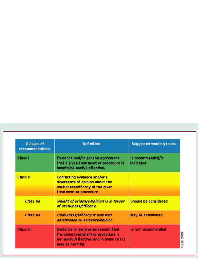
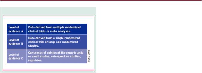
strength of the recommendation of particular management options were weighed and graded according to predefined scales, as outlined in Tables 1 and 2.
The experts of the writing and reviewing panels provided declaration of interest forms for all relationships that might be perceived as real or potential sources of conflicts of interest. These forms were compiled into one file and can be found on the ESC website (http://www.escardio.org/guidelines). Any changes in declarations of interest that arise during the writing period were notified to the ESC and updated. The Task Force received its entire financial support from the ESC without any involvement from the healthcare industry.
The ESC CPG supervises and coordinates the preparation of new Guidelines. The Committee is also responsible for the endorsement process of these Guidelines. The ESC Guidelines undergo extensive review by the CPG and external experts. After appropriate revisions the Guidelines are approved by all the experts involved in the Task Force. The finalized document is approved by the CPG for publication in the European Heart Journal. The Guidelines were developed after careful consideration of the scientific and medical knowledge and the evidence available at the time of their dating.
.
.
.
.
.
.
.
.
.
.
.
.
.
.
.
.
.
.
.
.
.
.
.
.
.
.
.
.
.
.
.
.
.
.
.
.
.
.
.
.
.
.
.
.
.
.
.
.
.
.
.
.
.
.
.
.
.
.
.
.
.
.
.
.
.
.
.
.
.
ESC Guidelines
The task of developing ESC Guidelines also includes the creation of educational tools and implementation programmes for the recommendations including condensed pocket guideline versions, summary slides, booklets with essential messages, summary cards for nonspecialists and an electronic version for digital applications (smartphones, etc.). These versions are abridged and thus, if needed, one should always refer to the full text version, which is freely available via the ESC website and hosted on the EHJ website. The National Societies of the ESC are encouraged to endorse, translate and implement all ESC Guidelines. Implementation programmes are needed because it has been shown that the outcome of disease may be favourably influenced by the thorough application of clinical recommendations.
Surveys and registries are needed to verify that real-life daily practice is in keeping with what is recommended in the guidelines, thus completing the loop between clinical research, writing of guidelines, disseminating them and implementing them into clinical practice.
Health professionals are encouraged to take the ESC Guidelines fully into account when exercising their clinical judgment, as well as in the determination and the implementation of preventive, diagnostic or therapeutic medical strategies. However, the ESC Guidelines do
Table 1 Classes of recommendation
ESC Guidelines
Table 2 Level of evidence
not override in any way whatsoever the individual responsibility of health professionals to make appropriate and accurate decisions in consideration of each patient’s health condition and in consultation with that patient or the patient’s caregiver where appropriate and/or necessary. It is also the health professional’s responsibility to verify the rules and regulations applicable to drugs and devices at the time of prescription.
2. Introduction
2.1 Why do we need new Guidelines on the management of cardiovascular diseases in pregnancy?
Since the previous version of these Guidelines was published in 2012, new evidence has accumulated, particularly on diagnostic techniques, risk assessment, and the use of cardiovascular drugs. This made a revision of the recommendations necessary.
2.2 New format of the Guidelines
The new Guidelines have been adapted to facilitate their use in clinical practice and to meet readers’ demands by focusing on condensed, clearly presented recommendations. At the end of each section, ‘recommendations’ summarize the essentials. ‘Gaps in the evidence’ are listed in section 13 to propose topics for future research. The Guidelines document is harmonized with the simultaneously published chapter on the management of cardiovascular diseases (CVDs) in pregnancy of the ESC Textbook of Cardiology (http://oxfordmedi cine.com/view/10.1093/med/9780199566990.001.0001/med-978019
.
.
.
.
.
.
.
.
.
.
.
.
.
.
.
.
.
.
.
.
.
.
.
.
.
.
.
.
.
.
.
.
.
.
.
.
.
.
.
.
.
.
.
.
.
.
.
.
.
.
.
.
.
.
.
.
.
.
.
.
.
.
.
.
.
.
.
.
.
.
.
.
.
.
.
.
.
.
.
.
.
.
.
.
.
.
.
.
.
.
.
.
.
.
.
.
.
.
.
.
.
.
.
.
.
.
.
.
.
.
.
.
.
.
.
.
.
.
.
.
.
.
.
.
.
.
.
.
.
.
.
.
.
.
.
.
.
.
.
.
.
.
3171
9566990-chapter-33). Background information and a detailed discussion of the data that have provided the basis for the recommendations can be found in the relevant book chapter.
2.3 Why these Guidelines are important
Pregnancy is complicated by maternal disease in 1–4% of cases. New data about the prevalence and incidence of pregnancy-related heart disease are limited from most parts of the world. Sudden adult death syndrome, peripartum cardiomyopathy (PPCM), aortic dissection, and myocardial infarction (MI) were the most common causes of maternal death in the UK over the period 2006–08.1–5 Knowledge of the risks associated with CVDs during pregnancy and their management in pregnant women who suffer from serious pre-existing conditions is of pivotal importance for advising patients before pregnancy.6 Since all measures concern not only the mother but the foetus as well, the optimum treatment of both must be targeted. A therapy favourable for the mother can be associated with potential harm to the developing child and, in extreme cases, treatment measures that protect the survival of the mother can cause the death of the foetus. On the other hand, therapies to protect the child may lead to a suboptimal outcome for the mother. Because prospective or randomized studies are frequently absent, recommendations in these Guidelines mostly correspond to evidence level C. Therefore, registries and prospective studies are urgently needed to improve current knowledge.4,7 At the European level, the Registry Of Pregnancy And Cardiac disease (ROPAC) registry of the ESC and the European Surveillance of Congenital Anomalies network are providing data on epidemiology and drug exposure in pregnancy.4,8
2.4 Methods
The current Guidelines are based on the previously published ESC Guidelines on the management of CVDs during pregnancy,9 the literature found in a systematic search from 2011–16 in the National Institutes of Health database (PubMed), and on recent publications and recommendations from the American Heart Association and the American College of Cardiology.10 Furthermore, we considered related Guidelines of the ESC published in 2012–15 on the topics of congenital heart disease, aortic disease, valvular heart disease, cardiomyopathies and heart failure (HF), coronary artery disease (CAD), hypertension, pericardial diseases, pulmonary hypertension (PH), infective endocarditis (IE), ventricular arrhythmias, and acute coronary syndromes, and on the topics of cancer treatment and cardiovascular toxicity, dyslipidaemias, atrial fibrillation (AF), and CVD prevention published in 2016 (https://www.escardio.org/Guidelines/ Clinical-Practice-Guidelineshomepage).
3172 |
ESC Guidelines |
2.5 What is new in the 2018 CVD in Pregnancy Guidelines? (Figure 1)
Selected revised recommendations and selected new recommendations
Comment/comparison with 2011 version
Strengthening mWHO classification of maternal risk.
Upgrade in class of recommendation; patients with severe MS should undergo intervention before pregnancy.
In 2011, OACs were recommended during the second and third trimesters until the 36th week. Now, separate recommendations for women with low and high dose are given for VKA use during the second and third trimesters.
Sotalol deleted.
Changed in high-risk patients from UFH to LMWH. Dosing based on body weight introduced.
Changes: dose adjustment of UFH or LMWH dose within 36 h now recommended.
Upgrade of recommendation: IIb to IIa.
Change from D-dimers to imaging as the first line of investigation, as D-dimers are unreliable in pregnancy.
FDA categories A–X were used for all drugs in 2011.
‘Pre-pregnancy surgery’ is now deleted. Now also information on Turner syndrome with aortic diameter corrected for BSA
 Selected new recommendations
2018
It is recommended to perform risk assessment in all women with cardiac diseases of childbearing age and before conception, using the mWHO classification of maternal risk11 (IC).
Intervention is recommended before pregnancy in patients with MS and valve area <1.0 cm² (IC).
During the second and third trimesters until the 36th week, VKAs are recommended in women needing a low dose (low-dose VKA: warfarin <5 mg/day, phenprocoumon <3 mg/day, or acenocoumarol <2 mg/day) (IC).
Flecainide or propafenone are recommended for prevention of SVT in patients with WPW syndrome12 (IC).
LMWH is the drug of choice for the prevention and treatment of VTE in all pregnant patients13 (IB).
It is recommended that the therapeutic dose of LMWH is based on body weight14 (IC).
In pregnant women on LMWH or UFH, it is recommended to perform weekly anti-Xa level monitoring or aPTT monitoring with dose adjustment (within 36 h) (IC).
Catheter ablation with electroanatomical systems should be considered in experienced centres in case of drug-refractory and poorly tolerated SVT15–17 (IIaC).
If compression ultrasound is negative, magnetic resonance venography should be considered to diagnose VTE18 (IIaC).
Decision-making based on former FDA categories is no longer recommended (IIIC).
Pregnancy is not recommended in patients with severe dilatation of the aorta (heritable thoracic aortic disease such as Marfan syndrome >45 mm, bicuspid aortic valve >50 mm, >27 mm/m2
BSA, or Turner syndrome ASI >25 mm/m2 BSA)19,20 (IIIC).
Right heart catheterization is recommended to confirm the diagnosis of PAH. This can be performed during pregnancy but with very strict indications10 (IC).
LMWH in therapeutic dose is recommended in pregnant patients with chronic thrombo-embolic pulmonary hypertension (IC).
Figure 1 Selected revised and new recommendations.

ESC Guidelines |
3173 |
In patients with pulmonary embolism, thrombolytic therapy is recommended only in severe hypotension or shock21 (IC).
In women at high risk for thrombo-embolism, it is recommended to convert LMWH to UFH at least 36 h prior to delivery and stop the UFH infusion 4–6 h prior to anticipated delivery. aPTT should be normal before regional anaesthesia22 (IC).
In women at low risk for thrombo-embolism on therapeutic LMWH, induction or caesarean section is recommended to be performed 24 h after the last dose of LMWH22 (IC).
In women considering pregnancy and requiring heart valve surgery, it is recommended to choose the prosthesis in consultation with a pregnancy heart team (IC).
It is recommended to manage pregnancy in women with mechanical heart valves in a centre with a pregnancy heart team (IC).
 In treatment-naive pregnant PAH patients, initiating treatment should be considered23 (IIaC).
In patients with (history of) aortic dissection, caesarean delivery should be considered (IIaC). Beta-blocker therapy throughout pregnancy should be considered in women with Marfan syndrome and other heritable thoracic aortic diseases (IIaC).
Induction of labour should be considered at 40 weeks gestation in all women with cardiac disease (IIaC).
In patients with PPCM, bromocriptine treatment may be considered to stop lactation and enhance recovery (LV function)24,25 (IIbB).
 Pregnancy is not recommended in patients with vascular Ehlers–Danlos syndrome26 (IIIC).
Breastfeeding is not recommended in mothers who take antiplatelet agents other than low-dose aspirin (from section 7, see section 12) (IIIC).
 New concepts
Enforcing mWHO classification of maternal risk.
 Introduction of the pregnancy heart team.
More attention for assisted reproductive therapy.
 Discussion of the use of bromocriptine in PPCM. Introduction of specific levels of surveillance based on low/medium/high risk for arrhythmia with haemodynamic compromise at delivery.
New information on pharmacokinetics in pregnancy, more detailed information on pharmacodynamics in animal experiments on all drugs (Supplementary Data)
 Perimortem caesarean section is discussed. Advice on contraception and the termination of pregnancy in women with cardiac disease is now provided.
Figure 1 Continued.
3. General considerations
3.1 Epidemiology
In the western world, the risk of CVD in pregnancy has increased due to increasing age at first pregnancy. According to World Atlas,27 the 10 countries where mean age at first birth is highest record a mean age between 28.8–31.2 years. The mild increase in maternal age does not justify an increase in CVD during pregnancy because of maternal age. However, pregnancies in the late reproductive years (or between ages of 40–50 years) are more frequently associated with an increasing prevalence of cardiovascular risk factors, especially diabetes, hypertension, and obesity. Additionally, an increasing number of women with congenital heart disease reach childbearing age.5
.
.
.
.
.
.
.
.
.
.
.
.
.
.
.
.
.
.
.
.
.
.
.
.
.
.
.
.
.
.
.
.
.
.
.
.
.
.
.
In western countries, maternal heart disease is the major cause of maternal death during pregnancy.2,28
Hypertensive disorders are the most frequent cardiovascular disorders during pregnancy, occurring in 5–10% of all pregnancies (see section 10). Among the other disease conditions, congenital heart disease is the most frequent CVD present during pregnancy in the western world (75–82%).29,30 Rheumatic valvular disease dominates in non-western countries, comprising 56–89% of all CVDs in pregnancy.29,31
Peripartum intensive care unit (ICU) admissions are increasing in frequency, with affected women-who suffer from serious preexisting conditions, are older, and present with multiple comorbidities and also congenital heart disease-being more frequently

3174
admitted than in previous years.6 The admission rate to ICUs was 6.4 per 1000 deliveries, corresponding to 1 admission per 156 deliveries, in Vienna, Austria during the period 2011–14. A 5% mortality rate was also observed in the study and is considered as appropriate in comparison to the literature.6
Cardiomyopathies are rare, but represent severe causes of cardiovascular complications in pregnancy.32
3.2 Physiological adaptations to pregnancy
Pregnancy induces changes in the cardiovascular system to meet the increased metabolic demands of the mother and foetus. Plasma volume and cardiac output (CO) reach a maximum of 40–50% above baseline at 32 weeks of gestation, while 75% of this increase has occurred by the end of the first trimester. The increase in CO is achieved by an increase in stroke volume in the first-half of pregnancy and a gradual increase in heart rate thereafter. Atrial and ventricular diameters increase while ventricular function is preserved. In women with heart disease, left ventricular (LV) and right ventricular (RV) adaptation to pregnancy can be suboptimal.33–36 Maternal cardiac dysfunction is related to impaired uteroplacental flow and suboptimal foetal outcome.35–37 Systemic and pulmonary vascular resistances decrease during pregnancy.
Pregnancy is a hypercoagulable state associated with increased risk of thrombo-embolism. Increased activity of liver enzyme systems, glomerular filtration rate, and plasma volume, protein binding changes, and decreased serum albumin levels contribute to changes in the pharmacokinetics of many drugs.36,38 Uterine contractions, positioning (left lateral vs. supine), pain, anxiety, exertion, haemorrhage, and uterine involution cause significant haemodynamic changes during labour and post-partum. Anaesthesia, haemorrhage, and infection may induce additional cardiovascular stress. Blood pressure (BP) and CO increase during labour and post-partum. In conclusion, the physiological adaptations to pregnancy influence the evaluation and interpretation of cardiac function and clinical status.
3.3 Pre-pregnancy counselling
All women with known cardiac or aortic disease who wish to embark on pregnancy require timely pre-pregnancy counselling.39 Informed maternal decision-making is crucial and there is a clear need for individualized care, taking into account not only the medical condition but also the emotional and cultural context, psychological issues, and ethical challenges. Especially in patients with a high-risk or possible contraindication for pregnancy, the risk of pregnancy and the necessity of careful planning of pregnancy should be discussed at a young age. However, it is also important to explain that many women can go through pregnancy with low-risks.
For risk estimation, as a minimum, an electrocardiogram (ECG), echocardiography, and an exercise test should be performed. In case of aortic pathology, complete aortic imaging by computed tomography (CT) scanning or magnetic resonance imaging (MRI) is necessary for appropriate pre-conception counselling. Peak heart rate and peak oxygen uptake are both known to be predictive of maternal cardiac events in pregnancy.40 A pregnancy exercise capacity >80% is associated with a favourable pregnancy outcome.
.
.
.
.
.
.
.
.
.
.
.
.
.
.
.
.
.
.
.
.
.
.
.
.
.
.
.
.
.
.
.
.
.
.
.
.
.
.
.
.
.
.
.
.
.
.
.
.
.
.
.
.
.
.
.
.
.
.
.
.
.
.
.
.
.
.
.
.
.
.
.
.
.
.
.
.
.
.
.
.
.
.
.
.
.
.
.
.
.
.
.
.
.
.
.
.
.
.
.
.
.
.
.
.
.
.
.
.
.
.
.
.
.
.
.
.
.
.
.
.
.
.
.
.
.
.
.
.
.
.
.
.
.
.
.
.
.
.
.
.
.
.
.
.
.
.
.
.
.
.
.
.
.
.
.
.
.
.
.
.
.
.
.
.
.
.
.
.
.
.
.
.
.
ESC Guidelines
Several aspects must be discussed, including long-term prognosis, fertility and miscarriage rates, risk of recurrence of congenital disease, drug therapy, estimated maternal risk and outcome, expected foetal outcomes, and plans for pregnancy care and delivery. A multidisciplinary management plan should be constructed and discussed with the patient. In addition, attention to unhealthy habits including being overweight, smoking, and consuming alcohol is important, as these can have a clear impact on maternal and foetal outcomes. Pregnancy is a very suitable time for recommending a healthy lifestyle, including smoking cessation.
3.3.1Risk of maternal cardiovascular complications
The risk of complications in pregnancy depends on the underlying cardiac diagnosis, ventricular and valvular function, functional class, presence of cyanosis, pulmonary artery pressures, and other factors. Comorbidities, including for example rheumatoid and musculoskeletal diseases as well as mental disorders, should also be taken into account. Therefore, risk estimation should be individualized.
To assess the maternal risk of cardiac complications during pregnancy, the condition of the woman should be assessed, taking into account medical history, functional class, oxygen saturation, natriuretic peptide levels, echocardiographic assessment of ventricular and valvular function, intrapulmonary pressures and aortic diameters, exercise capacity, and arrhythmias. Disease-specific risk should be assessed using the modified World Health Organization (mWHO) classification (Table 3) and as described in the respective sections dealing with specific diseases in these Guidelines. Risk estimation should be further refined by taking into account predictors that have been identified in studies that included large populations with various diseases, such as the CARPREG (CARdiac disease in PREGnancy),
ZAHARA, and ROPAC (Registry Of Pregnancy And Cardiac disease) studies (Table 4).29,41–43
The mWHO classification is currently the most accurate system of risk assessment, although it is probably more appropriate for developed, rather than developing, countries.4,11,44 The general principles of this classification, and follow-up and management during pregnancy according to this mWHO classification, are presented in Table 3. Indications for intervention (surgical or catheter) do not differ in women who contemplate pregnancy compared with other patients. The few exceptions to this rule are women with at least moderate mitral stenosis and women with aortic dilatation. See also the disease-specific sections of these Guidelines. Fertility treatment is contraindicated in women with mWHO class IV, and should be carefully considered in those who have mWHO class III disease or who are anticoagulated.45
The risk estimation needs to be re-evaluated during each prepregnancy visit, because the risk of complications may change over time. Natriuretic peptide levels are associated with the occurrence of cardiac events, with N-terminal pro B-type natriuretic peptide (NTproBNP) >128 pg/mL at 20 weeks pregnancy being predictive of events later in the pregnancy.46,47 Pre-eclampsia is associated with HF in women with heart disease.43
3.3.2 Risk of obstetric and offspring complications
Women with cardiac disease have an increased risk of obstetric complications, including premature labour, pre-eclampsia, and postpartum haemorrhage.
Offspring complications occur in 18–30% of patients with heart disease, with neonatal mortality between 1–4%.29 Maternal and