

Materials Today Volume 19, Number 6 July/August 2016 |
RESEARCH |
Review RESEARCH:
Osteoimmunomodulation for the development of advanced bone biomaterials
Zetao Chen1,3, Travis Klein1,3, Rachael Z. Murray1, Ross Crawford1,3, Jiang Chang2,3, Chengtie Wu2,3,* and Yin Xiao1,3,*
1Institute of Health and Biomedical Innovation, Queensland University of Technology, Brisbane, 60 Musk Ave, Kelvin Grove, Brisbane, Queensland 4059, Australia
2State Key Laboratory of High Performance Ceramics and Superfine Microstructure, Shanghai Institute of Ceramics, Chinese Academy of Sciences, 1295 Dingxi Road, Shanghai 200050, People’s Republic of China
3Australia-China Centre for Tissue Engineering and Regenerative Medicine, Queensland University of Technology, Brisbane, 60 Musk Ave, Kelvin Grove, Brisbane, Queensland 4059, Australia
As direct effector cells for osteogenesis, osteoblastic cells are commonly used for evaluating the in vitro osteogenic capacity of bone biomaterials, and the traditional biological principle for developing bone biomaterials is to directly stimulate osteogenic differentiation. With this principle, most efforts are currently spent on optimizing the bio-mechanical and physicochemical properties to induce osteogenic differentiation of mesenchymal stem cells. This strategy has achieved certain success in the development of bone biomaterials; however, inconsistencies between in vitro and in vivo studies are not uncommon, implying the mechanisms that govern the material’s capacity to mediate osteogenesis is not wellunderstood. Osteoimmunology has revealed the vital role of immune cells in regulating bone dynamics. Neglecting the importance of the immune response is a major shortcoming of the traditional strategy, and may explain inconsistencies between in vitro and in vivo conditions. Here, we proposed osteoimmunomodulation (OIM) in recognition of the importance of the immune response during biomaterial-mediated osteogenesis. Accordingly, we proposed the paradigm shift of bone biomaterials to an osteoimmunomodulatory material and discussed the evaluation strategy for the osteoimmunomodulation property of bone biomaterials. It is the ambition of authors that this review will change traditional methods for bone biomaterials assessment and assist in developing new bone biomaterials with the osteoimmunomodulatory property for orthopedic and dental applications.
Introduction
Bone defects caused by tumor resection, traumatic fracture, aseptic necrosis, osteolysis, osteomyelitis, periodontitis, and spinal fusion, to name but a few, typically require surgical remediation using bone biomaterials [1–4]. Due to the direct relationship between osteoblastic lineage and bone formation, the major principle for developing bone biomaterials is to manipulate the in vitro osteogenic differentiation of the osteoblastic lineage and then investigating the potential osteogenic biomaterials in an in vivo model. With this principle, many strategies have been developed
*Corresponding authors: Wu, C. (chengtiewu@mail.sic.ac.cn), Xiao, Y. (yin.xiao@qut.edu.au)
to fabricate an ideal bone biomaterial that can gain desired osseointegration and osteogenesis. The fabrication techniques are quite advanced that materials scientists can somehow prepare bone biomaterials with the desired physicochemical and mechanical properties (from nano to particle size, from 2D to customized 3D structure, from hydrophilic to hydrophobic, etc.).
However, this principle often does not lead to clinically useful bone implant materials, with many candidates failing to make it beyond the confines of the laboratory. When analyzing the possible reasons, we focus on optimizing the compositions, the bio-physicochemical and mechanical properties of the candidate materials, but we rarely think that perhaps the basic biological
1369-7021/ 2015 The Authors. Published by Elsevier Ltd. This is an open access article under the CC BY-NC-ND license (http://creativecommons.org/licenses/by-nc-nd/4.0/). http://dx.doi.org/10.1016/
304 |
j.mattod.2015.11.004 |

Materials Today Volume 19, Number 6 July/August 2016 |
RESEARCH |
principle also needs optimization. Bone biology has made great progress and the mechanisms underlying osteogenesis have been much better understood. We now know that osteogenesis is not simply accomplished by bone cells from skeleton system, but a collaboration of multiple systems. This indicates that the traditional biological principle is outdated and insufficient, which could be a leading reason for the failure of trials. To fabricate an ideal bone biomaterial, we need to keep the pace with the development of bone biology and keep on modifying the basic biological principle.
Among all the achievements made in the area of bone biology, the development of osteoimmunology is one of the greatest. The immune and skeletal systems are found to be closely related, sharing a number of cytokines, receptors, signaling molecules and transcription factors [5,6]. Immune cells play a key role in bone homeostasis. Being a foreign body, an implant is recognized by the immune system and triggers a significant immune reaction that affects the biological behavior of bone cells. Such an event may eventually determine the in vivo fate of bone biomaterials [7,8]. The immune response may, therefore, be a key factor that is neglected when evaluating the osteogenic capacity of bone biomaterials. Accordingly, the design paradigm for advanced bone biomaterials should be shifted from being relatively inert to having immunomodulatory properties, emphasizing the important role of immune responses [7]. A new generation of bone biomaterials should be able to modulate the local immune environment such that it favors osteogenesis and the osseointegration of the implant.
Developing such biomaterials would require an in-depth understanding of a number of important issues. Firstly, it is important to understand the relationship between immune cells and bone cells and what effect the immune environment induced by implanted biomaterials has on osteogenesis. Secondly, the mechanisms underlying the material-mediated immune response must be understood in order to aid the design and preparation of biomaterials to induce an immune environment that provides conditions that balance osteogenesis and osteoclastogenesis for optimal osseointegration. Finally, determining whether or not a biomaterial can induce a favorable immune response should be a routine screening process and part of a standard evaluation protocol when developing advanced immunomodulatory bone biomaterials. In this review, we define such a capacity as osteoimmunomodulation (OIM) – a novel property of bone biomaterials. Favorable OIM properties are of great importance when attempting to produce advanced bone biomaterials for clinical application with optimal osteogenesis and osseointegration.
Overview of the integration between bone tissue and implants
The mechanism underlying bone biomaterial-mediated osteogenesis involves at least three interactive components: the host immune cells, the host bone cells and the materials. Following implantation, the host body will first undergo a universal response to the materials, which is an extension of the mammalian response following tissue injury [9]. Proteins from blood and interstitial fluids, such as fibrinogen, vitronectin, complement, and fibronection [7], will adsorb to the material’s surfaces within seconds and then form a transient surface matrix. In response, the coagulation
cascade and complement systems are activated, leading to thrombus formation and activation of other cell populations.
After the initial blood/material interaction, an acute inflammation is initiated, which features the recruitment and activation of neutrophils, or polymorphonuclear leukocytes (PMNs). PMNs, in an effort to degrade the materials, release proteolytic enzymes and reactive oxygen species (ROS) [10], which will corrode the surface of the material. The PMNs rapidly become exhausted, undergoing apoptosis and disappearing from the implantation sites within the first two days [11]. Mast cells are also active participants in the acute inflammatory reaction and degranulation of these cells leads to the release of inflammation-enhancing cytokines and histamine, which amplify the immune reaction [12].
Chemoattractants and activation cytokines released in the previous phase result in monocyte recruitment to the implant, where the cells differentiate into macrophages. Macrophages are able to engulf particles of up to 5 mm [7,13]; if the particle size is larger, the macrophages will coalesce to form foreign body giant cells (FBGCs), driven by stimulation with IL-4 and IL-13 [14]. The foreign components and molecules released during the hostbody/implant interaction can positively regulate osteogenic differentiation to form new bone on the surface of the implant and entrap it. In the bone remodeling phase newly formed bone undergoes functional remodeling and some entrapped implant materials, such as Ca–P based bioceramics, can be further degraded. Functional loading and the mechanical strain is the main cause for the remodeling. Osteocytes are known to translate signals related to mechanical strain into biochemical signals and regulate osteoblasts and osteoclasts and, therefore, may play a regulatory role in this late stage [15–17] (Fig. 1a).
The close relationship that exists between the immune and skeletal systems makes the proposition that stimulated immune cells may contribute to both the success and failure of an implant seem feasible (Fig. 1). The immune cells would exert this effect by releasing cytokines that regulate osteogenesis, in addition to their well-known effects on inflammation, thus inducing or inhibiting bone formation. The effects of the immune response to bone biomaterials in regulating osteogenesis are, therefore, ‘a doubleedged sword’. A favorable immune reaction creates an osteogenic microenvironment that can improve osteogenesis, whereas an inappropriate immune reaction may lead to the chronic inflammation and the formation of a fibrous capsule around the implant.
The capsule formed by the foreign body reaction (FBR) effectively separates the implant from the surrounding environment, such that it can remain safely in the host body throughout its lifetime [18]. However, this renders the implant an ‘inert’ mechanical support, a scenario which fails to meet the demands of a bone substitute material since it is intended to induce new bone formation and fill the defect space with fully functional bone. A fibrous encapsulation prevents direct interaction between bone marrow and the implants such that bone cells cannot attach to the surface of implants to form new bone. Instead, the defect will be filled by a fibrous tissue, resulting in the failure of bone reconstruction. This illustrates the importance of creating a local immune environment that favors bone regeneration and osseointegration; manipulating the immune response by targeted modifications of the bone biomaterials is, therefore, a good strategy to swing the balance toward this direction.
RESEARCH: Review
305

RESEARCH |
Materials Today Volume 19, Number 6 July/August 2016 |
Review RESEARCH:
 FIGURE 1
Schematic illustration of bone biomaterials mediated de novo bone formation (a) and the failure of bone biomaterials that lead to the fibrous encapsulation
(b). (a) This process can be divided into three phases: early, bone formation and remodeling phase, with the involvement of multiple systems: coagulation system, immune system, skeleton system. The most likely macrophage phenotype switch pattern is also presented. The early stage of the repair response is dominated by the inflammatory phase, when the majority of macrophages would be of the inflammatory M1 phenotype. An efficient and timely switch from M1 to M2 macrophage phenotype results in an osteogenic cytokine release and with it the formation of new bone tissue. (b) It is also divided into three phases: acute, chronic inflammation and fibrous encapsulation. The likely macrophage phenotype switch pattern is shown. The early stage is dominated by the inflammatory phase, during which the majority of macrophages would be of the inflammatory M1 phenotype. However, a prolonged M1 polarization phase leads to an increase in fibrosis-enhancing cytokine release pattern by the M2 macrophages, which results in the formation of a fibrocapsule. Adapted from [182].
Immune cells regulation of osteoclastogenesis and |
regulatory molecules which elicit significant effects on osteoclas- |
osteogenesis |
togenesis and osteogenesis (Fig. 2). Abnormal functioning of |
The field of osteoimmunology seeks to understand the interaction |
immune cells can lead to an imbalance between osteoclasts and |
between the immune and skeletal systems [6]. Immune cells |
osteoblasts and result in conditions such as osteolysis, osteoporo- |
participate actively in bone physiology and pathology by releasing |
sis, osteoarthritis and rheumatoid arthritis. In this part, we will |
306
Materials Today Volume 19, Number 6 July/August 2016 |
RESEARCH |
RESEARCH: Review
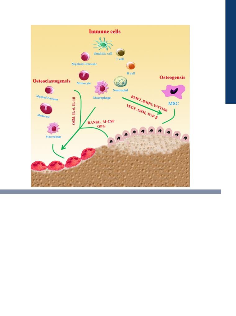
 FIGURE 2
Schematic illustration of the role of immune cells in bone dynamics. Immune cells participate actively in osteoclastogenesis and osteogenesis by releasing regulatory molecules.
review the role of immune cells in osteoclastogenesis and osteogenesis and possible molecular mechanisms at play.
Immune cells and osteoclastogenesis
Immune cells regulate osteoclastogenesis by three major cytokines: macrophage-colony stimulating factor (M-CSF), receptor activator of NF-kB ligand (RANKL) and osteoprotegerin (OPG). M-CSF binds to its cognate receptor c-FMS on osteoclast precursors and signals through the Akt and MAP kinase pathway [19]. RANKL binds to RANK, a receptor on the surface of osteoclast precursors, thereby transducing via TNF receptor associated factor 6 (TRAF6), NF-kB, activator protein 1 (AP-1) and nuclear factor of activated T cells 2 (NFAT2) to upregulate expression of genes for the survival and differentiation of osteoclasts [5,20]. RANKL is expressed, not only by osteoblastic cells that maintain normal osteoclastogenesis in bone tissue, but also by activated T cells and neutrophils, indicating the involvement of these immune cells during osteoclastogenesis [21,22]. Macrophages are the precursor of osteoclasts, which under the stimulation of M-CSF and RANKL can differentiate into osteoclasts during bone remodeling.
IL-6 and oncostatin M (OSM) are important mediators of osteoclast formation and function. IL-6 is known to induce the expression of RANKL, and utilize the RANKL/RANK-OPG system to elicit indirect effects on promoting osteoclastogenesis and osteoclast activation [23,24]. IL-6 is also found to participate in the TNF-a and IL-l induced osteoclast formation [25]. OSM uses gp130, the same receptor subunit as does IL-6, for signaling and these two cytokines often have similar and overlapping functions [26]. OSM can also stimulate the production of RANKL by osteoblasts and enhance the formation of osteoclasts in a dose dependent manner, which might be related to its synergistic effects with IL-6 [27,28]. By contrast, interferon-g (IFN g) promotes the degradation of TRAF6, a key intermediate in RANKL/RANK pathway, thereby preventing massive bone destruction during inflammation [5].
OPG, a decoy receptor for RANKL, interrupts the interaction of RANKL/RANK, thereby inhibiting both differentiation and the function of osteoclasts [29,30]. B cells have been shown to be a major source of bone marrow-derived OPG [31,32], which implies that B cells are one of the main inhibitor of osteoclastogenesis in normal physiology. Depletion of CD4 and CD8 T lymphocyte
307