

Биоинженерия / Биомеханика_микрофлюидные_устройства / статьи / Копия c3lc41393d
.pdf2013. Downloaded on 10/19/2019 12:57:40 PM. |
Creative Commons Attribution 3.0 Unported Licence. |
Article. Published on 04 April |
This article is licensed under a |
Open Access |
|
Critical Review
experiment. In one study, the authors found that tumor cells migrated downstream as a result of autocrine chemokine gradients.102 In a subsequent study, it was shown that there are two competing mechanisms, the one just mentioned and another that drives cells to migrate in the opposite direction, against the flow (Fig. 2). Evidence suggest that the latter is mediated by a mechanotransduction response.103 Haessler et al. developed a modified microfluidic device that allows introduction of two hydrogels in series and maintenance of an interstitial flow field with a peristaltic pump. By modifying the permeability of the upstream hydrogel, the interstitial flow speed could be tuned between 0.1 and 4.7 mm s21 (Table 1) for a given permeability in the downstream hydrogel and constant 7 Pa pressure drop across the device. Experiments within this platform demonstrate that cell migration within a population is heterogeneous, with cells migrating either upstream or downstream, with upstream migration characterized by lower migration velocities and increased persistence.104
ECM topography
The stromal ECM presents a host of mechanical and chemical signals to migrating tumor cells (for review, see: Polacheck et al.105). In particular, it has been demonstrated that tumor cells migrate through tracks created by migrating fibroblasts.106 To investigate how tumor cell confinement within microtracks might alter cell migration, Irimia fabricated a series of parallel microchannels on one microfluidic chip, and found that confinement of tumor cells dramatically influenced migratory characteristics. For breast cancer cells cultured in 12 6 15 6 600 mm channels, more than 80% of tumor cells migrated from one end of the channel to the other (600 mm) without stopping or changing direction. The migration velocity was more than 2 fold that found for cells cultured in chambers much larger than the cell diameter, where migration was characterized by stopping and much lower persistence.107 The confined channels were fabricated from PDMS, which represents a key limitation to this study, as PDMS is much stiffer than the tumor stroma. Recently, a two-photon laser ablation technique was used to generate microtracks in a more physiologically relevant collagen gel. The authors found that cells migrated through microtracks even without MMP, Rho, or ROCK activity.108 These results suggest that tracks laid down by migrating stromal cells such as fibroblasts and macrophages are crucial regulators of the metastatic cascade.
Shear stress
Circulating tumor cells (CTCs) in the blood stream are exposed to shear stress, and extravasation requires tumor cells to arrest on the endothelium and traverse the endothelial barrier layer (a recent review highlights, in part, the mechanics of extravasation3). In one of the first steps in metastatic disease, tumor cells break loose from the primary tumor in a process termed the epithelial-mesenchymal transition (EMT), characterized by a loss of E-cadherin and overexpression of N-cadherin. Fueled by this change and motivated by previous devices that allow differential separation of cell types based on adhesive strength to specific ligands,109 Cheung et al. coated the surface of a microchannel with N-cadherin antibodies to trap and collect circulating PC3N prostate cancer cells and
View Article Online
Lab on a Chip
MDA-MB-231-N breast cancer cells. The authors found that fluid speed influenced CTC capture, and when CTCs were exposed to a time-varying flow, the rate of fluid acceleration influenced CTC deformation and capture.110 Recently, a device similar to that introduced by Rossi et al. has been developed that implements a single channel with cross-sectional area that varies along the channel length. This system allows a range of shear stresses (0.25–10 Pa) to be applied within a channel, and the device has been employed to study the effect of shear stress on detachment of human breast cancer cells from collagen-coated substrates.111
Extravasation is thought to occur at sites of vessel confinement,112 and a recent device was developed to explore the synergistic effects of mechanical confinement and shear stress on tumor cells. The device was fabricated by bonding PDMS microchannels to a flexible, soft PDMS substrate in which fluorescent beads are embedded. By flowing fluid through the channel, shear stress was applied to cells cultured within the channels, and the cells deformed the underlying substrate. The deformations were measured by traction force microscopy, and related to focal adhesion disassembly, while membrane fluidization was measured using fluorescence recovery after photobleaching (FRAP). By measuring the time lag between focal adhesion disassembly and membrane fluidization, the authors developed a metric that measures the time required for cellular adaptation to shear stress (the time required for the feedback element in Fig. 1b). The authors found that the time lag is a function of shear stress and channel cross-sectional area and suggested the dependence on cross-sectional area is due to modulation in autocrine EGF concentration (smaller cross-sectional area increases autocrine concentration). Furthermore, tumor cells are more sensitive to fluid shear stress and confinement than non-cancerous cells, suggesting that tumor cells might be more likely to rearrange their cytoskeleton for migration and extravasation.113
Mechanically active tissues
Mechanically active tissues are defined as those comprised of cells equipped with specialized contractile apparatus such as skeletal muscle, smooth muscle, cardiac muscle and myofibroblasts. Due to their contractile nature, these tissues are inherently exposed to various types and levels of mechanical stimuli, which in turn are responsible for the tissue functionality (Fig. 1b). For example, myogenesis is influenced by cyclic strain and substrate stiffness,114,115 while smooth muscle cell and myofibroblast migration are influenced by shear stress116 and interstitial flow117. However, much of these data were generated in micropatterned or bulk assays and few microfluidic devices have been used to investigate mechanotransduction in these mechanically active tissues. The existing microfluidic platforms developed to date primarily focus on differentiation, force generation and measurement, and biomimetic fluid pumps.
2262 | Lab Chip, 2013, 13, 2252–2267 |
This journal is The Royal Society of Chemistry 2013 |
2013. Downloaded on 10/19/2019 12:57:40 PM. |
Creative Commons Attribution 3.0 Unported Licence. |
Article. Published on 04 April |
This article is licensed under a |
Open Access |
|
Lab on a Chip
Differentiation
Studies show that myocyte differentiation is affected by mechanical stimuli, in addition to electrical and biochemical influences. Stem cells preferentially differentiate into myocytes and cardiomyocytes when cultured on substrates with stiffness levels that match that of the corresponding differentiated tissue.45,114 However, the role of other mechanical stimuli such as mechanical strain remain unclear, with studies showing that cyclic mechanical loading enhances cardiac differentiation,115,118 and seemingly conflicting studies showing that cyclic loading favors pluripotency in human embryonic stem cells.119
Despite considerable efforts to elucidate the effect of stretch on cardiomyogenesis, only one device has been developed to apply cyclic strain to embryoid bodies of mouse embryonic stem cells plated on a deformable membrane or embedded within a collagen matrix.120 In the device, the cells contained in a 1 6 0.5 6 0.2 mm gel region were exposed to 10% uniaxial strain at a 1 Hz for 24 h. Differentiation into cardiomyocytes was assessed by measuring expression of a-MHC (a late-stage cardiomyogenesis marker), which was found to decrease with the application of stretch. Compared to traditional strain application assays, an advantage of this device is the possibility to apply controlled chemical environ-
View Article Online
Critical Review
ment and the reduction in the number of cells required for the differentiation experiment.
Force generation
One critical aspect when studying mechanically active tissue is the ability to quantify the force generated by the cells or tissue constructs. Most common techniques for contractile adherent cells rely on traction force microscopy, where cells are cultured on an array of microscopic pillars28 or on a compliant gel embedded with fluorescent beads.121 Recently, several microengineered devices have emerged that share a common feature: two microscopic compliant PDMS pillars fabricated within millimeter-scale wells are used as supports for 3D muscle tissue, made of either fibroblasts,122 cardiomyocytes or skeletal muscle cells differentiated from myoblasts. Due to passive or active contraction, the muscle tissue bends the compliant pillars to which they are attached. Using beam theory and the mechanical properties of the polymer, the force generated by the muscle construct can be inferred from the
deflection of the tip of the pillar. Active contraction can be triggered either via electric stimulation123,124 or via light on
channelrhodopsin-transduced muscle cells.125 The forces generated by these constructs range from a few mN to tens of mN depending on the construct geometry, the mechanical properties of the polymer and the cell type. Also, Boudou et al.
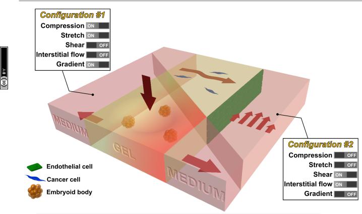
Fig. 3 Future intergrated microfluidic system that allows the study of multiple mechanical stimuli on multiple cell types. For example, a microfluidic device can be used to study the effect of compression, stretch, and chemical gradient on embryoid bodies seeded in the collagen gel (configuration #1). In addition, a microlfuidic device can be used to study the effect of shear stress and interstitial flow on cancer cells (seeded in collagen gel) in the vicinity of endothelial cells (seeded as a monolayer in the channel) (configuration #2).
This journal is The Royal Society of Chemistry 2013 |
Lab Chip, 2013, 13, 2252–2267 | 2263 |

2013. Downloaded on 10/19/2019 12:57:40 PM. |
Creative Commons Attribution 3.0 Unported Licence. |
Article. Published on 04 April |
This article is licensed under a |
Open Access |
|
Critical Review
demonstrated that the post stiffness influenced the tissue morphology and the amount of tension generated by the construct (Table 1, Fig. 2), illustrating how cellular tissue can
adapt their behavior in response to various mechanical stimuli (Fig. 1b).124
Pumps
Various strategies have been developed to use the contractile force generated by mechanically active tissue to power mechanical devices (reviewed in Pilarek et al.126). A common feature of these devices is a monolayer of cardiomyocytes that, by its contraction, causes the deflection of a PDMS membrane. Tanaka and colleagues implemented a check valve system powered by cardiomyocytes in which the fluid flow rates of 2 nL min21 were generated,127 and Park et al. developed a diffuser/nozzle system to produce 0.226 nL min21 flow rates.128 Finally, an updated system by Tanaka et al. involved plating cardiomyocytes onto a spherical-shaped membrane and fluid within the membrane is displaced in a similar manner to a beating heart,129 producing flow rates up to 0.5 mL min21. In all these cases, however, the cardiomyocytes beat at their own, intrinsic frequency, providing little control over the generated flow rates. To further regulate the function of these pumps, it is crucial to understand how cardiomyocyte function is influenced by the mechanical stimuli associated with coordinated actuation and fluid flow within the pump itself. Consequently, functional fluid pumps are an excellent example of the feedback involved with mechanotransduction (Fig. 1b) and highlight current shortcomings in our understanding of tissue-level mechanotransduction.
Prospective
Here we review the various microfluidic platforms that have been developed to investigate the effects of mechanical forces on the constitutive cells of various tissues; however, our understanding of the molecular mechanisms underlying the ability of these cells to sense and react to mechanical stimuli are still largely based on traditional, macro-assays. The limited mechanistic insight provided by microfluidic platforms is due, in part, to difficulty in performing standard quantitative biochemical assays for measuring gene and protein expression on-chip. New experimental approaches, however, allow for the extraction of cells after incubation within a microfluidic device for qRT-PCR,130 and platforms for on-chip DNA extraction and PCR131 and ELISA132 are becoming more readily available and advancing the capabilities of microfluidics.
Although many of the molecular mechanisms require further elucidation, certain elements of in vivo physiology have been captured in microfluidic platforms that have not been observed in traditional macro-assays, such as gut epithelial cell co-culture with bacteria,79 neutrophil diapedesis through lung epithelium,78 and apical-basal polarity in IMCDs.67 Other phenomena such as the differential responses of ECs to basal-to-apical and apical-to-basal transendothelial flows and the migratory responses of cancer cells to interstitial flow, are better studied in a microfluidic assay where
View Article Online
Lab on a Chip
hydrostatic pressures and resulting fluid flows can be more tightly regulated than in traditional platforms. Because these cell behaviors have only been observed on-chip or because microfluidic platforms are required for precise control of the applied stimulus, future advances to microfluidic technology that integrate quantitative biochemical techniques will allow investigation of these cell behaviors and tissue properties such as the more in vivo-like permeability in gut epithelial cells cultured under shear stress.
While innovative methods and systems will no doubt continue to be developed, we propose that the next major advance lies in the combination of existing technologies, creating progressively more realistic in vitro models of mechanotransduction in living systems. For example, methods that produce tissue compression might be combined with matrix stretch and chemical gradients (configuration #1 in Fig. 3), to mimic processes that occur during development using embryoid bodies embedded in a 3D hydrogel. Alternatively, systems can be produced allowing for simultaneous shear flow and interstitial flow (configuration #2), perhaps with multiple cell types present, in the context of studies examining the combined effects on tumor cell in the vicinity of a vascularized tumor. Such compound systems could retain the tight control and real-time visualization capabilities characteristic of in vitro models, while also offering a degree of in vivo realism.
Finally, while the addition of new functionalities or combinations of existing ones is needed, this should be done with an eye toward ease of use by the broader community. The value of these systems will be optimally enhanced if the assays can be incorporated into the numerous labs that study mechanobiology. For some, the technology is intimidating, and added capabilities are not required for many of the fundamental studies that are needed.
Acknowledgements
Funding from The National Cancer Institute (R21CA140096), the National Science Foundation Science and Technology Center for Emergent Behaviors of Integrated Cellular Systems (CBET-0939511), and Janssen Pharmaceuticals, Inc. is greatly appreciated. W. J. P. and R. L. are supported by National Science Foundation Graduate Research Fellowships. The authors would like to thank the members of the Kamm Lab for the helpful discussion and Serge Roux for the valuable suggestions in figure design.
References
1 Y. Xia and G. Whitesides, Annu. Rev. Mater. Sci., 1998, 28,
153–84.
2S. W. Moore, P. Roca-Cusachs and M. P. Sheetz, Dev. Cell, 2010, 19, 194–206.
3D. Wirtz, K. Konstantopoulos and P. C. Searson, Nat. Rev. Cancer, 2011, 11, 512–22.
4 G. Bao and S. Suresh, Nat. Mater., 2003, 2, 715–25.
2264 | Lab Chip, 2013, 13, 2252–2267 |
This journal is The Royal Society of Chemistry 2013 |

2013. Downloaded on 10/19/2019 12:57:40 PM. |
Creative Commons Attribution 3.0 Unported Licence. |
Article. Published on 04 April |
This article is licensed under a |
Open Access |
|
Lab on a Chip
5 F. Kurth, K. Eyer, A. Franco-Obrego´n and P. S. Dittrich,
Curr. Opin. Chem. Biol., 2012, 1–9.
6Kshitiz, D.-H. Kim, D. J. Beebe and A. Levchenko, Trends Biotechnol., 2011, 29, 399–408.
7D. Huh, G. A. Hamilton and D. E. Ingber, Trends Cell Biol., 2011, 21, 745–54.
8 Y. Zheng and Y. Sun, Micro Nano Lett., 2011, 6, 327.
9 S. a. Vanapalli, M. H. G. Duits and F. Mugele,
Biomicrofluidics, 2009, 3, 12006.
10 H. W. Hou, W. C. Lee, M. C. Leong, S. Sonam, S. R. K. Vedula and C. T. Lim, Cell. Mol. Bioeng., 2011, 4, 591–602.
11D.-H. Kim, P. K. Wong, J. Park, A. Levchenko and Y. Sun,
Annu. Rev. Biomed. Eng., 2009, 11, 203–33.
12C. F. Dewey, S. R. Bussolari, M. A. Gimbrone and P.
F.Davies, J. Biomech. Eng., 1981, 103, 177–85.
13Y.-S. J. Li, J. H. Haga and S. Chien, J. Biomech., 2005, 38, 1949–1971.
14B. L. Gray, D. K. Lieu, S. D. Collins, R. L. Smith and A.
I.Barakat, 2002, 9–16.
15J. W. Song, W. Gu, N. Futai, K. A. Warner, J. E. Nor and
S.Takayama, Anal. Chem., 2005, 77, 3993–3999.
16L. Chau, M. Doran and J. Cooper-White, Lab Chip, 2009, 9, 1897–1902.
17A. D. van der Meer, A. A. Poot, J. Feijen and I. Vermes,
Biomicrofluidics, 2010, 4, 11103.
18J. Wang, J. Heo and S. Z. Hua, Lab Chip, 2010, 10, 235–239.
19M. Rossi, R. Lindken, B. P. Hierck and J. Westerweel, Lab Chip, 2009, 9, 1403–1411.
20R. Estrada, G. A. Giridharan, M.-D. Nguyen, S. D. Prabhu and P. Sethu, Biomicrofluidics, 2011, 5, 32006–3200611.
21L. K. Chin, J. Q. Yu, Y. Fu, T. Yu, A. Q. Liu and K. Q. Luo, Lab Chip, 2011, 11, 1856–1863.
22G. M. Price, K. H. K. Wong, J. G. Truslow, A. D. Leung,
C.Acharya and J. Tien, Biomaterials, 2010, 31, 6182–9.
23X. Lin and B. P. Helmke, Biophys. J., 2008, 95, 3066–3078.
24X. Lin and B. P. Helmke, Cell. Mol. Bioeng., 2009, 2, 231–243.
25A. M. Ross, Z. Jiang, M. Bastmeyer and J. Lahann, Small, 2012, 8, 336–55.
26Y. Yang, K. Kulangara, J. Sia, L. Wang and K. W. Leong, Lab Chip, 2011, 11, 1638–46.
27R. H. W. Lam, Y. Sun, W. Chen and J. Fu, Lab Chip, 2012, 12, 1865–1873.
28J. L. Tan, J. Tien, D. M. Pirone, D. S. Gray, K. Bhadriraju and C. S. Chen, Proc. Natl. Acad. Sci. U. S. A., 2003, 100, 1484–9.
29M. A. Swartz and M. E. Fleury, Annu. Rev. Biomed. Eng., 2007, 9, 229–256.
30J. W. Song and L. L. Munn, Proc. Natl. Acad. Sci. U. S. A., 2011, 108, 15342–15347.
31V. Vickerman and R. D. Kamm, Integr. Biol., 2012, 4, 863–74.
32J. W. Song, J. Daubriac, J. M. Tse, D. Bazou and L.
L.Munn, Lab Chip, 2012.
33D. H. Ausprunk and J. Folkman, Microvasc. Res., 1977, 14, 53–65.
34Y.-H. Hsu, M. L. Moya, P. Abiri, C. C. W. Hughes, S.
C.George and A. P. Lee, Lab Chip, 2012.
35J. Zhou and L. E. Niklason, Integr. Biol., 2012, 4, 1487–97.
View Article Online
Critical Review
36 W. Zheng, B. Jiang, D. Wang, W. Zhang, Z. Wang and X. Jiang, Lab Chip, 2012, 12, 3441–3450.
37 T. Herricks, K. B. Seydel, G. Turner, M. Molyneux, R. Heyderman, T. Taylor and P. K. Rathod, Lab Chip, 2011, 11, 2994–3000.
38 J. W. Song, S. P. Cavnar, A. C. Walker, K. E. Luker, M. Gupta, Y.-C. Tung, G. D. Luker and S. Takayama, PLoS One, 2009, 4, e5756.
39U. Y. Schaff, M. M. Q. Xing, K. K. Lin, N. Pan, N. L. Jeon and S. I. Simon, Lab Chip, 2007, 7, 448–456.
40E. Gutierrez, B. G. Petrich, S. J. Shattil, M. H. Ginsberg,
A.Groisman and A. Kasirer-Friede, Lab Chip, 2008, 8, 1486–1495.
41M. J. Moehlenbrock, A. K. Price and R. S. Martin, Analyst, 2006, 131, 930–937.
42J. Wan, W. D. Ristenpart and H. A. Stone, Proc. Natl. Acad. Sci. U. S. A., 2008, 105, 16432–16437.
43A. M. Forsyth, J. Wan, W. D. Ristenpart and H. A. Stone,
Microvasc. Res., 2010, 80, 37–43.
44B. Yap and R. D. Kamm, J. Appl. Physiol., 2005, 98, 1930–9.
45A. J. Engler, S. Sen, H. L. Sweeney and D. E. Discher, Cell, 2006, 126, 677–89.
46L. A. Flanagan, Y.-E. Ju, B. Marg, M. Osterfield and P.
A.Janmey, NeuroReport, 2002, 13, 2411–5.
47M. A. Hemphill, B. E. Dabiri, S. Gabriele, L. Kerscher,
C.Franck, J. A. Goss, P. W. Alford and K. K. Parker, PLoS One, 2011, 6, e22899.
48S. Siechen, S. Yang, A. Chiba and T. Saif, Proc. Natl. Acad. Sci. U. S. A., 2009, 106, 12611–6.
49H. G. Sundararaghavan, G. A. Monteiro, B. L. Firestein and D. I. Shreiber, Biotechnol. Bioeng., 2009, 102, 632–643.
50B. S. Elkin, E. U. Azeloglu, K. D. Costa and B. Morrison, J. Neurotrauma, 2007, 24, 812–822.
51S. K. W. Dertinger, X. Jiang, Z. Li, V. N. Murthy and G.
M.Whitesides, Proc. Natl. Acad. Sci. U. S. A., 2002, 99, 12542–12547.
52L. J. Millet, M. E. Stewart, R. G. Nuzzo and M. U. Gillette, Lab Chip, 2010, 10, 1525–1535.
53A. M. Taylor, S. W. Rhee, C. H. Tu, D. H. Cribbs, C.
W.Cotman and N. L. Jeon, Langmuir, 2003, 19, 1551–1556.
54A. M. Taylor, M. Blurton-Jones, S. W. Rhee, D. H. Cribbs,
C.W. Cotman and N. L. Jeon, Nat. Methods, 2005, 2, 599–605.
55S. Hosmane, A. Fournier, R. Wright, L. Rajbhandari,
R.Siddique, I. H. Yang, K. T. Ramesh, A. Venkatesan and
N.Thakor, Lab Chip, 2011, 11, 3888–3895.
56M. H. Magdesian, F. S. Sanchez, M. Lopez, P. Thostrup,
N.Durisic, W. Belkaid, D. Liazoghli, P. Gru¨tter and D.
R.Colman, Biophys. J., 2012, 103, 405–14.
57A. N. Hellman, B. Vahidi, H. J. Kim, W. Mismar,
O.Steward, N. L. Jeon and V. Venugopalan, Lab Chip, 2010, 10, 2083–92.
58Y. Yang, O. Gozen, A. Watkins, I. Lorenzini, A. Lepore,
Y.Gao, S. Vidensky, J. Brennan, D. Poulsen, J. Won Park,
N.Li Jeon, M. B. Robinson and J. D. Rothstein, Neuron, 2009, 61, 880–94.
59H. J. Kim, J. W. Park, J. H. Byun, W. W. Poon, C.
W.Cotman, C. C. Fowlkes and N. L. Jeon, ACS Chem. Neurosci., 2012, 3, 433–438.
60K. Zhang, Y. Osakada, M. Vrljic, L. Chen, H. V Mudrakola and B. Cui, Lab Chip, 2010, 10, 2566–2573.
This journal is The Royal Society of Chemistry 2013 |
Lab Chip, 2013, 13, 2252–2267 | 2265 |

2013. Downloaded on 10/19/2019 12:57:40 PM. |
Creative Commons Attribution 3.0 Unported Licence. |
Article. Published on 04 April |
This article is licensed under a |
Open Access |
|
Critical Review
61 S. Hosmane, I. H. Yang, A. Ruffin, N. Thakor and A. Venkatesan, Lab Chip, 2010, 10, 741–747.
62J.-M. Peyrin, B. Deleglise, L. Saias, M. Vignes, P. Gougis,
S.Magnifico, S. Betuing, M. Pietri, J. Caboche,
P.Vanhoutte, J.-L. Viovy and B. Brugg, Lab Chip, 2011, 11, 3663–3673.
63K. Rambani, J. Vukasinovic, A. Glezer and S. M. Potter, J. Neurosci. Methods, 2009, 180, 243–254.
64M. R. H. Hill and S. A. Greenfield, J. Neurosci. Methods, 2011, 195, 15–23.
65Y. Huang, J. C. Williams and S. M. Johnson, Lab Chip, 2012, 12, 2103–2117.
66Z. Cai, J. Xin, D. M. Pollock and J. S. Pollock, Am. J. Physiol. Renal Physiol., 2000, 279, F270–F274.
67K.-J. Jang and K.-Y. Suh, Lab Chip, 2010, 10, 36–42.
68K. Jang, K. Sato, K. Igawa, U. Chung and T. Kitamori, Anal. Bioanal. Chem., 2008, 390, 825–32.
69R. Baudoin, L. Griscom, M. Monge, C. Legallais and
E.Leclerc, Biotechnol. Prog., 2007, 23, 1245–53.
70E. M. Frohlich, X. Zhang and J. L. Charest, Integr. Biol., 2012, 4, 75–83.
71D. Carney, J. DiRocco and G. Nieman, Crit. Care Med., 2005, 33, S122–S128.
72S. S. Kay, A. M. Bilek, K. C. Dee and D. P. Gaver, J. Appl. Physiol., 2004, 97, 269–76.
73D. Huh, H. Fujioka, Y.-C. Tung, N. Futai, R. Paine, J.
B.Grotberg and S. Takayama, Proc. Natl. Acad. Sci. U. S. A., 2007, 104, 18886–91.
74A. J. Banes, J. Gilbert, D. Taylor and O. Monbureau, J. Cell Sci., 1985, 75, 35–42.
75C. Moraes, J.-H. Chen, Y. Sun and C. A. Simmons, Lab Chip, 2010, 10, 227–34.
76K. Svennersten, M. Berggren, A. Richter-Dahlfors and E.
W.H. Jager, Lab Chip, 2011, 11, 3287–93.
77N. J. Douville, P. Zamankhan, Y.-C. Tung, R. Li, B.
L.Vaughan, C.-F. Tai, J. White, P. J. Christensen, J.
B.Grotberg and S. Takayama, Lab Chip, 2011, 11, 609–19.
78D. Huh, B. D. Matthews, A. Mammoto, M. MontoyaZavala, H. Y. Hsin and D. E. Ingber, Science, 2010, 328, 1662–8.
79H. J. Kim, D. Huh, G. Hamilton and D. E. Ingber, Lab Chip, 2012, 12, 2165–74.
80J. H. Sung, J. Yu, D. Luo, M. L. Shuler and J. C. March, Lab Chip, 2011, 11, 389–92.
81S. Weinbaum, S. C. Cowin and Y. Zeng, J. Biomech., 1994, 27, 339–60.
82E. Leclerc, B. David, L. Griscom, B. Lepioufle, T. Fujii,
P.Layrolle and C. Legallaisa, Biomaterials, 2006, 27, 586–95.
83S. Kou, L. Pan, D. van Noort, G. Meng, X. Wu, H. Sun, J. Xu and I. Lee, Biochem. Biophys. Res. Commun., 2011, 408, 350–5.
84S. Akizuki, V. C. Mow, F. Mu¨ller, J. C. Pita, D. S. Howell and D. H. Manicourt, J. Orthop. Res., 1986, 4, 379–92.
85P. G. Chao, Z. Tang, E. Angelini, A. C. West, K. D. Costa and C. T. Hung, J. Biomech., 2005, 38, 1273–81.
86P.-H. G. Chao, A. C. West and C. T. Hung, Am. J. Physiol.: Cell Physiol., 2006, 291, C718–25.
87N. Ne`ve, S. S. Kohles, S. R. Winn and D. C. Tretheway, Cell. Mol. Bioeng., 2010, 3, 213–228.
88S. Suresh, Acta Mater., 2007, 55, 3989–4014.
View Article Online
Lab on a Chip
89M. J. Bissell, D. C. Radisky, A. Rizki, V. M. Weaver and O. W. Petersen, Differentiation, 2002, 70, 537–546.
90M. J. Paszek, N. Zahir, K. R. Johnson, J. N. Lakins, G.
I. Rozenberg, A. Gefen, C. A. Reinhart-King, S. S. Margulies, M. Dembo, D. Boettiger, D. A. Hammer and V. M. Weaver, Cancer Cell, 2005, 8, 241–254.
91 K. Funamoto, I. K. Zervantonakis, Y. Liu, C. J. Ochs, C. Kim and R. D. Kamm, Lab Chip, 2012, 1.
92Y.-A. Chen, A. D. King, H.-C. Shih, C.-C. Peng, C.-Y. Wu, W.-H. Liao and Y.-C. Tung, Lab Chip, 2011, 11, 3626–33.
93N. L. Jeon, S. K. W. Dertinger, D. T. Chiu, I. S. Choi, A.
D.Stroock and G. M. Whitesides, Langmuir, 2000, 16, 8311–8316.
94W. Saadi, S. J. Wang, F. Lin and N. L. Jeon, Biomed. Microdevices, 2006, 8, 109–118.
95V. V Abhyankar, M. W. Toepke, C. L. Cortesio, M. a Lokuta, A. Huttenlocher and D. J. Beebe, Lab Chip, 2008, 8, 1507–15.
96W. K. Raja, B. Gligorijevic, J. Wyckoff, J. S. Condeelis and
J.Castracane, Integr. Biol., 2010, 2, 696–706.
97B. J. Kim and M. Wu, Ann. Biomed. Eng., 2012, 40, 1316–27.
98E. T. Roussos, J. S. Condeelis and A. Patsialou, Nat. Rev. Cancer, 2011, 11, 573–587.
99R. K. Jain and L. T. Baxter, Cancer Res., 1988, 7022–7032. 100 T. Hompland, C. Ellingsen, K. M. Ovrebo and E.
K. Rofstad, Cancer Res., 2012, 72, 4899–4908.
101S. R. Chary and R. K. Jain, Proc. Natl. Acad. Sci. U. S. A., 1989, 86, 5385–5389.
102J. D. Shields, M. E. Fleury, C. Yong, A. A. Tomei, G.
J.Randolph and M. A. Swartz, Cancer Cell, 2007, 11, 526–538.
103W. J. Polacheck, J. L. Charest and R. D. Kamm, Proc. Natl. Acad. Sci. U. S. A., 2011, 108, 11115–11120.
104U. Haessler, J. C. M. Teo, D. Foretay, P. Renaud and M.
A.Swartz, Integr. Biol., 2012, 4, 401–9.
105W. J. Polacheck, I. K. Zervantonakis and R. D. Kamm, Cell. Mol. Life Sci., 2012.
106C. Gaggioli, S. Hooper, C. Hidalgo-Carcedo, R. Grosse, J.
F.Marshall, K. Harrington and E. Sahai, Nat. Cell Biol., 2007, 9, 1392–1400.
107D. Irimia and M. Toner, Integr. Biol., 2009, 1, 506–12.
108O. Ilina, G.-J. Bakker, A. Vasaturo, R. M. Hoffman and
P.Friedl, Phys. Biol., 2011, 8, 029501–029501.
109W. C. Chang, L. P. Lee and D. Liepmann, Lab Chip, 2005, 5, 64–73.
110L. S. L. Cheung, X. Zheng, A. Stopa, J. C. Baygents,
R.Guzman, J. a. Schroeder, R. L. Heimark and Y. Zohar, Lab Chip, 2009, 9, 1721–31.
111P. Rupprecht, L. Gole´, J.-P. Rieu, C. Ve´zy, R. Ferrigno, H.
C.Mertani and C. Rivie`re, Biomicrofluidics, 2012, 6, 14107–1410712.
112F. L. Miles, F. L. Pruitt, K. L. van Golen and C. R. Cooper,
Clin. Exp. Metastasis, 2008, 25, 305–24.
113T. Das, T. K. Maiti and S. Chakraborty, Integr. Biol., 2011, 3, 684–95.
114A. J. Engler, M. A. Griffin, S. Sen, C. G. Bo¨nnemann, H.
L.Sweeney and D. E. Discher, J. Cell Biol., 2004, 166, 877–887.
2266 | Lab Chip, 2013, 13, 2252–2267 |
This journal is The Royal Society of Chemistry 2013 |

2013. Downloaded on 10/19/2019 12:57:40 PM. |
Creative Commons Attribution 3.0 Unported Licence. |
Article. Published on 04 April |
This article is licensed under a |
Open Access |
|
Lab on a Chip
115 X. Huang, N. Yang, V. F. Fiore, T. H. Barker, Y. Sun, S. W. Morris, Q. Ding, V. J. Thannickal and Y. Zhou, Am. J. Respir. Cell Mol. Biol., 2012, 47(3), 340–348.
116 J. S. Garanich, M. Pahakis and J. M. Tarbell, Am. J. Physiol.: Heart Circ. Physiol., 2005, 288, H2244–52.
117Z. D. Shi, X. Y. Ji, H. Qazi and J. M. Tarbell, Am. J. Physiol.: Heart Circ. Physiol., 2009, 297, H1225–H1234.
118V. F. Shimko and W. C. Claycomb, Tissue Eng. A, 2008, 14, 49–58.
119S. Saha, L. Ji, J. J. de Pablo and S. P. Palecek, J. Cell. Physiol., 2006, 206, 126–137.
120C. Wan, S. Chung and R. D. Kamm, Ann. Biomed. Eng., 2011, 39, 1840–1847.
121K. A. Beningo and Y.-L. Wang, Trends Cell Biol., 2002, 12, 79–84.
122W. R. Legant, A. Pathak, M. T. Yang, V. S. Deshpande, R.
M.McMeeking and C. S. Chen, Proc. Natl. Acad. Sci. U. S. A., 2009, 106, 10097–10102.
123H. Vandenburgh, J. Shansky, F. Benesch-Lee, V. Barbata,
J.Reid, L. Thorrez, R. Valentini and G. Crawford, Muscle Nerve, 2008, 37, 438–447.
View Article Online
Critical Review
124 T. Boudou, W. R. Legant, A. Mu, M. A. Borochin, N. Thavandiran, M. Radisic, P. W. Zandstra, J. A. Epstein, K. B. Margulies and C. S. Chen, Tissue Eng. A, 2012, 18, 910–919.
125M. S. Sakar, D. M. Neal, T. Boudou, M. a. Borochin, Y. Li,
R.Weiss, R. Kamm, C. S. Chen and H. H. Asada, Lab Chip, 2012, 12(23), 4976–4985.
126M. Pilarek, P. Neubauer and U. Marx, Sens. Actuators, B, 2011, 156, 517–526.
127Y. Tanaka, K. Morishima, T. Shimizu, A. Kikuchi, M. Yamato, T. Okano and T. Kitamori, Lab Chip, 2006, 6, 362–368.
128J. Park, I. C. Kim, J. Baek, M. Cha, J. Kim, S. Park, J. Lee and B. Kim, Lab Chip, 2007, 7, 1367–1370.
129Y. Tanaka, K. Sato, T. Shimizu, M. Yamato, T. Okano and
T.Kitamori, Lab Chip, 2007, 7, 207–212.
130S. Han, K. Yang, Y. Shin, J. S. Lee, R. D. Kamm, S. Chung and S.-W. Cho, Lab Chip, 2012, 12, 2305–8.
131C.-M. Chang, L.-F. Chiu, P.-W. Wang, D.-B. Shieh and G.-
B.Lee, Lab Chip, 2011, 11, 2693–700.
132K. Eyer, S. Stratz, P. Kuhn, S. K. Ku¨ster and P. S. Dittrich, Anal. Chem., 2013, DOI: 10.1021/ac303628j.
This journal is The Royal Society of Chemistry 2013 |
Lab Chip, 2013, 13, 2252–2267 | 2267 |