Добавил:
Опубликованный материал нарушает ваши авторские права? Сообщите нам.
Вуз: Предмет: Файл:

книги студ / Color Atlas of Pharmacology

.pdf
Скачиваний:
117
Добавлен:
03.09.2020
Размер:
18.14 Mб
Скачать


Hormones

261


B


Time


L


D


B


S


L

Healthy


absorption


S


subject


Carbohydrate


B = Breakfast


sugar


elease


S = Snack


eas


L = Lunch


pancr


D


D = Dinner


r


N = Supper


B

absorption

Blood

Insulin


om


Carbohydrate


sugar


elease


N


fr


depot


L


Blood

r


Insulin


om


fr


Feast


B


S


Glucose


L


no


S


lunch


B


Feast


L


B


Diabetic


D


S


L


20


22


24


A. Control of blood sugar in healthy and diabetic subjects

Lüllmann, Color Atlas of Pharmacology © 2000 Thieme

All rights reserved. Usage subject to terms and conditions of license.

262 Hormones

Treatment of Maturity-Onset (Type II)

Diabetes Mellitus

In overweight adults, a diabetic metabolic condition may develop (type II or non-insulin-dependent diabetes) when there is a relative insulin deficiency— enhanced demand cannot be met by a diminishing insulin secretion. The cause of increased insulin requirement is a loss of insulin receptors or an impairment of the signal cascade activated by the insulin receptor. Accordingly, insulin sensitivity of cells declines. This can be illustrated by comparing concentration-binding curves in cells from normal and obese individuals

(A). In the obese, the maximum binding possible (plateau of curve) is displaced downward, indicative of the reduction in receptor numbers. Also, at low insulin concentrations, there is less binding of insulin, compared with the control condition. For a given metabolic effect a certain number of receptors must be occupied. As shown by the binding curves (dashed lines), this can still be achieved with a reduced receptor number, although only at a higher concentration of insulin.

Development of adult diabetes

(B). Compared with a normal subject, the obese subject requires a continually elevated output of insulin (orange curves) to avoid an excessive rise of blood glucose levels (green curves) during a glucose load. When the secretory capacity of the pancreas decreases, this is first noted as a rise in blood glucose during glucose loading (latent diabetes). Subsequently, not even the fasting blood level can be maintained (manifest, overt diabetes). A diabetic condition has developed, although insulin release is not lower than that in a healthy person (relative insulin deficiency).

Treatment. Caloric restriction to restore body weight to normal is associated with an increase in insulin receptor number or cellular responsiveness. The releasable amount of insulin is again adequate to maintain a normal metabolic rate.

Therapy of first choice is weight reduction, not administration of drugs! Should the diabetic condition fail to resolve, consideration should first be given to insulin replacement (p. 260).

Oral antidiabetics of the sulfonylurea type increase the sensitivity of B-cells towards glucose, enabling them to increase release of insulin. These drugs probably promote depolarization of the !-cell membrane by closing off ATP-gat- ed K+ channels. Normally, these channels are closed when intracellular levels of glucose, hence of ATP, increase. This drug class includes tolbutamide (500– 2000 mg/d) and glyburide (glibenclamide) (1.75–10.5 mg/d). In some patients, it is not possible to stimulate insulin secretion from the outset; in others, therapy fails later on. Matching dosage of the oral antidiabetic and caloric intake follows the same principles as apply to insulin. Hypoglycemia is the most important unwanted effect. Enhancement of the hypoglycemic effect can result from drug interactions: displacement of antidiabetic drug from plasma protein-binding sites by sulfonamides or acetylsalicylic acid.

Metformin, a biguanide derivative, can lower excessive blood glucose levels, provided that insulin is present. Metformin does not stimulate insulin release. Glucose release from the liver is decreased, while peripheral uptake is enhanced. The danger of hypoglycemia apparently is not increased. Frequent adverse effects include: anorexia, nausea, and diarrhea. Overproduction of lactic acid (lactate acidosis, lethality 50%) is a rare, potentially fatal reaction. Metformin is used in combination with sulfonylureas or by itself. It is contraindicated in renal insufficiency and should therefore be avoided in elderly patients.

Thiazolidinediones (Glitazones: rosiglitazone, pioglitazone) are insulinsensitizing agents that augment tissue responsiveness by promoting the synthesis or the availability of plasmalemmal glucose transporters via activation of a transcription factor (peroxisome proliferator-activated receptor- ).

Lüllmann, Color Atlas of Pharmacology © 2000 Thieme

All rights reserved. Usage subject to terms and conditions of license.


Hormones

263


Insulin binding


Normal receptor number


Insulin receptor

Normal


binding

diet


needed


for euglycemia

Decreased


receptor number


Obesity


Insulin concentration


A. Insulin concentration and binding in normal and overweight subjects


Oral


anti-


diabetic

Insulin release


Time


Glucose in blood


Diagnosis:


latent

overt


Diabetes mellitus

Therapy of

1st

choice

Therapy of 2nd choice

B. Development of maturity-onset diabetes

Membrane

K+

Blockade

Sulfonylurea derivatives

depolarization


Insulin


ATP


Glucose

Tolbutamide

B cell


C. Action of oral antidiabetic drugs

Lüllmann, Color Atlas of Pharmacology © 2000 Thieme

All rights reserved. Usage subject to terms and conditions of license.

264 Hormones

Drugs for Maintaining Calcium Homeostasis

At rest, the intracellular concentration of free calcium ions (Ca2+) is kept at 0.1 µM (see p. 128 for mechanisms involved). During excitation, a transient rise of up to 10 µM elicits contraction in muscle cells (electromechanical coupling) and secretion in glandular cells (electrosecretory coupling). The cellular content of Ca2+ is in equilibrium with the extracellular Ca2+ concentration (approx. 1000 µM), as is the plasma pro- tein-bound fraction of calcium in blood. Ca2+ may crystallize with phosphate to form hydroxyapatite, the mineral of bone. Osteoclasts are phagocytes that mobilize Ca2+ by resorption of bone. Slight changes in extracellular Ca2+ concentration can alter organ function: thus, excitability of skeletal muscle increases markedly as Ca2+ is lowered (e.g., in hyperventilation tetany). Three hormones are available to the body for maintaining a constant extracellular Ca2+ concentration.

Vitamin D hormone is derived from vitamin D (cholecalciferol). Vitamin D can also be produced in the body; it is formed in the skin from dehydrocholesterol during irradiation with UV light. When there is lack of solar radiation, dietary intake becomes essential, cod liver oil being a rich source. Metabolically active vitamin D hormone results from two successive hydroxylations: in the liver at position 25 (! calcifediol) and in the kidney at position 1 (! calcitriol = vit. D hormone). 1-Hydroxylation depends on the level of calcium homeostasis and is stimulated by parathormone and a fall in plasma levels of Ca2+ or phosphate. Vit. D hormone promotes enteral absorption and renal reabsorption of Ca2+ and phosphate. As a result of the increased Ca2+ and phosphate concentration in blood, there is an increased tendency for these ions to be deposited in bone in the form of hydroxyapatite crystals. In vit. D deficiency, bone mineralization is inadequate (rickets, osteomalacia). Therapeutic

use aims at replacement. Mostly, vit. D is given; in liver disease calcifediol may be indicated, in renal disease calcitriol. Effectiveness, as well as rate of onset and cessation of action, increase in the order vit. D. < 25-OH-vit. D < 1,25-di-OH-vit. D. Overdosage may induce hypercalcemia with deposits of calcium salts in tissues (particularly in kidney and blood vessels): calcinosis.

The polypeptide parathormone is released from the parathyroid glands when plasma Ca2+ level falls. It stimu- lates osteoclasts to increase bone resorption; in the kidneys, it promotes calcium reabsorption, while phosphate excretion is enhanced. As blood phosphate concentration diminishes, the tendency of calcium to precipitate as bone mineral decreases. By stimulating the formation of vit. D hormone, parathormone has an indirect effect on the enteral uptake of Ca2+ and phosphate. In parathormone deficiency, vitamin D can be used as a substitute that, unlike parathormone, is effective orally.

The polypeptide calcitonin is secreted by thyroid C-cells during imminent hypercalcemia. It lowers plasma Ca2+ levels by inhibiting osteoclast activity. Its uses include hypercalcemia and osteoporosis. Remarkably, calcitonin injection may produce a sustained analge- sic effect that is not restricted to bone pain.

Hypercalcemia can be treated by

(1) administering 0.9% NaCl solution plus furosemide (if necessary) ! renal excretion "; (2) the osteoclast inhibitors calcitonin, plicamycin, or clodronate (a bisphosphonate) ! bone calcium mobilization #; (3) the Ca2+ chelators EDTA sodium or sodium citrate; as well as (4) glucocorticoids.

Lüllmann, Color Atlas of Pharmacology © 2000 Thieme

All rights reserved. Usage subject to terms and conditions of license.


Hormones

265

Electrical


excitability


Bone trabeculae


~1 x 10⁻⁷M


Hydroxyapatite crystals


function


Ca2+

1 x 10⁻³M

Ca2+ + PO43-

Ca10(PO4)6(OH)2


Muscle cell

Gland cell

cell


on


~1 Ca


Osteoclast


Effect


~10-5M

x


10 Ca


-


3


M


Albumin Globulin


Ca2+


Contraction

Secretion


Skin


Parathyroid hormone, Ca2+

, PO3-


4


25


25


1


7


7-Dehydrocholesterol


1


Cholecalciferol

25-Hydroxychole-

1,25-Dihydroxychole-


(vitamin D3)

calciferol

calciferol (calcitriol)


50-5000 g/day

(calcifediol)

0,5-2 g/day


Cod liver oil


Vit. D-Hormone


Parafollicular


Ca2+


Parathyroid


cells of


glands


thyroid


Calcitonin


Parathyroid


hormone


Ca2+ + PO3-


4


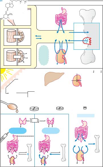
A. Calcium homeostasis of the body

Lüllmann, Color Atlas of Pharmacology © 2000 Thieme

All rights reserved. Usage subject to terms and conditions of license.

266 Antibacterial Drugs

Antibacterial Drugs

Drugs for Treating Bacterial Infections

When bacteria overcome the cutaneous or mucosal barriers and penetrate body tissues, a bacterial infection is present. Frequently the body succeeds in removing the invaders, without outward signs of disease, by mounting an immune response. If bacteria multiply faster than the body’s defenses can destroy them, infectious disease develops with inflammatory signs, e.g., purulent wound infection or urinary tract infection. Appropriate treatment employs substances that injure bacteria and thereby prevent their further multiplication, without harming cells of the host organism (1).

Apropos nomenclature: antibiotics are produced by microorganisms (fungi, bacteria) and are directed “against life” at any phylogenetic level (prokaryotes, eukaryotes). Chemotherapeutic agents originate from chemical synthesis. This distinction has been lost in current usage.

Specific damage to bacteria is particularly practicable when a substance interferes with a metabolic process that occurs in bacterial but not in host cells. Clearly this applies to inhibitors of cell wall synthesis, because human and animal cells lack a cell wall. The points of attack of antibacterial agents are schematically illustrated in a grossly simplified bacterial cell, as depicted in (2).

In the following sections, polymyxins and tyrothricin are not considered further. These polypeptide antibiotics enhance cell membrane permeability. Due to their poor tolerability, they are prescribed in humans only for topical use.

The effect of antibacterial drugs can be observed in vitro (3). Bacteria multiply in a growth medium under control conditions. If the medium contains an antibacterial drug, two results can be discerned: 1. bacteria are killed—bacte- ricidal effect; 2. bacteria survive, but do not multiply—bacteriostatic effect. Al- though variations may occur under therapeutic conditions, different drugs

can be classified according to their respective primary mode of action (color tone in 2 and 3).

When bacterial growth remains unaffected by an antibacterial drug, bacterial resistance is present. This may occur because of certain metabolic characteristics that confer a natural insensitivity to the drug on a particular strain of bacteria (natural resistance). Depending on whether a drug affects only a few or numerous types of bacteria, the terms narrow-spectrum (e.g., penicillin G) or broad-spectrum (e.g., tetracyclines) antibiotic are applied. Naturally susceptible bacterial strains can be transformed under the influence of antibacterial drugs into resistant ones (acquired resistance), when a random genetic alteration (mutation) gives rise to a resistant bacterium. Under the influence of the drug, the susceptible bacteria die off, whereas the mutant multiplies unimpeded. The more frequently a given drug is applied, the more probable the emergence of resistant strains (e.g., hospital strains with multiple resistance)!

Resistance can also be acquired when DNA responsible for nonsusceptibility (so-called resistance plasmid) is passed on from other resistant bacteria by conjugation or transduction.

Lüllmann, Color Atlas of Pharmacology © 2000 Thieme

All rights reserved. Usage subject to terms and conditions of license.


Antibacterial Drugs

267


Anti-


bacterial


Bacterial


drugs


invasion:


infection


Selective


antibacterial


Immune

toxicity


Body cells

Bacteria


1.

defenses


Penicillins

Bacitracin

Polymyxins


Cephalosporins

Vancomycin

Tyrothricin


Cell wall


DNA

RNA


Cell


Tetrahydro-


membrane


folate


Protein


synthesis


Sulfonamides

Rifampin


Tetracyclines


Bacterium

Trimethoprim


Aminoglycosides


Chloramphenicol


"Gyrase-inhibitors"


Erythromycin


Nitroimidazoles


Clindamycin


2.


1 day


Resistance


Antibiotic


Insensitive strain


Bactericidal


Bacteriostatic

Sensitive strain with

Selection


3.

resistant mutant


A. Principles of antibacterial therapy

Lüllmann, Color Atlas of Pharmacology © 2000 Thieme

All rights reserved. Usage subject to terms and conditions of license.

268 Antibacterial Drugs

Inhibitors of Cell Wall Synthesis

In most bacteria, a cell wall surrounds the cell like a rigid shell that protects against noxious outside influences and prevents rupture of the plasma membrane from a high internal osmotic pressure. The structural stability of the cell wall is due mainly to the murein (peptidoglycan) lattice. This consists of basic building blocks linked together to form a large macromolecule. Each basic unit contains the two linked aminosugars N-acetylglucosamine and N-acetyl- muramyl acid; the latter bears a peptide chain. The building blocks are synthesized in the bacterium, transported outward through the cell membrane, and assembled as illustrated schematically. The enzyme transpeptidase cross-links the peptide chains of adjacent aminosugar chains.

Inhibitors of cell wall synthesis are suitable antibacterial agents, because animal and human cells lack a cell wall. They exert a bactericidal action on growing or multiplying germs. Members of this class include !-lactam antibiotics such as the penicillins and cepha- losporins, in addition to bacitracin and vancomycin.

Penicillins (A). The parent substance of this group is penicillin G (benzylpenicillin). It is obtained from cultures of mold fungi, originally from Pen- icillium notatum. Penicillin G contains the basic structure common to all penicillins, 6-amino-penicillanic acid (p. 271, 6-APA), comprised of a thiazolidine and a 4-membered !-lactam ring. 6- APA itself lacks antibacterial activity. Penicillins disrupt cell wall synthesis by inhibiting transpeptidase. When bacteria are in their growth and replication phase, penicillins are bactericidal; due to cell wall defects, the bacteria swell and burst.

Penicillins are generally well tolerated; with penicillin G, the daily dose can range from approx. 0.6 g i.m. (= 106 international units, 1 Mega I.U.) to 60 g by infusion. The most important ad- verse effects are due to hypersensitivity

(incidence up to 5%), with manifestations ranging from skin eruptions to anaphylactic shock (in less than 0.05% of patients). Known penicillin allergy is a contraindication for these drugs. Because of an increased risk of sensitization, penicillins must not be used locally. Neurotoxic effects, mostly convulsions due to GABA antagonism, may occur if the brain is exposed to extremely high concentrations, e.g., after rapid i.v. injection of a large dose or intrathecal injection.

Penicillin G undergoes rapid renal elimination mainly in unchanged form

(plasma t1/2 ~ 0.5 h). The duration of the effect can be prolonged by:

1.Use of higher doses, enabling plasma levels to remain above the minimally effective antibacterial concentration;

2.Combination with probenecid. Re- nal elimination of penicillin occurs chiefly via the anion (acid)-secretory system of the proximal tubule (-COOH of 6-APA). The acid probenecid (p. 316) competes for this route and thus retards penicillin elimination;

3.Intramuscular administration in depot form. In its anionic form (-COO-) penicillin G forms poorly water-soluble salts with substances containing a positively charged amino group (procaine, p. 208; clemizole, an antihistamine; benzathine, dicationic). Depending on the substance, release of penicillin from the depot occurs over a variable interval.

Lüllmann, Color Atlas of Pharmacology © 2000 Thieme

All rights reserved. Usage subject to terms and conditions of license.


Antibacterial Drugs


269

Cell wall


Cell membrane


Bacterium


Cross-linked


by


transpeptidase

Inhibition of


Amino acid


chain


cell wall synthesis


Cell wall


Sugar


building block


Human


Antibody


Penicillin


allergy


Neurotoxicity


at very


Fungus


high dosage


Penicillium notatum


Plasma concentration


Penicillin


+


ocaine

-


Pr


3 x Dose


Penicillin


~1


Minimal


+


Clemizole


bactericidal


-


concentration


(d)


Probenecid


Penicillin


action


+


~2


+


-


Anion

BenzathinePenicillins


of


secretory


Duration


system


2


Time


~7-28


Increasing the dose

Combination with probenecid

Depot preparations

A.Penicillin G: structure and origin; mode of action of penicillins; methods for prolonging duration of action

Lüllmann, Color Atlas of Pharmacology © 2000 Thieme

All rights reserved. Usage subject to terms and conditions of license.

270 Antibacterial Drugs

Although very well tolerated, peni- cillin G has disadvantages (A) that limit its therapeutic usefulness: (1) It is inactivated by gastric acid, which cleaves the !-lactam ring, necessitating parenteral administration. (2) The !-lactam ring can also be opened by bacterial enzymes (!-lactamases); in particular, penicillinase, which can be produced by staphylococcal strains, renders them resistant to penicillin G. (3) The antibacterial spectrum is narrow; although it encompasses many gram-positive bacteria, gram-negative cocci, and spirochetes, many gram-negative pathogens are unaffected.

Derivatives with a different substituent on 6-APA possess advantages (B): (1) Acid resistance permits oral administration, provided that enteral absorption is possible. All derivatives shown in (B) can be given orally. Penicil- lin V (phenoxymethylpenicillin) exhibits antibacterial properties similar to those of penicillin G. (2) Due to their penicillinase resistance, isoxazolylpen- icillins (oxacillin dicloxacillin, flucloxacillin) are suitable for the (oral) treatment of infections caused by penicillinaseproducing staphylococci. (3) Extended activity spectrum: The aminopenicillin amoxicillin is active against many gramnegative organisms, e.g., coli bacteria or Salmonella typhi. It can be protected from destruction by penicillinase by combination with inhibitors of penicillinase (clavulanic acid, sulbactam, tazobactam).

The structurally close congener am- picillin (no 4-hydroxy group) has a similar activity spectrum. However, because it is poorly absorbed (<50%) and therefore causes more extensive damage to the gut microbial flora (side effect: diarrhea), it should be given only by injection.

A still broader spectrum (including Pseudomonas bacteria) is shown by car- boxypenicillins (carbenicillin, ticarcillin) and acylaminopenicillins (mezclocillin, azlocillin, piperacillin). These substances are neither acid stable nor penicillinase resistant.

Cephalosporins (C). These !-lac- tam antibiotics are also fungal products and have bactericidal activity due to in- hibition of transpeptidase. Their shared basic structure is 7-aminocepha- losporanic acid, as exemplified by cephalexin (gray rectangle). Cephalosporins are acid stable, but many are poorly absorbed. Because they must be given parenterally, most—including those with high activity—are used only in clinical settings. A few, e.g., cephalexin, are suitable for oral use. Cephalosporins are penicillinase-resistant, but cephalosporinase-forming organisms do exist. Some derivatives are, however, also resistant to this !-lactamase. Cephalosporins are broad-spectrum antibacterials. Newer derivatives (e.g., cefotaxime, cefmenoxin, cefoperazone, ceftriaxone, ceftazidime, moxalactam) are also effective against pathogens resistant to various other antibacterials. Cephalosporins are mostly well tolerated. All can cause allergic reactions, some also renal injury, alcohol intolerance, and bleeding (vitamin K antagonism).

Other inhibitors of cell wall synthesis. Bacitracin and vancomycin interfere with the transport of peptidoglycans through the cytoplasmic membrane and are active only against gram-positive bacteria. Bacitracin is a polypeptide mixture, markedly nephrotoxic and used only topically. Vancomy- cin is a glycopeptide and the drug of choice for the (oral) treatment of bowel inflammations occurring as a complication of antibiotic therapy (pseudomembranous enterocolitis caused by Clos- tridium difficile). It is not absorbed.

Lüllmann, Color Atlas of Pharmacology © 2000 Thieme

All rights reserved. Usage subject to terms and conditions of license.

Соседние файлы в папке книги студ