

книги студ / Color Atlas of Pathophysiology (S Silbernagl et al, Thieme 2000)
.pdfA. The Erythrocyte Parameters MCH, MCV, and MCHC
Centrifugation
Blood sample
a
b
b/a = hematocrit (Hct)
(LRBC/Lblood)
Hemoglobin (Hb) concentration (g/L blood)
Erythrocytes (RBC)
(number/L blood)
MCH (mean Hb mass/RBC)
= |
Hb conc. |
(g/RBC) |
|
|
No. RBC |
|
Normal: |
|
|
|
~30 pg |
|
|
|
|
MCV (mean volume of one RBC)
= |
Hct |
(L/RBC) |
|
|
No. RBC |
Normal: |
|||
|
|
|||
|
|
|
~90 fl |
|
|
|
|
|
MCHC (mean Hb conc. in RBCs)
= |
|
Hb conc. |
|
|
(g/L RBC) |
|
|
|||
|
|
|
Hct |
|
|
|
|
|
Normal: |
|
|
|
|
|
|
|
|
|
|
~320 g/L |
|
|
|
|
|
|
||||||
|
|
|
|
|
|
|
|
|
|
|
|
|
|
|
|
|
|
|
|
|
|
|
|
|
|
|
|
|
|
|
|
|
|
|
|
MCH |
/ |
MCV |
= |
MCHC |
|
||
|
|
|
|
|
|
|
|
|
|
|
|
|
|
|
|
|
|
|
|
|
|
|
|
|
|
|
|
|
|
|
|
|
|
|
|
|
|
|
Erythrocyte |
|
|
|
|
|
|
|
|
|
|
|
|
|
|
|
|
|
|
|
|
|
|
|
|
|
|
|
|
|
|
|
|
|
|
|
|
|
|
|
|
|
|
|
|
|
|
|
|
|
|
|
|
|
|
|
|
|
|
|
|
|
|
|
|
|
|
|
|
|
|
|
|
|
|
|
|
|
|
|
|
|
|
|
|
|
|
|
|
|
|
|
|
|
||
|
|
|
|
|
|
|
|
|
|
|
|
|
|
|
|
|
|
|
|
|
|
|
|
|
|
|
|
|||
|
|
|
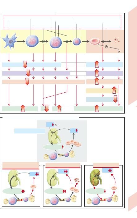
B. Forms of Anemia |
|
|
|
|
|
|
|
|
|
|
|
|
|
|
|
|
|
|
|
|
|
|
|
|
|||
|
|
|
|
|
|
|
|
|
|
|
|
|
|
|
|
|
|
|
|
|
|
|
|
|
|
|||||
|
|
|
|
|
|
|
|
|
|
|
|
|
|
|
|
|
|
|
|
|
|
|
|
|
|
|
|
|
|
|
|
|
|
Defect of |
|
|
|
|
|
|
|
|
|
|
|
|
|
|
|
|
|
|
|
|
|
|
|
|
Erythrocytes |
|
|
|
|
|
differentiation |
|
Virus infection |
|
|
Gene defect |
|
|
|
|
|
|
|
|
|
– defects |
|
|||||||||||
|
|
|
|
|
|
|
|
|
|
|
|
|
|
|
||||||||||||||||
|
|
|
|
|
|
|
|
|
|
|
|
|
|
|
|
|
|
|
|
|
Iron deficiency |
|
|
|
(membrane, |
|
||||
|
|
|
|
|
|
|
Autoimmune |
|
|
|
|
|
Folic acid |
|
|
|
|
|
|
|
|
metabolism) |
|
|||||||
|
|
|
Bone |
|
|
|
|
reaction |
|
|
|
|
|
deficiency |
|
|
|
Defect of |
|
|
– damage |
|
||||||||
|
|
|
marrow |
|
|
|
|
|
|
|
|
|
|
B12 deficiency |
|
globin synthesis |
|
|
(mechanical, |
|
||||||||||
|
|
|
|
|
|
|
|
Renal failure |
|
|
|
|
|
|
|
|
Defect of |
|
immunological, |
|
||||||||||
|
|
|
|
|
|
|
|
|
|
|
|
|
|
|
|
|
|
|
|
|
|
|||||||||
|
|
|
|
|
|
|
|
|
|
|
|
|
|
|
heme synthesis |
|
toxic) |
|
||||||||||||
|
|
|
|
|
|
|
|
|
|
|
|
|
|
|
|
|
|
|
|
|
|
|
|
|
||||||
|
|
|
|
|
|
|
|
|
|
|
|
|
|
|
|
|
|
|
|
|
|
|
|
|
|
|
|
– parasites |
|
|
|
|
|
|
|
|
|
|
|
|
|
|
|
|
|
|
|
|
|
|
|
|
|
|
|
|
|
|
|
||
|
|
|
|
|
|
|
|
|
|
|
|
|
|
|
|
|
|
|
|
|
|
|
|
|
|
|
|
(malaria etc.) |
|
|
|
|
|
|
|
|
|
|
|
|
|
|
|
|
|
|
|
|
|
|
|
|
|
|
|
|
|
|
|
||
|
|
|
|
|
|
|
|
|
|
|
|
|
|
|
|
|
|
|
|
|
|
|
|
|
|
|
|
Blood |
|
|
|
|
|
|
|
|
|
|
|
|
|
|
|
|
|
|
|
|
|
|
|
|
|
|
|
|
|
|
|
||
|
|
|
Stem cell |
|
|
|
|
|
|
|
|
|
|
|
|
|
|
|
|
|
|
|
|
|
|
|
|
|
|
|
|
|
|
|
Erythrocytic |
|
|
|
|
|
|
|
|
|
|
|
|
|
|
|
|
|
|
|
|
|
|||||
|
|
|
|
precursor |
|
|
|
|
|
|
|
|
|
|
|
|
|
|
|
|
|
|
|
|
|
|||||
|
|
|
Proerythroblast |
|
Panmyelo- |
|
|
|
Erythroblast |
|
|
|
|
|
pathy |
Aplastic |
|
|
Erythrocyte |
|
|
|
||
|
anemia |
Renal |
Megalo- |
Hemolysis |
|
|
anemia |
||
|
|
|
||
|
|
|
blastic |
Microcytic hypo- |
|
|
|
anemia |
|
|
|
|
chromic anemia |
|
|
|
|
|
|
|
|
|
|
Hemolytic anemia |
Silbernagl/Lang, Color Atlas of Pathophysiology © 2000 Thieme
All rights reserved. Usage subject to terms and conditions of license.
Plate 3.2 Erythrocytes; Erythropoiesis, Anemia
31

Erythrocyte Turnover: Abnormalities, Compensation, and Diagnosis
|
Proliferation and differentiation of the ery- |
|
throid precursor cells up to the mature eryth- |
|
rocytes takes barely a week. This time can be |
|
shortened to a few days if erythropoiesis is |
|
stimulated, for example, by an increase in cell |
|
loss (hemolysis or bleeding). As the average |
|
life-span of RBCs in peripheral blood is more |
|
than 100 days, a brief disorder of cell forma- |
|
tion is not detectable, but increased cell loss |
|
quickly results in anemia. (With neutrophil |
|
leukocytes, whose differentiation time is |
|
roughly as long, the reverse is the case, be- |
|
cause their life-span in peripheral blood is |
|
only about 10 hours: neutropenia occurs if |
|
there is an acute disorder of cell formation, |
|
but not after cell loss.) |
|
With a survival time of ca. 107 sec and a total |
Blood |
RBC count of ca. 1.6 × 10¹³ in blood, the rate |
second. If necessary, this production rate in- |
|
|
of formation is 1.6 million erythrocytes per |
3 |
creases up to tenfold without causing bone |
|
marrow exhaustion. Life-long hemolytic ane- |
|
mia, for example, can thus largely be compen- |
|
sated. |
|
Disorders of erythrocyte metabolism, be it |
abnormal erythropoiesis in its various steps (→ A), a shortened life-span, or chronic blood loss, can be differentiated by means of a number of diagnostic parameters:
Stem cells obtained by bone marrow puncture can be stimulated to proliferate and differentiate by erythropoietin in a cell culture. Colonies of more or less differentiated, hemoglo- bin-containing cells (E) are formed in this way (burst-forming units [BFU-E] or colony-forming units [CFU-E]). Their number is decreased if the anemia is caused by abnormal cell formation; it is increased if the cells are lost in a late stage of differentiation (erythroblast, erythrocyte) (→ A1).
Erythroblasts can be morphologically identified and quantified in a stained bone marrow sample. They decrease in number in aplasia and in defects of stem cell differentiation; they increase if erythropoiesis is stimulated, for example, by increased hemolysis (→ A2).
The efficiency of the entire erythropoiesis can be measured by determining the number of re-
32 ticulocytes (→ p. 30). If the number of reticulocytes is reduced, one must assume an abnor-
mality of cell formation (→ A3) because the second, theoretically possible cause, a prolongation of RBC life-span, does not occur. On the other hand, a longer lasting increase in reticulocyte numbers (reticulocytosis) is evidence for a chronically shortened life-span in the circulation on the part of the RBCs (chronic bleeding or hemolysis). Transitory reticulocytosis is a sign of stimulated erythropoiesis, for example, after acute blood loss, after acute hemolysis, or after correction of abnormal cell formation (with a high level of erythropoietin;
→ B2,3).
When erythrocytes are broken down in macrophages (→ p. 30), bilirubin, formed from liberated heme, is excreted in the bile after conjugation in the liver. The concentration of unconjugated (“indirect”) bilirubin in serum is increased in hemolysis (→ A4 and p.164ff.), but in some circumstances also if hemoglobin turnover is increased as a result of ineffective erythropoiesis.
The life-span of RBCs (shortened in hemolytic anemia; → A5) as well as their total volume can be measured by marking the erythrocytes in vitro with radioactive 51Cr (binding Cr to the Hb-β chain) and then re-infusing them. As 51Cr is released in hemolysis and then excreted by the kidneys, the erythrocyte life-span can be calculated from the loss of radioactivity measured daily. Total erythrocyte volume can be determined from the amount of 51Cr injected and the initial 51Cr concentration
in blood, using the principle of indicator dilution.
Measuring erythropoietin (→ A6). Lowered concentration of plasma erythropoietin suggests the anemia is caused nephrogenically (→ B4). However, most anemias are associated with a (compensatory) increase in erythropoietin concentration (→ B2,3).
Silbernagl/Lang, Color Atlas of Pathophysiology © 2000 Thieme
All rights reserved. Usage subject to terms and conditions of license.
A. Diagnostic Parameters in Anemia |
|
|
|
|
|
|
||
Defect of |
Virus infection |
Gene defect |
Iron deficiency |
|
|
Erythrocytes |
|
|
differentiation |
Autoimmune |
Folic acid deficiency |
Globin synthesis |
(defects, damage, |
|
|||
|
reaction |
B12 deficiency |
|
defect |
|
|
parasites) |
|
|
Renal |
|
|
Heme synthesis |
Chronic |
|
||
|
failure |
|
|
defect |
|
|
bleeding |
|
Bone marrow |
|
|
|
|
|
Blood |
|
|
|
|
|
|
|
|
|
Hemolysis |
|
Stem cell |
Erythrocytic |
Proerythroblast |
Erythroblast |
|
Erythrocyte |
Turnover |
||
precursor cell |
|
|||||||
|
|
|
|
|
|
Chronic |
||
|
|
|
|
|
|
|
||
|
|
|
|
|
|
|
blood loss |
|
BFU-E, CFU-E |
|
1 |
|
|
1 |
|
|
|
|
|
|
|
|
Erythrocyte |
|||
No. of erythroblasts |
2 |
|
|
2 |
|
|
||
No. of retikulocytes |
3 |
|
|
3 |
|
|
||
|
|
|
|
|
||||
|
|
|
|
|
4 |
Unconj. bilirubin |
3.3 |
|
|
|
|
|
|
Plate |
|||
|
|
|
|
|
5 |
|
|
|
|
|
|
|
|
Erythrocyte life-span |
|
||
Erythropoietin |
6 |
|
|
|
6 |
|
|
|
B. Erythropoietin Concentration as Anemia Indicator |
|
|||
|
PO2 |
e. g. |
|
|
|
when at high altitude |
|
||
1 Normal regulation cycle |
|
PO2 |
|
|
Kidney |
|
|
||
|
Erythropoietin |
|
||
|
|
Bone marrow |
|
|
2 Abnormality of erythrocyte |
3 Hemolysis, blood failure |
4 Renal failure |
||
or hemoglobin formation |
||||
|
|
|
||
|
|
PO2 |
PO2 |
|
PO2 |
|
|
||
|
Hemolysis |
|
||
|
|
|
||
|
|
Loss |
|
|
Erythropoietin |
Erythropoietin |
Erythropoietin |
||
Bone marrow |
|
|
33 |
|
|
|
|
||
Silbernagl/Lang, Color Atlas of Pathophysiology © 2000 Thieme
All rights reserved. Usage subject to terms and conditions of license.

3 Blood
34
Megaloblastic Anemia Due to Abnormalities in DNA Synthesis
Some acquired forms of anemia are due to abnormalities in the absorption or metabolism of folate or cobalamine (vit. B12) (→ A). The result is that DNA synthesis is inhibited and the cell cycle is slowed down during erythropoiesis. However, hemoglobin synthesis in the cytoplasm continues unchanged so that the erythroblasts increase in size (megaloblasts) and over-large, oval erythrocytes pass into the blood (megalocytes: MCV > 100 fL). The formation of granulocytes and megakaryocytes is also disturbed. In addition to the delay in proliferation, the anemia is aggravated by the premature destruction of megaloblasts in bone marrow (increased inefficient erythropoiesis;
→ p. 38) as well as by the shortened life-span of the megalocytes that have passed into the blood (premature hemolysis).
Folate. The folate metabolite N5, N10-methy- lene-tetrahydrofolate is necessary for the synthesis of deoxythymidylate (→ A3), the only source of thymine, which is in turn necessary for DNA synthesis. Thus, a folate deficiency inhibits DNA synthesis. This particularly affects the rate of formation of rapidly proliferating cells, for example, during erythropoiesis and tumor formation. The folate requirement for two to four months is stored in the liver. Folate is largely present in food in the form of pteroylpolyglutamate, from which excess glutamate residues must be split off before it can be absorbed in the form of pteroylmonoglutamate in the upper small intestine (→ A1). N5- methyltetrahydrofolate, the substrate for tetrahydrofolate formation (→ A2), is then formed in the intestinal mucosa. Methyl-co- balamine is essential for this step (see below). N5,N10-methyltetrahydrofolate is formed from tetrahydrofolate, the former together with deoxyuridylate being metabolized through the action of thymidylate synthase to deoxythymidylate and 7,8-dihydrofolate. Finally, the used up tetrahydrofolate is regenerated from 7,8-dihydrofolate (→ A3).
The following disorders of folate absorption or metabolism impair DNA synthesis, and thus erythropoiesis:
Too little folate uptake with food (< 50 µg/d; overcooking food destroys folate);
Increased requirement (pregnancy);
Malabsorption, for example, in diseases of the small intestine, or inhibition of the folate carrier caused by methotrexate (→ A1);
Cobalamine deficiency (→ A4);
Inhibition of thymidylate synthase by the fluorouracil metabolite fluordeoxyuridylate;
Inhibition of dihydrofolate reductase by aminopterin or methotrexate, whose affinity for the enzyme is 100 times that of the natural substrate 7,8-dihydrofolate (→ A3).
As inhibition of folate metabolism also retards tumor growth, the drugs fluorouracil, methotrexate, and aminopterin are used as cytostatic chemotherapeutics. Their side effect on erythropoiesis is usually undesirable and therefore often limits their dosage.
Cobalamine (vitamin B12) must be taken up by humans in their food (daily requirement: 3 –5 µg). About a thousand times this amount is stored in the liver. Bound to different proteins, it is transported inside the organism from food to the site of its action where, in the form of methylcobalamine, it serves as coenzyme in demethylating N5-methyltetrahydro- folate (→ A2). Among possible causes of cobal-
amine deficiency are (→ A4):
Too little uptake with food (e.g., a strict vegetarian diet);
Intrinsic factor (IF) deficiency (in atrophic gastritis etc.; see p.142): IF is essential for the binding and absorption of cobalamine. It is freed from its binding to salivary proteins in the lumen of the small intestine;
Competition for cobalamine and splitting of
IF from bacteria (blind-loop syndrome; → p.148), or broad fish tapeworms in the intestinal lumen;
Absence (congenital, after resection) or inflammation of the terminal ileum, i.e., at the site of absorption of cobalamine (→ p.152f.);
Defective transcobalamine II (TCII), which is responsible for cobalamine transport in plasma and for its uptake into cells.
Because of the great store of cobalamine in the liver, the symptoms of cobalamine deficiency (pernicious anemia, neurological abnormalities) occur only after years of blocked supply.
Silbernagl/Lang, Color Atlas of Pathophysiology © 2000 Thieme
All rights reserved. Usage subject to terms and conditions of license.
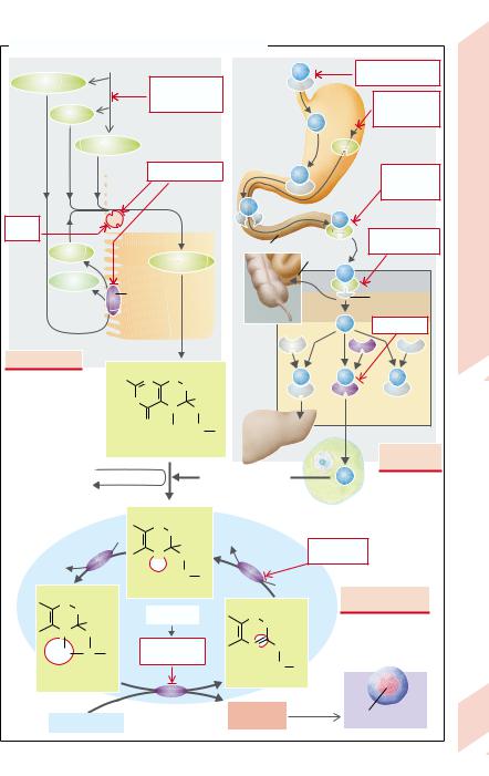
A. Anemias Caused by Disorders of DNA Synthesis |
|
|
|
|
|
|
||||||||||
|
|
|
Folate |
|
|
|
|
|
|
|
B12 |
B12 deficiency in |
|
|||
Folate–Glu–Glu... |
|
Folate deficiency |
Cobalamine |
food (< 3 |
g/d) |
|
||||||||||
|
|
|
|
|||||||||||||
|
|
|
|
|
|
|
||||||||||
|
|
|
|
|
|
|
in food |
(vitamin B12) |
|
Stomach |
Gastrectomy, |
|
||||
|
|
|
|
|
|
(< 50 g/d) |
|
|
|
|
||||||
|
Folate |
|
|
|
|
|
|
atrophic |
|
|||||||
|
|
|
|
|
|
|
|
|
|
|
|
|
||||
|
|
|
|
|
|
|
|
|
|
|
|
gastritis, etc. |
|
|||
|
|
|
|
|
|
|
|
|
|
|
|
|
|
|
||
|
|
Me–H4–folate |
|
|
|
|
|
|
|
|
IF |
|
Intrinsic |
4 |
|
|
|
|
|
|
|
|
|
|
|
|
|
|
|
|
factor |
|
|
|
|
|
|
Malabsorption |
|
|
|
|
|
|
Bacteria, |
Anemia |
||||
|
|
|
|
|
|
|
|
|
|
|
|
|
|
broad tape- |
||
|
|
|
|
|
Mucosal cell |
|
|
|
|
|
|
worm |
||||
|
|
|
|
|
(jejunum) |
|
|
|
|
|
|
|
|
|||
|
|
|
|
|
|
|
|
|
|
|
|
|
|
|
|
|
Metho- |
|
|
|
|
|
|
|
|
|
|
|
|
|
|
Megaloblastic |
|
trexate |
|
|
|
|
|
|
|
|
|
|
|
|
Gut resection, |
|||
|
Folate |
|
|
|
|
|
|
Duodenum |
|
Terminal |
|
ileitis |
|
|||
1 |
|
Me–H4–folate |
|
|
|
|
ileum |
|
|
|
||||||
|
|
|
|
|
|
|
|
|
|
|||||||
|
|
|
|
|
|
|
B12 |
|
|
|||||||
|
Glu–Glu... |
|
|
|
|
|
|
|
|
|
|
|
||||
|
|
|
|
|
|
|
|
|
|
|
|
|
|
|||
|
|
|
Pteroylpoly- |
|
|
|
|
|
|
|
|
|
|
|||
|
|
|
|
|
|
|
|
|
|
|
Receptor of |
|
||||
|
|
|
glutamate |
|
|
|
|
|
|
|
|
|
ileum mucosa |
3.4 |
||
|
|
|
hydrolase |
|
|
|
|
|
|
|
|
|
|
|
||
|
|
|
|
|
|
|
|
|
|
|
|
|
|
TC-II defect |
Plate |
|
|
|
|
|
|
|
|
|
|
|
TC III |
|
TC II |
TC I |
|||
|
|
|
|
|
|
|
|
|
|
|
|
|||||
Folate deficiency |
|
|
H |
|
|
|
|
|
|
|
|
|
|
|
||
|
|
|
|
N |
|
|
|
|
|
|
|
|
|
Plasma |
|
|
|
|
|
H2N |
N |
CH2 |
|
|
|
|
|
|
|
|
|||
|
|
|
|
|
|
|
|
|
|
|
|
|
|
|||
|
|
|
HN |
|
5 |
|
H |
|
|
|
|
|
|
|
|
|
|
|
|
|
|
|
|
|
|
|
|
Plasma |
|
||||
|
|
|
|
N |
|
CH2 |
|
|
|
|
|
|
|
|||
|
|
|
|
|
|
|
|
|
|
|
|
|
||||
|
|
|
|
O |
|
|
|
|
|
Liver |
|
|
‘store’ |
|
||
|
|
|
|
CH3 |
N |
R |
|
|
|
|
|
|
||||
|
|
|
|
|
|
Storage |
|
|
|
|
|
|||||
|
|
|
|
|
|
|
H |
|
|
|
|
|
|
|
||
|
|
|
|
|
|
|
|
(3 – 5 mg) and |
|
|
|
|
|
|||
|
|
|
N5-methyltetrahydrofolate |
|
|
Cobalamine |
|
|||||||||
|
|
|
|
|
|
|
|
|
|
excretion |
|
|
|
|||
|
Homocysteine |
|
|
|
|
|
|
|
|
|
|
|
deficiency |
|
||
|
|
|
|
|
Methylcobalamine |
|
B12 |
|
|
|
|
|||||
|
Methionine |
|
|
|
2 |
|
|
|
|
|
||||||
|
|
|
|
|
|
|
|
Cellular metabolism |
|
|
||||||
|
|
|
|
|
|
|
|
|
|
|
|
|
||||
|
|
|
|
H |
|
|
|
|
|
|
|
|
|
|
|
|
|
|
|
|
N |
CH2 |
|
|
|
|
|
|
|
|
|
|
|
|
|
|
Serine |
|
|
NADP |
|
|
|
|
|
|
|
|||
|
|
|
|
|
H |
|
|
|
Aminopterin, |
|
|
|
||||
|
|
|
|
|
|
|
|
|
|
|
|
|||||
|
|
|
|
N |
|
|
|
Dihydrofolate |
|
|
|
|||||
|
|
|
|
|
CH2 |
|
methotrexate |
|
|
|
||||||
|
|
|
|
|
|
reductase |
|
|
|
|
||||||
|
Glycine |
|
|
H |
|
|
|
|
|
|
|
|
|
|
||
|
|
|
|
HN |
R |
|
|
|
|
|
|
|
|
|
||
|
|
|
|
|
|
|
NADPH + H+ |
|
|
|
|
|||||
|
H |
|
|
Tetrahydrofolate |
|
|
Impaired folate |
|
||||||||
|
|
|
|
|
|
|
|
|
||||||||
|
N |
CH2 |
3 |
|
|
|
|
|
H |
|
|
|
metabolism |
|
||
|
|
Fluoruracil |
|
N |
|
|
|
|
|
|
|
|||||
|
|
H |
|
7CH2 |
|
|
|
|
|
|
||||||
|
5 |
|
|
|
|
|
|
8 |
|
|
|
|
|
|
||
|
N |
CH2 |
|
|
|
|
|
|
|
|
|
|
|
|
|
|
|
H2C |
10 |
|
Fluordesoxy- |
|
N |
CH2 |
|
|
|
|
|
|
|||
|
N |
R |
uridylate |
|
|
|
HN |
R |
|
|
Erythroblast |
|
||||
|
N5, N10-Methylen- |
|
|
|
|
|
|
|
|
|
||||||
|
|
|
|
|
|
|
|
|
|
|
|
|||||
|
tetrahydrofolate |
|
|
|
|
|
7,8-Dihydrofolate |
|
|
|
|
|
||||
|
|
|
|
|
|
|
|
|
|
|
|
|
|
|||
|
|
|
|
Thymidylate |
|
Desoxy- |
|
|
|
|
|
|
||||
|
|
|
|
synthase |
|
|
|
|
DNA synthesis |
|
||||||
|
Desoxyuridylate |
|
|
thymidylate |
|
|
35 |
|||||||||
|
|
|
|
|
|
|
|
|||||||||
Silbernagl/Lang, Color Atlas of Pathophysiology © 2000 Thieme
All rights reserved. Usage subject to terms and conditions of license.

Anemias Due to Disorders of Hemoglobin Synthesis
Erythrocytes (RBCs) serve to transport O2 and CO2 and also as a buffer. Hemoglobin (Hb) is essential for all three functions. It is composed of
|
four subunits (2α, 2β in HbA; see below), each |
||||
|
of which is formed from three components: |
||||
|
protoporphyrin, iron (Fe2+) and globin (α or β). |
||||
|
When Fe2+ is built into protoporphyrin, heme |
||||
|
is formed. If there is a deficiency or defect in |
||||
|
one of the components, Hb synthesis is im- |
||||
|
paired. In this case the RBCs are usually small |
||||
|
(MCV ↓) |
and |
their |
Hb content |
decreased |
|
(MCH ↓) (microcytic hypochromic anemia). |
||||
|
Disorders of protoporphyrin synthesis are |
||||
|
due to inherited enzyme defects (→ p. 254), as |
||||
|
for example, in hereditary sideroblastic anemia, |
||||
|
in which the formation of δ-aminolevulinic |
||||
|
acid (δ-ALA) from glycine and succinyl-CoA is |
||||
Blood |
reduced, |
and |
thus |
also heme |
synthesis |
(→ A1). Heme inhibits δ-ALA synthase in a |
|||||
negative feedback loop. If heme concentration |
|||||
3 |
is now reduced, inhibition of the enzyme is re- |
||||
|
versed and, despite the defect, sufficient |
||||
|
amounts of heme are formed. Defects in sub- |
||||
|
sequent enzymes lead to an increase in the |
||||
concentration of intermediary products. While the rate of heme production is thus increased, these metabolites cause other disorders, namely porphyrias (→ p. 254).
Disorders of globin synthesis. Normally Hb is made up of 2 α chains of 141 amino acids each and 2 β chains of 146 amino acids (HbA1 = HbAα2β2). Only 2– 3% of Hb contains so-called δ-chains (HbA2 = Hbα2δ2) instead of the β-chains. Before birth a form of Hb is formed that has a higher O2 affinity (adaptation to a lower Po2 in the placenta). This fetal Hb (HbF) contains so-called γ-chains (Hbα2γ2) instead of the β-chains.
The properties of Hb (solubility, O2 affinity, oxidizability, etc.) are dependent upon the particular amino acid sequence. However, most of the over 300 genetically-determined Hb variants which have been indentified so far do not signficantly impair function. On the other hand, even a single “false” amino acid (valine instead of glutamate in position 6 in the β- chain = HbS; → A2) can lead to extensive functional disorders, as seen in sickle cell anemia,
36 which is caused by a homozygous gene defect. In the deoxygenated form, HbS aggregates in a
way that results in sickle-shaped erythrocytes (→ A). These sickle cells cannot be further deformed and get stuck inside the capillaries, causing occlusion of smaller blood vessels. Aggregation of HbS takes a few minutes so that it is especially those capillaries through which the blood flows slowly which are affected (spleen; vasa recta of the renal medulla; → p.106). If blood flow is slowed in general (shock) or if hypoxia occurs (at high altitude, during a flight, anesthesia), the abnormalities can spread to other organs (e.g., to the heart). Occlusion of the blood vessels further slows down blood supply in the affected regions and the Po2 is further reduced, so that a vicious circle results (crisis). Sickle cell anemia occurs nearly exclusively in blacks who themselves, or whose forbears, come from regions of Central Africa with a high prevalence of malaria. “Survival” of the defective gene in 40% of the population in Central Africa, despite the fact that until recently the disease was fatal in homozygous children, can be explained by the fact that heterozygous gene carriers are protected against the dangerous forms of malaria (selective advantage).
In β-thalassemia the production of β-chains is restricted, thus leading to a deficiency of HbA. It can be only partly compensated by an increased production of HbA2 and HbF. The incorporation of Fe2+ is diminished so that it remains in the erythrocytes (sideroachresia) and may accumulate excessively in the body (secondary hemochromatosis; → p. 252). Although the RBCs’ osmotic resistance (→ p. 40) is actually increased, their mechanical vulnerability is increased (rapid breakdown in the spleen, early hemolysis). While the heterozygous form (T. minor) causes few symptoms, the homozygous form (T. major) may be fatal even before puberty. The rare α-thalassemia usually causes death of the fetus, because without α- chains no HbF can be formed either. Hbγ4, produced in the fetus, and Hbβ4, occurring postnatally, are apparently inadequate substitutes for the normal Hb forms.
Silbernagl/Lang, Color Atlas of Pathophysiology © 2000 Thieme
All rights reserved. Usage subject to terms and conditions of license.
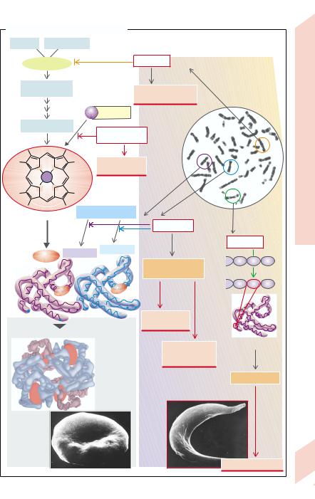
A. Defects of Hemoglobin Synthesis |
|
|
|
|
Synthesis |
||||
Glycine |
Succinyl-CoA |
|
|
|
|
|
|||
α-ALA synthase |
|
|
Gene defect |
|
|
|
|||
1 |
|
|
|
|
|
|
|
|
Hemoglobin |
δ-Aminolevulinic |
|
|
|
|
|
|
|||
acid (δ-ALA) |
|
|
|
Hereditary sidero- |
|
|
|
||
|
|
|
|
|
blastic anemia |
|
|
|
|
|
|
|
Fe |
Transferrin |
|
|
|
|
|
|
|
|
|
|
|
|
of |
||
|
|
|
|
|
|
|
|
|
|
Protoporphyrine |
|
Iron deficiency etc. |
|
|
|
Disorders |
|||
|
|
|
|
|
|
|
|||
|
|
|
|
(see next page) |
|
|
|
||
|
|
|
|
|
|
|
|
|
|
N |
N |
|
|
Iron deficiency |
|
|
|
Anemias: |
|
|
|
anemia |
|
|
|
|
|||
Fe |
|
|
|
|
|
|
|||
|
|
|
|
|
|
|
|||
N |
N |
|
|
|
|
|
|
|
|
|
|
|
|
|
|
|
|
||
|
|
|
|
|
|
|
|
|
3.5 |
Heme |
|
Globin synthesis |
|
|
|
|
Plate |
||
|
|
|
|
|
|
|
|||
|
|
|
|
|
Gene defects |
|
|
|
|
|
|
|
|
|
|
|
|
|
|
|
|
|
|
2 α-chains |
|
Gene defect |
|
|
|
Heme |
2 β-chains |
|
|
|
|
|
|||
|
|
5 |
Glu |
7 |
|
||||
|
|
|
|
|
|||||
|
|
|
|
|
Hemoglobin A |
|
|||
|
|
|
|
|
2 |
|
|
|
|
|
|
|
|
|
deficiency |
|
|
|
|
|
|
|
|
|
|
5 |
Val |
7 |
|
1 |
|
146 |
1 |
141 |
α-Thalassemia |
|
|
|
|
|
|
|
|
|
|
||||
|
|
|
|
|
|
|
|
||
|
|
|
|
|
(with Hbγ4; Hbβ4) |
6 |
|
|
|
Hemoglobin A1 (= Hbα2β2) |
|
|
|
|
|
|
|||
β |
|
|
α |
|
β-Thalassemia |
False β-chain |
|
||
|
|
|
|
|
|
|
|||
|
|
|
|
|
(with Hb F = Hbα2γ2 |
|
|
|
|
|
|
|
|
|
and Hb A2 = Hbα2δ2) |
|
|
|
|
|
|
|
|
|
|
Hemoglobin S |
|
||
α |
|
|
β |
|
|
|
|
|
|
|
|
|
|
|
|
|
|
|
|
|
|
|
|
|
|
Sickle |
|
|
|
|
|
|
|
|
|
erythrocyte |
|
|
|
Normal |
|
|
|
|
|
|
|
|
|
erythrocyte |
|
|
|
|
|
Sickle-cell anemia |
37 |
||
|
|
|
|
|
|
||||
Silbernagl/Lang, Color Atlas of Pathophysiology © 2000 Thieme
All rights reserved. Usage subject to terms and conditions of license.

3 Blood
Iron Deficiency Anemias
Of the iron (Fe) content in the body (2 g in females, 5 g in males) ca. 2⁄3 is bound to hemoglobin (Hb), 1⁄4 is stored iron (ferritin, hemosiderin), the rest is iron with diverse functions (myoglobin, Fe-containing enzymes). Loss of iron is ca. 1 mg/d in males and up to 2 mg/d in females (menstruation, pregnancy, birth). Of Fe taken up in food, 3– 15% is absorbed in the duodenum (→ A); in cases of Fe deficiency it can be up to 25% (see below). Iron intake with food should therefore be at least 10– 20 mg/d (women > children > men).
Iron absorption (→ A1). Fe can be absorbed relatively efficiently as heme-Fe2+ (found in meat and fish). The Fe (split off from heme) gets into the blood or remains in the mucosa as ferritin-Fe3+ and returns to the lumen on mucosal cell disintegration. Non-heme Fe can be absorbed only in the form of Fe2+, which is absorbed by a Fe2+-H+-symport carrier (DCT1) (in competition with Mn2+, Co2+, Cd2+, etc.). A low pH of the chyme is essential for absorption, because it will 1) increase the H+ gradient that drives Fe2+ into the cell via DCT1, and 2) release Fe from compounds in food. Non-heme Fe3+ in food must be reduced by ferrireductase (+ascorbate) to Fe2+ on the surface of the luminal mucosa (→ A1, FR). Fe uptake by blood is regulated by the intestinal mucosa: in Fe deficiency mucosal ferritin translation is inhibited by binding the Fe-regulating protein IRP1 to ferritin-mRNA, so that more of the absorbed Fe2+ can reach the blood. There it is oxidized by ceruloplasmin (+copper) to Fe3+ and bound to apotransferrin, which transports Fe in plasma (→ A). Transferrin (= apotransferrin with 2 Fe3+) is taken up, via transferrin receptors, endocytotically in erythroblasts and in hepatic, placental, and other cells. After Fe has transferred to the target cells, apotransferrin again becomes available for Fe absorption from the intestine and macrophages (see below).
Iron storage (→ A2). Ferritin (in the intestinal mucosa, liver, bone marrow, erythrocytes,
and plasma), which has a “pocket” for 4500 Fe3+ ions, is a rapidly available iron reserve (ca. 600 mg), while Fe from hemosiderin is more difficult to mobilize (250 mg Fe in macrophages from liver and bone marrow). Hb-Fe and heme-Fe, released from malformed erythroblasts (so-called inefficient erythropoiesis) and hemolyzed erythroblasts, is bound to haptoglobin and hemopexin respectively, and taken up by the macrophages in bone marrow or by liver and spleen by endocytosis, 97% being reused.
Iron deficiency (serum Fe < 0.4 mg/L; serum ferritin ↓) inhibits Hb synthesis (→ p. 36) so that hypochromic microcytic anemia develops: MCH < 26 pg, MCV < 70 fL, Hb < 110 g/L. Its causes are (→ A and Table):
Blood loss (gastrointestinal tract, increased menstrual bleeding) in adults is the most common cause of iron deficiency (0.5 mg Fe lost with each mL of blood).
Fe recycling is decreased; this form of anemia (the second most common worldwide) occurs with chronic infections. In this situation the Fe regained by the macrophages is no longer adequately released and thus cannot be reused.
Fe uptake is too low (malnutrition, especially in the developing countries).
Fe absorption is reduced due to: 1) achlorhydria (atrophic gastritis, after gastrectomy; → p.142, 148); and 2) malabsorption in diseases of the upper small intestine or in the presence of Fe-binding food components (phytate in cereals and vegetables; tannic acid in tea, oxalates, etc.).
There is increased Fe requirement (growth, pregnancy, breast-feeding).
An apotransferrin defect (rare).
If Fe overloading occurs in the body, damage
is caused mainly to the liver, pancreas and myocardium (hemochromatosis) (→ p. 252).
|
|
Normal |
Fe deficiency Apotrans- |
Fe utilization |
Fe recycling |
|
|
|
|
|
ferrin defect |
defect |
defect |
|
|
|
|
|
|
|
38 |
Serum Fe : Fe binding capacity |
1 mg/L:3.3 mg/L |
↓ : ↑ |
↓ : ↓ |
↑: normal |
↓ : ↓ |
|
Transferrin saturation |
ca. 33% |
< 10% |
0 |
> 50% |
> 10% |
|
|
|
|
|
|
|
Silbernagl/Lang, Color Atlas of Pathophysiology © 2000 Thieme
All rights reserved. Usage subject to terms and conditions of license.
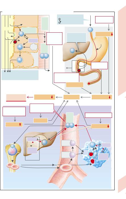
A. Iron (Fe) Deficiency Inhibits Hemoglobin Synthesis |
|
|
|
||||||||||
Lumen |
|
|
|
Mucosal cells |
Blood Apotransferrin |
Normal Fe uptake: |
|
Malnutrition |
|
||||
|
Heme |
(duodenum) |
|
10 – 20 mg/d |
|
|
|||||||
|
|
|
etc. |
|
|||||||||
|
Fe2+ |
Heme |
|
5 –10 mg/d |
|
|
|||||||
2+ |
Fe |
3+ |
|
Mucosal |
|
|
|
|
Fe |
|
|
||
Fe |
|
FR |
|
|
|
|
|
|
|
||||
|
|
|
transferrin |
|
|
|
|
|
|
|
|
||
|
Fe2+ |
Fe |
|
Transferrin |
|
Fe absorption: |
|
Fe uptake |
|
||||
|
|
|
deficiency, |
|
|
|
|
||||||
|
|
|
|
|
|
|
3 –15% of |
|
|
|
|||
|
|
|
|
|
Ferritin |
transferrin |
|
Fe uptake |
|
|
Anemias |
||
|
|
H+ |
|
|
defect |
|
|
|
|
|
HCI |
||
|
|
3+ |
Lyso- |
Transferrin |
|
|
|
|
|
|
|||
|
|
|
|
Fe |
some |
|
|
|
|
|
|
||
|
|
|
|
|
|
|
|
Fe |
|
|
|||
|
|
|
|
|
|
|
|
|
|
Stomach |
|||
|
|
|
|
|
|
|
|
|
|
|
|||
|
|
|
|
|
Cell |
Fe3+ Fe3+ |
|
|
|
|
|
Deficiency |
|
|
|
|
|
|
|
Liver |
|
|
|
|
|||
|
|
|
|
Fe3+ |
turnover |
|
|
|
|
|
|
||
|
|
|
|
|
|
|
|
|
|
|
|
||
|
|
|
|
|
|
|
|
|
|
|
Achlorhydria, |
||
|
|
|
|
|
|
|
|
|
|
|
gastrectomy |
||
|
Nonabsorbed Fe in stool |
To liver |
Disease in upper |
|
|
Iron |
|||||||
|
|
|
|
|
|||||||||
|
normal: 85 – 97 % of uptake |
|
|
small intestine, |
|
|
3.6 |
||||||
|
|
|
|
|
1 |
|
Fe-binding food |
|
|
||||
|
|
|
|
|
|
|
Malabsorption |
|
|
Plate |
|||
|
|
|
|
|
Absorption |
|
|
|
|||||
|
|
|
|
|
|
|
|
|
|
|
|
|
|
|
|
Fe deficiency |
Hb synthesis |
|
Fe deficiency |
Fe absorption |
|
||||||
|
|
|
anemia |
|
|
|
|
|
|
|
|
|
|
|
|
|
|
|
Blood loss |
|
|
|
|
|
|
|
|
|
|
|
Growth, |
(GI-tract, |
|
|
|
|
|
|
|
||
|
|
pregnancy, |
menstruation) |
|
|
|
|
|
|
|
|||
|
|
breast feeding |
|
|
|
- |
|
Chronic infections |
|
||||
|
|
|
|
|
|
|
|
Trans ferrin |
|
||||
|
|
|
|
|
Fe loss |
|
|
|
|
||||
|
|
|
|
|
|
|
|
|
|
|
|
||
|
|
Fe demand |
|
|
|
|
|
Fe |
Fe recycling |
|
|||
|
|
|
|
|
|
|
|
|
|
|
|
||
|
|
|
|
|
Liver |
|
|
Systemic |
|
|
|
||
|
|
|
|
|
|
|
|
|
|
|
|||
Bone |
|
|
Ferritin |
|
|
blood |
|
|
|
||||
|
|
Fe |
|
Hemo- Heme |
|
|
|
||||||
marrow |
|
|
|
|
|
|
|||||||
|
|
|
|
|
|
|
|
pexin |
|
|
Fe |
Ferritin |
|
|
|
|
|
|
Hemo- |
|
|
|
|
|
|
|
|
|
|
|
|
|
siderin |
|
|
Hapto- |
Hb |
|
|
|
|
|
|
|
|
|
|
|
|
|
Hemo- |
|
|||
|
|
|
|
|
|
|
|
globin |
|
|
|
||
|
|
|
|
|
Fe storage |
|
|
|
|
|
siderin |
|
|
|
|
|
|
|
|
|
|
|
|
|
|
||
|
|
|
|
|
Already in bone marrow |
|
Ery- |
|
|
|
|
|
|
|
|
|
|
|
|
|
|
|
|
|
|
|
|
|
|
|
|
|
|
|
|
throcytes |
Macrophages |
|
|||
|
|
|
|
|
|
|
|
|
|
|
in spleen, liver |
|
|
|
|
|
|
|
|
|
|
|
|
|
and bone marrow |
|
|
|
|
|
|
2 |
|
|
|
|
|
|
(extravasal) |
|
|
|
|
|
|
Storage, loss |
|
|
|
|
|
|
|
|
|
|
|
|
|
and recycling |
|
|
|
|
|
|
|
39 |
|
|
|
|
|
|
|
|
|
|
|
|
|
|
|
Silbernagl/Lang, Color Atlas of Pathophysiology © 2000 Thieme
All rights reserved. Usage subject to terms and conditions of license.

3 Blood
40
Hemolytic Anemias
Erythrocytes can only attain their normal lifespan when their flexibility, their ability to withstand osmotic and mechanical stress, their reductive potential, and their energy supply are normal (→ p. 30). Defects in these properties lead to a shorter life-span (in some cases to just a few days [corpuscular hemolytic anemia]). There are, however, many other causes that shorten the life-span of normal erythrocytes (extracorpuscular hemolytic anemia). A common feature of these anemias is an increased concentration of erythropoietin, which provides compensatory stimulation of erythropoiesis (→ p. 33, A and B3).
Causes of corpuscular hemolytic anemia
(→ A) are usually genetic defects:
One of the membrane diseases is hereditary spherocytosis (spherocyte anemia). It is caused by a functional abnormality (defective ankyrin) or deficiency of spectrin, which, as an important constituent of the cytoskeleton, is essential for its stability (→ A1). The volume of spherocytes is normal, but the defect in the cytoskeleton results in erythrocytes being spherical, instead of having a normal flexible discoid shape. The osmotic resistance of these cells is reduced, i.e., they hemolyse when the hypotonicity of the external medium is still low. As they are prematurely segregated in the spleen, splenectomy is therefore therapeutically effective.
Enzyme defects disturb the glucose metabo-
lism of erythrocytes (→ A2): 1) if pyruvate kinase is affected, ATP to Na+-K+-ATPase supply is stopped, the cells swell up so that they become vulnerable and hemolyse early; 2) defective glucose-6-phosphate dehydrogenase (gluc- 6-PDH; → A3) slows the pentose phosphate cycle, so that oxidized glutathione (GSSG), formed under oxidative stress, can no longer be adequately regenerated to the reduced form (GSH). As a result, free SH groups of enzymes and membrane proteins as well as phospholipids are no longer sufficiently protected against oxidation, leading to premature hemolysis. Eating horsebeans (vicia faba major, causing favism) or certain drugs (e.g., primaquin or sul-
fonamides) increase oxidative stress and thus aggravate the situation; 3) a defect of hexokinase results in a deficiency of both ATP and GSH (→ A2,3).
Sickle cell anemia and thalassemias (→ p. 36) also have a hemolytic component (→ A4).
In (acquired) paroxysmal nocturnal hemoglobinuria (PNH) some of the erythrocytes (derived from somatically mutated stem cells) have increased complement sensitivity. It is based on a defect of certain membrane proteins that are involved in regulating the complement system (especially the decay accelerating factor [DAF]; → A5). Complement activation then leads to perforation of the erythrocyte membrane. It is not clear why this usually occurs during sleep.
Examples of the causes of extracorpuscular hemolytic anemia are:
Mechanical causes, such as damage to the erythrocytes by collision with artificial heart valves or vascular prostheses, especially if cardiac output (CO) is raised;
Immunological causes, for example, in ABO blood group transfusion mismatches, or Rh incompatibility between mother and fetus;
Toxins, for example, certain snake poisons. In most hemolytic anemias the erythrocytes
will, as would occur normally, be phagocytized and “digested” in bone marrow, the spleen and liver (extravascular hemolysis), and Fe is reused (→ p. 38). A small amount of Hb released intravascularly is bound to haptoglobin (→ p. 38). In massive acute intravascular hemolysis
(→ B) haptoglobin is, however, overloaded and free Hb is filtered in the kidneys. This results not only in hemoglobinuria, but can also through tubular occlusion lead to acute renal failure (→ p.108). Chronic hemoglobinuria additionally causes Fe deficiency anemia, cardiac output rises and the resulting mechanical hemolysis creates a vicious circle (→ B). Finally, the erythrocytic fragments produced in intravascular hemolysis may cause thrombi and emboli, which can result in ischemia in the brain, cardiac muscle, kidneys, and other organs.
Silbernagl/Lang, Color Atlas of Pathophysiology © 2000 Thieme
All rights reserved. Usage subject to terms and conditions of license.