

биохимия атеросклероза
.pdf472 P.K. Shah and Behrooz Sharifi
148.Shah PK, Yano J, Reyes O, et al: High-dose recombinant apolipoprotein A-I [Milano] mobilizes tissue cholesterol and rapidly reduces plaque lipid and macrophage content in apolipoprotein E-deficient mice: potential implications for acute plaque stabilization. Circulation 103: 3047–3050, 2001.
149.Crisby M, Nordin-Fredriksson G, Shah PK, Yano J, Zhu J, Nilsson J: Pravastatin treatment increases collagen content and decreases lipid content, inflammation, metalloproteinases, and cell death in human carotid plaques: implications for plaque stabilization. Circulation 103: 926–933, 2001.
150.Shah PK, Kaul S, Nilsson J, Cercek B: Exploiting the vascular protective effects of high-density lipoprotein and its apolipoproteins: an idea whose time for testing is coming, part I. Circulation 104: 2376–2383, 2001.
151.Shah PK, Kaul S, Nilsson J, Cercek B: Exploiting the vascular protective effects of high-density lipoprotein and its apolipoproteins: an idea whose time for testing is coming, part II. Circulation 104: 2498–2502, 2001.
152.Forrester J, Makkar R, Shah PK: Increasing HDL cholesterol in dyslipidemia by cholesterol ester transfer protein inhibition: an update for clinicians. Circulation 111: 1847–1854, 2005.
153.Li X, Chyu KY, Neto JR, Yano J, Nathwani N, Ferreira C, Dimayuga PC, Cercek B, Kaul S, Shah PK: Differential effects of apolipoprotein A-I-mimetic peptide on evolving and established atherosclerosis in apolipoprotein E-null mice. Circulation 110(12): 1701–1705, 2004.
154.Claudel T, Leibowitz MD, Fievet C, et al: Reduction of atherosclerosis in apolipoprotein E knockout mice by activation of the retinoid X receptor. Proc Natl Acad Sci U S A 98: 2610–2615, 2001.
155.Kirkhamm TC, Williams CM: Endocannabinoid receptor antagonists: potential for obesity treatment. Treat Endocrinol 3(6): 345–360, 2004.
Section V
Management of Atherosclerosis
Biochemistry of Atherosclerosis edited by S.K. Cheema, Springer, New York, 2006
22
Modification of Biochemical and Cellular Processes in the Development of Atherosclerosis by Red Wine
HARJOT K. SAINI, PARAMBIR DHAMI, YAN-JUN XU, SUKHINDER KAUR CHEEMA, AMARJIT S. ARNEJA, AND NARANJAN S. DHALLA
Abstract
Atherosclerosis is commonly associated with unstable angina and acute myocardial infarction. It occurs as a result of a cascade of events caused by various environmental, dietary, genetic, and inflammatory factors. Different epidemiological studies have suggested that moderate amounts of red wine consumption reduce the risk of complications associated with atherosclerosis. This contention is further supported by a variety of experimental investigations demonstrating that both alcoholic and phenolic components are responsible for the apparent protective effects of red wine. The maintenance of endothelial function, augmentation in the levels of high-density lipoproteins (HDLs), prevention of low-density lipoprotein (LDL) oxidation, attenuation of smooth muscle proliferation and migration, inhibition of platelet aggregation and adhesion, as well as reduction in inflammatory mediators are major mechanisms linked with the protective effects of red wine. Despite these beneficial effects, insufficient information is available to recommend red wine as a therapeutic strategy to prevent atherosclerosis. Particularly, in view of high alcoholic content, excessive consumption of red wine can be seen to produce harmful effects. Therefore, a large-scale clinical trial is needed to determine the exact amount of red wine required for the beneficial effects and to categorize it as a future antiatherosclerotic agent.
Keywords: endothelial function; inflammation; LDL oxidation; lipoproteins; nitric oxide; red wine; thrombosis; vascular smooth muscle cells
Abbreviations: CAD, coronary artery disease; eNOS, endothelial NO synthase; ET-1, endothelin-1; HDL, high-density lipoprotein; hs-CRP, high-sensitivity C-reactive protein; HUVEC, human umbilical vein endothelial cells; ICAM, intercellular adhesion molecule; IL, interleukin; iNOS, inducible NO synthase; LDL, low-density lipoprotein; MCP, monocyte chemoattractant protein; NFkB, nuclear factor kappa B; NO, nitric oxide; Ox-LDL, oxidized LDL; PDGF, platelet-derived growth factor; SMC, smooth muscle cell; TNF, tumor necrosis factor; VCAM, vascular cell adhesion molecule; VSMC, vascular smooth muscle cell
475
476 Harjot K. Saini et al.
Introduction
Atherosclerosis, manifested by coronary artery disease (CAD), peripheral arterial disease, and cerebrovascular disease, is the major problem of global concern [1]. Multiple Risk Factor Intervention Trial and Framingham Studies have identified that hypercholesterolemia, hypertension, diabetes, as well as lifestyle factors such as stress, physical inactivity, and cigarette smoking are the major risk factors associated with the development of atherosclerosis [2, 3]. Over the past decades, primary and secondary prevention trials with hydroxyl-methyl glutaryl coenzyme A (HMG-CoA) inhibitors/statins have supported the contention that lowering low-density lipoprotein (LDL) cholesterol is the primary target for the prevention of atherosclerotic disease development [4]. However, a significant gap exists between the current statin therapy and the third report of the National Cholesterol Education Program (NCEP) Expert Panel on Detection, Evaluation, and Treatment of High Blood Cholesterol in Adults (Adult Treatment Panel III) guidelines [5]. It has been suggested that treatment goal for aggressive lipid lowering for LDL for high-risk patients of CAD should be <100 mg/dL [5]. To achieve this goal, higher doses of statins are needed; however, such higher doses have greater risk of adverse effects and each two-, three-, and fourfold increase causes 6%, 12%, and 18% decrease (only a minor change) in LDL concentration, respectively [6]. In addition, for every 1% fall in serum LDL level only 2% reduction in the incidence of CAD has been reported [7]. Because of the pleiotropic effects of statins (beyond LDL lowering) including the maintenance of endothelial function, attenuation of smooth muscle cell (SMC) proliferation, reduction of inflammatory response, stabilization of plaque and prevention of oxidative stress [8], the relationship between reducing cholesterol and atherosclerotic plaque development remains questionable. Therefore, new treatment strategies, especially the modification of lifestyle factors are considered necessary, which can be opted in combination with the moderate doses of statins to prevent the development and progression of the disease.
Epidemiological studies have demonstrated that despite the consumption of high saturated fat intake, the prevalence of death due to CAD in France is relatively less as compared to USA and other Western countries [9]. An inverse relationship between intake of red wine and mortality due to CAD has been revealed as the basis for the French paradox [9]. In addition, a significant reduction (70%) in the risk of newer coronary events has been reported by the intake of Mediterranean diet, which includes moderate consumption of red wine [10]. Since red wine is a combination of ethanol and other polyphenolic substances (Table 22.1) [11, 12], the participation of these individual components in the prevention of atherosclerotic plaque development is not completely elucidated. Accordingly, this review has attempted to describe the effects of red wine on the modification of biochemical and cellular events occurring during the disease process. In addition, the pathophysiology of atherosclerosis has been discussed to have a

Chapter 22. Modification of Biochemical and Cellular Processes |
477 |
TABLE 22.1. Different components of red wine.
Components
Ethanol
Polyphenolic substances
Flavonoids
Flavonols (Myricetin, Quercetin, Kaempferol)
Flavan-3-ols (Tannins, Proanthocyanidines, Gallocatechin, Catechin, Epicatechin) Anthocyanins (Cyanidin, Malvidin)
Nonflavonoids
Hydroxycinnamic acid derivatives (Caffeic acid, Ferulic acid, p-Coumaric acid) Hydroxy benzoic acid derivatives (Gallic acid, Vanillic acid)
Stilbenes and Stilbene glycosides (Resveratrol)
better understanding of its therapeutic targets in general and red wine in particular.
Biochemical and Cellular Events in the Development
of Atherosclerosis
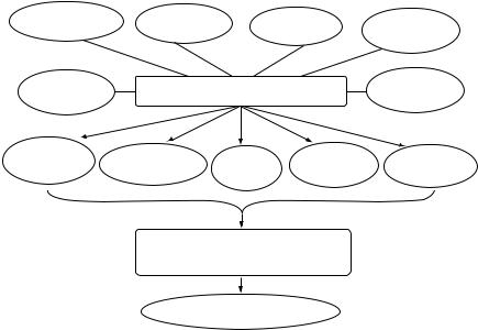
Atherosclerosis is multifactorial and polygenic in origin and develops decades earlier than its clinical manifestation [13]. The clinical silent phase of the disease initiates in the form of an inflammatory response, associated with fatty streak formation subsequent to intimal thickening, leading to the development of atherosclerotic plaque, which consists of cholesterol crystals, lymphocytes, proliferating SMCs, foam cells, proteoglycans, and cell debris [13, 14]. The later apparent stage of the disease is manifested by arterial calcification, thinning of fibrous cap, plaque rupture producing microemboli, hemorrhage, and intravascular thrombosis [14, 15]. Different biochemical and cellular events underlying the disease processes due to various pathophysiological factors including diabetes, hypertension, and hypercholesterolemia are shown in Fig. 22.1.
Intimal Thickening and Plaque Formation
The classic response-to-injury hypothesis has postulated that alteration in endothelial function is the initial step of disease process [16]. The potential causes of endothelial dysfunction include oxidative stress, mechanical stress, genetic alterations, elevated plasma homocysteine levels, and infectious microorganisms [17]. In spite of the different means of endothelial injury, the end result occurs as an increase in endothelial permeability, imbalance between endothelium-derived relaxing factors and endothelium-derived contracting factors, expression of adhesion molecules, release of growth factors and chemotactic factors leading to intimal thickening and plaque formation [18]. It is important to note that under physiological conditions, the secretion
478 Harjot K. Saini et al.
Hypercholesterolemia |
Hypertension |
|
Diabetes |
Genetic |
|
|
|
|
|
alterations |
|
|
|
|
|
|
|
Homocysteine |
|
Pathophysiological factors |
Infectious |
||
|
microorganisms |
||||
|
|
|
|
|
|
Oxidative stress |
Leukocyte adhesion |
Platelet |
Endothelial |
Smooth muscle |
|
|
|
and migration |
dysfunction |
dysfunction |
dysfunction |
Inflammation, Ox LDL, smooth muscle cell migration and proliferation,
foam cell formation
Atherosclerosis
FIGURE 22.1. Different biochemical and cellular processes participating in the development of atherosclerosis.
of adhesion molecules such as vascular cell adhesion molecule-1 (VCAM-1), intercellular adhesion molecule-1 (ICAM-1), E-selectin, and endothelial leukocyte adhesion molecule is regulated by proinflammatory cytokines such as tumor necrosis factor-α (TNF-α), interleukin (IL)-1, IL-4, IL-6, and inter- feron-γ [19]. However, in the presence of endothelial dysfunction, the levels of these cytokines have been shown to be elevated, which in turn cause more production of adhesion molecules and thus favor the monocyte recruitment and adhesion to the endothelium [20]. The recruited monocytes differentiate to macrophages in the subendothelial space and further aggravate the development of atherosclerosis [21].
Formation of Fatty Streak
The alteration in endothelial permeability triggers the transmigration of LDL particles into the intima through the endothelial layer, where it is modified by oxidative stress to oxidized LDL (Ox-LDL) [22]. Macrophage lipoxygenase, myeloperoxidase, and NADPH oxidase are the major sources of reactive oxygen species production [23]. Oxidation of polyunsaturated fatty acids within the LDL causes the release of aldehyde and ketones such as malondialdehyde and 4-hydroxynonenal, which can alter the lysine residues on apolipoproteins B-100 (apoB-100), the major protein of LDL [24]. The
Chapter 22. Modification of Biochemical and Cellular Processes |
479 |
modified apoB-100 is no longer recognized by the macrophage apoB receptor, but instead is taken by the macrophage scavenger receptor leading to the formation of foam cells and results in fatty streak [24].
Formation of Lipid Core
The uptake of Ox-LDL by macrophages is the major stimulus for the production and release of various cytokines such as TNF-α, IL-1β, and IL-8 as well as cytotoxic substances leading to an inflammatory response [25]. These cytokines cause the recruitment of macrophages, T cells, and SMCs, as well as upregulate the expression of endothelial adhesion molecules [17]. The cytotoxic substances released by macrophages further oxidize LDL particles in the intima and promote their uptake by macrophages. The continuation of this process converts the foam cells into lipid pools and ultimately forms the lipid core of the atherosclerotic plaque [17].
Stabilization of the Plaque
The proliferation and migration of SMCs from media to intima form the fibrous cap, which causes stabilization of the plaque [15]. Once in the intima, SMCs cause the production of cytokines, growth factors, and extracellular matrix consisting of collagen and proteoglycans [15]. In addition, lysophosphatidylcholine, a product formed by LDL oxidation and a potent chemoattractant for monocytes and T-lymphocytes, induces the expression of VCAM-1 and ICAM-1, increases the level of platelet-derived growth factor (PDGF) and heparin-binding epidermal growth factor in endothelial cells and SMCs [15]. Furthermore, lysophosphatidic acid is released from OxLDL, which stimulates vascular smooth muscle cell (VSMC) proliferation as well as platelet aggregation, increases intracellular Ca2+ concentration and thus promotes the formation of atherosclerotic plaque [26, 27]. Ox-LDL itself is toxic to macrophages and therefore contributes to the amplification of inflammatory process and triggers the formation of necrotic core in the advanced atherosclerotic lesions [23]. The lipid droplets released from the dead macrophages are phagocytized by SMCs leading to the production of SMC foam cells [28]. It is emphasized that the degree of SMC proliferation, migration, extracellular matrix formation, and extent of mature plaque formation is determined by the mutual interaction between platelets, endothelium, SMCs, and macrophages.
Progression of the Disease and Destabilization
of Atherosclerotic Plaque
The slow progression of disease leads to ischemia by reduction in the lumen size; this condition is clinically manifested as angina [29]. Chronic inflammatory reaction and induction of calcification-mediated changes in the
480 Harjot K. Saini et al.
mechanistic characteristics of arteries predisposes the plaque to rupture [29]. The increased expression of matrix metalloproteases is mainly responsible for atherosclerotic plaque rupture [15]. In addition, thinning of the fibrous cap triggers the plaque rupturing process and exposes the thrombogenic contents of plaque to blood stream leading to the formation of thrombus [17]. Furthermore, production of Ox-LDL, platelet accumulation, local increase in tissue factor, thromboxane A2, serotonin, ADP, and platelet-activating factor aggravate the formation of thrombosis [17], which is manifested by acute coronary syndromes such as acute myocardial infarction and unstable angina [29]. Because of the complex acquaintance between environmental, genetic, cellular, and biochemical factors, the sequential characterization of atherosclerotic lesions in individual patients is far from clear. Therefore, evidence-based therapies as a result of epidemiological studies through risk factor modification are considered to provide significant clinical benefits.
Antiatherosclerotic Effects of Red Wine
Various experimental studies have demonstrated that both the alcoholic and polyphenolic components of red wine prevent the development and progression of atherosclerosis by modifying the following biochemical and cellular events during the disease process.
Modification of Nitric Oxide (NO)-Mediated Vasoprotection by Red Wine
NO plays an important role in maintaining the vascular tone because of its strong vasorelaxing ability [30]. NO-induced protection in the early phase of atherosclerosis is mainly mediated by prevention of leukocyte migration and adhesion to the vascular endothelium by a decrease in monocyte chemoattractant protein-1 (MCP-1) [31], surface adhesion molecules such as CD11/CD18, P selectin, VCAM-1, and ICAM-1 [32, 33]. The protection by NO in the latter stages of atherosclerosis is manifested by inhibition of DNA synthesis, mitogenesis, VSMC proliferation, and migration [34]. In addition, NO is a potent inhibitor of platelet aggregation and adhesion to the vascular wall [35] and prevents the release of PDGFs, which are known to stimulate SMC proliferation [36]. Furthermore, the antiatherosclerotic properties of NO are associated with its ability to decrease endothelial permeability, reduce influx of LDL into the intima and inhibit LDL oxidation [37]. Red wine has been shown to cause the endothelium-dependent relaxation of blood vessels via enhanced generation and increased biological activity of NO [38]. Fitzpatrick et al. [39] have demonstrated that this effect of red wine is due to its polyphenolic components such as quercitin and tannic acid, unlike resveratrol and malvidin, which have the
Chapter 22. Modification of Biochemical and Cellular Processes |
481 |
ability to generate NO from the vascular endothelium as seen in phenyle- phrine-contracted rat aortic rings. Similarly, NO-mediated vasodilation after incubation with red wine polyphenols has been observed in human coronary arteries [40].
Andriambeloson et al. [41] have demonstrated that in rat aortic rings preconstricted with norepinephrine, red wine polyphenols-induced endotheliumdependent relaxation to acetylcholine is linked with enhanced NO synthesis instead of increasing the biological effectiveness of NO or via protecting its breakdown by superoxide anions. On the other hand, Zenebe et al. [42] have reported that polyphenols in red wine prolong the half-life and increase the bioavailability of NO by preventing its degradation by reactive oxygen species. Although the molecular mechanisms associated with red wine polyphenolsinduced vasoprotection are not completely elucidated, recent studies have shown the involvement of endothelial NO synthase (eNOS) activation in the presence of polyphenols [43]. An increase in the activity of eNOS promoter with eNOS mRNA stabilization has been demonstrated in human endothelial cells by red wine polyphenol treatment [43]; resveratrol has been reported to be the major red wine polyphenol responsible for such an effect [44]. In contrast, the ethanol present in red wine has not been associated with upregulation of eNOS expression under similar experimental conditions [43]. However, the alterations in eNOS expression in the presence of red wine are still controversial as results vary with the variety of red wine. Exposure of French red wine independent of their maturation in oak barrels or steel tanks, significantly enhances eNOS activity, mRNA, and protein content in cultured human umbilical vein endothelial cells (HUVEC) both in a time and concentrationdependent manner [43]. On the other hand, no such effect on eNOS expression was observed in the presence of German red wines [43]. The difference in the polyphenol content may be the reason for these controversial findings because the French red wine has higher contents of polyphenols than other wines [45].
Additional mechanisms underlying NO-dependent vasoprotection have been demonstrated in different cell types after red wine polyphenol treatment. An increase in the intracellular Ca2+ concentration, which causes augmentation of NO biosynthesis, has been reported in rat thoracic aorta and bovine aortic endothelial cells upon red wine polyphenol treatment [41, 46]. In addition, an increase in the expression of cyclooxgenase-2, release of endothelial thromboxane A2 and the expression of inducible NO synthase (iNOS) have been observed in rat aorta after treatment with dry powder of red wine [47]. These investigators have suggested that increased expression of iNOS may compensate the extra endothelial NO-induced hyporeactivity in response to norepinephrine in order to maintain the agonist-induced contraction [47]. Besides NO production, the formation of other mediators of vascular tone such as prostacyclin (PGI2), a potent vasodilator, and antiplatelet agent [48] as well as endothelium-derived hyperpolarizing factor, a redox-sensitive vasodilator [49], are also increased by red wine polyphenols. In addition, the synthesis of endothelin-1 (ET-1), a potent vasoconstrictor
482 Harjot K. Saini et al.
substance, is reduced by polyphenol treatment [50]. Furthermore, red wine polyphenols decrease the levels of high-sensitivity C-reactive protein (hsCRP), which is a marker of early inflammatory disease and a potential risk predictor for future atherosclerosis [51].
Modification of High-Density Lipoprotein (HDL) Content by Red Wine
The antiatherosclerotic properties of HDL are related with the role of HDL in reverse cholesterol transport as HDL removes excess cholesterol from peripheral tissues and transports it to the liver [52]. In addition, HDLinduced prevention of LDL oxidation plays an important role in the prevention of atherosclerotic plaque development [53]. Conversion of bioactive lipid peroxidation products into inactive compounds by HDL associated enzymes such as paraoxonase [54], platelet activating factor acetylhydrolase [55] and lecithin-cholesterol acyltransferase [56] appear to be the major mechanisms for such an effect. Furthermore, shift of lipid peroxidation products from LDL to HDL followed by conversion of cholesteryl ester hydroperoxides to stable cholesteryl ester hydroxides are implicated in the protective effects of HDL [57]. Red wine consumption increases the level of HDL [58], which is linked with an increase in HDL cholesterol and apolipoprotein A-I [59]. Enrichment of HDL particles with polyunsaturated phospholipids such as arachidonic acid and eicosapentaenoic acid, especially those containing omega-3 (C20:5) are associated with the antiatherosclerotic effects of red wine [59]. Furthermore, recent studies have shown that HDL causes an increase in the production of NO [60], a known vasoprotective agent [30–37]. Red wine-induced increase in the plasma HDL content along with an increase in NO and attenuation of ET-1 as well as hs-CRP play an important role in maintenance of endothelial function in the initial stages of atherosclerosis as shown in Fig. 22.2.
Modification of LDL Oxidation by Red Wine
Polyphenols from red wine such as catechin and quercetin have the capability to protect the LDL from oxidation [61]. It has been demonstrated that polyphenols bind to LDL and polyphenol-enriched LDL is resistant to oxidation in contrast to native LDL [62]. Reduction in LDL oxidation followed by a significant decrease in atherosclerotic lesion area was also observed in apolipoprotein E (apoE) deficient mice in which accelerated atherosclerosis is associated with increased lipid peroxidation of plasma, LDL and very lowdensity lipoproteins (VLDL), as well as increased susceptibility of these lipoproteins to undergo lipid peroxidation under oxidative stress [63, 64]. In addition, LDL aggregation which is directly related with LDL oxidation, and shown to be taken up by macrophages at an enhanced rate leading to foam cell