

surgical knot tying manual covidien
.pdfWhen the tension is reapplied in equal and opposing directions, the slip knots can usually be converted into either the square or granny knots. A simple code has been devised to describe a knot’s configuration (Figure 2).21 The number of wraps for each throw is indicated by the appropriate Arabic number. The relationship between each throw being either crossed or parallel is signified by the symbols X or =, respectively. In accordance with this code, the square knot is designated 1=1,
and the granny knot 1x1. The presence of a slip knot construction is indicated by the letter S. This method of describing knots facilitates their identification and reproduction. It is, for example, perfectly obvious what is meant by 2x2x2, without giving the knot a name, and all surgical knots can be defined unequivocally in this international language.
Sutures remain the most common method of approximating the divided edges of skin. Skin closure can be achieved by either percutaneous suture closure, dermal suture closure, or a combination of both techniques. Dermal or percutaneous suture closures are accomplished by either interrupted suture or a continuous suture closure technique.
When utilizing a continuous suture, the first suture loop is constructed by using the single strand of the fixed suture end attached to the needle and a single strand of the free suture end. However, the knot of the last suture loop at the end of the continuous suture is constructed by a suture loop containing two strands and the single strand of the fixed suture end attached to the needle.
27
IV. components of a knotted suture loop (cont’d)
Figure 6. Construction of a square knot with two single strands.
Figure 7. Construction of a square knot with a single strand and suture loop.
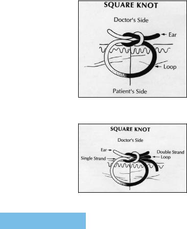
The first knot in a percutaneous and dermal suture closure usually has a square knot construction. A tied square knot suture has three components (Figure 6). The loop created by the knot maintains the apposition of tissue. The knot is composed of a number of throws secured against each other. The ears are the cut ends of the suture, which add security to the knot by preventing untying during slippage.
At the end of a percutaneous and dermal closure, te knot must be constructed as a square knot with a single strand and a suture loop. A square knot of an interrupted suture loop is formed by two single throws in which the right ear and loop come out in a position that is directly opposite to that of the left ear and loop. The configuration of a knot using a looped suture end and a fixed suture end attached to a needle is markedly different from the interrupted knotted suture loop in which knot construction is accomplished by two separate strands of sutures. The interrupted suture has a knot constructed by two single strands. In contrast, knots constructed with a suture loop have two suture strands that
28

are intertwined with the single strand of the fixed suture end. When knot construction is complete, the interrupted percutaneous suture has two cut suture ends. Knots constructed by a double-strand suture loop and a single strand of the fixed suture end has three separate knot ears (Figure 7). The square knot is formed when the right ear and the loop of a two-throw knot exit on the same side of the knot and are parallel to each other.
During wound closure, knot construction involves two steps. The first secures precise approximation of the wound edges by advancing either a one-throw or a two-throw knot to the wound surface. The second step is the construction of additional throws until knot security is attained and slippage is prevented.22
Our biomechanical performance studies demonstrate that secure knot configuration of the interrupted suture loop created at the beginning of the wound is quite different from secure knot configuration of the knot constructed at the end of the wound. These differences in secure knot configurations are related to the types of suture strands used in knot construction. The first knot in a continuous suture is constructed by two single strands, and the second knot is created by a single strand
and a suture loop. Knot construction with a suture loop predisposes the knot to suture slippage and requires additional throws for knot security. When constructing the
first interrupted suture loop with either absorbable or nonabsorbable monofilament sutures, knot security is achieved with square knot construction by using three or four throws. The last suture loop and single strand involves at least five or six throws for knot security. If the trauma surgeon fails to construct secure knots at the beginning or the end of the laceration, knot slippage will occur, resulting in wound dehiscence.
29

V. mechanical performance
The mechanical performance of a suture is an important consideration in the selection of a surgical suture and can be measured by reproducible, biomechanical parameters.23 The suture’s stiffness reflects its resistance to bending. Its coefficient of friction is a measure of the resistive forces encountered by contact of the surfaces of the suture material during knot construction. Strength is a key performance parameter that indicates the
suture’s resistance to breakage. The knot breakage load for a secure knot that fails by breakage is a reliable measure of strength. During these tests, forces are applied to the divided ends of the suture loop, the patient’s side of the knot. As the suture is subjected to stress, it will elongate. The load elongation properties of a suture have important clinical implications. Ideally, the suture should elongate under low loads to accommodate for the developing wound edema, but return to its original length after resolution of the edema. Although it should exhibit an immediate stretch under low loads, it should not elongate any further while continuously maintaining the load, exhibiting a resistance to creep.
These biomechanical parameters play important roles in the clinical performance of the suture.24 Surgeons consider the handling characteristics of the suture to be one of the most important parameters in their selection of sutures. Surgeons evaluate the handling characteristics of sutures by constructing knots using manual and instrument-tie techniques. The surgeon prefers a suture which permits two-throw knots to be easily advanced to the wound edges, providing a preview of the ultimate
30

apposition of the wound edges. The force required to advance the knot is called knot rundown force. Once meticulous approximation of the wound edges is achieved, the surgeon prefers to add one more throw to the two-throw knot so that it does not fail by slippage.
The magnitude of the knot rundown force is influenced considerably by the configuration of two-throw knots. 24 Knot rundown of the surgeon’s knot square (2=1) generates sufficient forces to break the knot. In contrast, knot rundown of square (1=1), granny (1x1) and slip (S=S, SxS) knots occurs by slippage. For comparable sutures, the mean knot rundown force for square knots is the greatest, followed by that for the granny (1x1) knots, and then the slip (S=S, SxS) knots.
Failure of the knotted suture loop may be the result of either knot slippage or breakage, suture cutting through tissues, and mechanical crushing of the suture by surgical instruments. Initially, the knotted suture fails by slippage, which results in untying of the knot. All knots slip to some degree regardless of the type of suture material. When slippage is encountered, the cut ends (“ears”) of the knot must provide the additional material to compensate for the enlarged suture loop. When the amount of knot slippage exceeds the length of the cut “ears,” the throws of the knot become untied. In general, surgeons recommend that the length of the knot ”ears” be 3mm to accommodate for any knot slippage. 3 Dermal sutures are, however, an exception to this rule. Because the “ears” of dermal suture knots may protrude through the divided skin edges, surgeons prefer to cut their dermal suture “ears” as they exit from the knot. It must be emphasized that knot security is achieved in a knot with “ears”with one more throw than in a comparable knot whose “ear” length is 3mm.
31

knot slippage |
Knot slippage is counteracted by the frictional forces of the knots. The |
|
degree to which a knot slips can be influenced by a variety of factors |
|
including the coefficient of friction of the suture material, suture diameter, |
|
moisture, knot type and final geometry. Knots of the granny type (crossed) |
|
usually exhibit more slippage than do knots with a square-type (parallel) |
|
construction. |
|
With each additional throw, incrementally greater forces are required for |
|
knot untying. After a specified number of throws, failure will occur by knot |
|
breakage, after which the knot breakage force will not be enhanced by the |
|
addition of more throws. Consequently, these additional throws offer no |
|
mechanical advantage and represent more foreign bodies in the wound that |
|
damage host defenses and resistance to infection. |
|
The human element in knot tying has considerable influence on the |
|
magnitude of knot slippage.19 The amount of tension exerted by the |
|
surgeon on the “ears” of the knot significantly alters the degree of slippage. |
|
The careless surgeon who applies minimal tension (10% of knot break |
|
strength) to the “ears” of the knot constructs knots that fail by slippage. |
|
Knot slippage can be minimized by applying more tensions (80% of knot |
|
break strength) to the “ears” of the knot. Another serious error often made |
|
by the inexperienced surgeon is not to change the position of his/her hands |
|
appropriately during construction of square and/or granny type knots. The |
|
resulting knot, a sliding or slip knot, will become untied regardless of the |
|
suture material. The risk of forming a slip knot is greatest when tying one- |
|
hand knots and/or with deep seated ligatures.20-23 |
32

When enough force is applied to the tied suture to result in breakage, the site of |
knot breakage |
disruption of the suture is almost always the knot. The force necessary to break |
|
a knotted suture is lower than that required to break an untied suture made |
|
of the same material.19 The forces exerted on a tied suture are converted into |
|
shear forces, by the knot configuration that break the knot. The percentage loss |
|
of tensile strength, as a result of tying a secure knot, is least with mono-filament |
|
and multifilament steel.25 This relationship between the tensile strength of |
|
unknotted and knotted suture which is designated knot efficiency, is described |
|
in the following equation: |
|
Knot efficiency (%) = tensile strength of a knotted suture |
|
tensile strength of unknotted suture |
|
Regardless of the type of suture material, the efficiency of the knot is enhanced |
|
with an increasing number of throws, although only up to a certain limit. |
|
The type of knot configuration that results in a secure knot that fails by |
|
breaking varies considerably with different suture material. The magnitude of |
|
force necessary to produce knot breakage is influenced by the configuration |
|
of the knotted suture loop, type of suture material, and the diameter of the |
|
suture.3 The tissue in which the suture is implanted also has considerable |
|
influence on the knot strength of suture. In the case of absorbable sutures, a |
|
progressive decline in knot breaking strength is noted after tissue implantation. |
|
In addition, the magnitude of knot breakage force is significantly influenced by |
|
the rate of application of forces to the “ears” of the knot.23 When a constant |
|
force is applied slowly to the knot “ears,” the knot breakage force is significantly |
|
greater than that for knots in which the same constant force is applied rapidly |
|
to the “ears.” The latter knot loading rate is often referred to as “the jerk at the |
|
end of the knot,” especially when the knotted suture breaks. |
|
33

suture cutting tissue
Suture failure also may occur if the knotted suture loop cuts through the tissue. The type of tissue has considerable influence on the magnitude of force required to tear the suture through the tissue. Howes and Harvey26 reported that the forces required to tear gut sutures through canine fascia was the greatest followed by muscle, peritoneum and then fat. Using cadaver specimens 6 to 93 days after death, Tera and °Aberg27 measured the magnitude of forces required for suture to tear through excised musculoaponeurotic layers of laparotomy incisions. The rationale for this study was that the forces required to tear sutures through a musculoaponeurotic layer would provide a basis for the choice of a suture whose strength is at least as strong as the forces required to tear the suture through the tissue. When the suture was passed lateral to the transition between the linea alba and the rectus sheath, the force required to tear the suture through the tissue was greater than that for any other musculoaponeurotic layer tested; the paramedian incision required the lowest forces to pull sutures through its sheaths. When they recorded the
forces needed for sutures to tear through structures involved in the repair of inguinal hernia, the structures making up the conjoined tendon and Cooper’s ligament were the strongest and exhibited twice the resistance to suture tearing than those of the other structures.
As expected, the force required for sutures to tear through tissue changes during healing. Aberg28 reported that the forces needed for sutures to tear through the aponeurotic muscle layer reduced significantly during the first week of healing. When the wound edges were approximated by suture tied tightly around this aponeurotic muscle layer, the reduction in force needed for the suture to pull through this tissue persisted for two weeks.
34

Mechanical trauma to the suture by surgical instruments can also result in suture failure. Nichols et al29 cautioned surgeons about the handling of sutures by surgical instruments. They indicated that either the application of clamps and forceps to the suture or rough handling of sutures could damage and weaken them. Stamp et al30 incriminated the teeth in the needle holder jaws as important causal factors of sutural damage. Compression of sutures between the needle holder jaws with teeth produced morphologic changes in sutures that resulted in a marked reduction in the suture breaking strength. Similarly, the sharp edges of needle holder jaws without teeth can even crush the suture and, thereby, decrease its strength.31 Finally application of large compressive loads by pinching polypropylene sutures with DeBakey forceps decreases the strength of the suture.32
mechanical trauma
35
VI. tying techniques
The surgeon may use an individual ligature (“free tie”) or a suture that is attached to a needle or ligature reel. The length of a free tie or suture attached to a needle is usually 18 inches. The longest strands of suture material are available on a reel or spool. When the suture is attached to a needle or reel, there is a free end and a fixed end; the fixed end is attached to either the needle or reel. The first throw of a knot is accomplished by wrapping the free end either once or twice (surgeon’s) around the fixed end. During practice, clamp one end of the suture with an instrument that serves to represent either the needle or the reel, which is the fixed end of the suture.
Formation of each throw of a knot is accomplished in three steps. The first step is the formation of a suture loop. In the second step, the free suture end is passed through the suture loop to create a throw. The final step is to advance the throw to the wound surface. For the first throw of a square, granny, and surgeon’s knot square, for each additional throw the direction in which tension is applied to the
suture ends is reversed. The surgeon should construct a knot by carefully snugging each throw tightly against another. The rate of applying tension to each throw should be relatively slow.
|
For either the square knot or surgeon’s knot square, the direction in which the |
|
|
free suture end is passed through the loop will be reversed for each additional |
|
|
throw. If the free suture end is passed down through the first suture loop, it must |
|
|
be passed up through the next suture loop. Reversal of the direction of passage of |
|
|
the free suture end through the loops does not alter either the knot’s mechanical |
|
|
performance or its configuration. It simply reverses the presentation of the knot |
|
|
(Figure 8). For granny knots, the direction in which the free suture end is passed |
|
36 |
||
through the loop is the same for each additional throw. |
||
|
|