


4brs Physiology
■Both na+ and K+ are transported against their electrochemical gradients.
■Energy is provided from the terminal phosphate bond of ATP.
■The usual stoichiometry is 3 na+/2 K+.
■Specific inhibitors of Na+, K+-ATPase are the cardiac glycoside drugs ouabain and digitalis.
b.Ca2+-aTPase (or Ca2+ pump) in the sarcoplasmic reticulum (SR) or cell membranes transports Ca2+ against an electrochemical gradient.
■Sarcoplasmic and endoplasmic reticulum Ca2+-ATPase is called serCa.
c.H+, K+-aTPase (or proton pump) in gastric parietal cells transports H+ into the lumen of the stomach against its electrochemical gradient.
■It is inhibited by proton pump inhibitors, such as omeprazole. e. secondary active transport
1.Characteristics of secondary active transport
a.The transport of two or more solutes is coupled.
b.One of the solutes (usually Na+) is transported “downhill” and provides energy for the “uphill” transport of the other solute(s).
c.Metabolic energy is not provided directly but indirectly from the na+ gradient that is maintained across cell membranes. Thus, inhibition of Na+, K+-ATPase will decrease transport of Na+ out of the cell, decrease the transmembrane Na+ gradient, and eventually inhibit secondary active transport.
d.If the solutes move in the same direction across the cell membrane, it is called cotransport or symport.
■Examples are na+-glucose cotransport in the small intestine and renal early proximal tubule and na+–K+–2Cl– cotransport in the renal thick ascending limb.
e.If the solutes move in opposite directions across the cell membranes, it is called countertransport, exchange, or antiport.
■Examples are na+-Ca2+ exchange and na+–H+ exchange.
2.example of na+–glucose cotransport (Figure 1.1)
a.The carrier for Na+–glucose cotransport is located in the luminal membrane of intestinal mucosal and renal proximal tubule cells.
b.Glucose is transported “uphill”; Na+ is transported “downhill.”
c.Energy is derived from the “downhill” movement of Na+. The inwardly directed Na+ gradient is maintained by the Na+–K+ pump on the basolateral (blood side) membrane. Poisoning the Na+–K+ pump decreases the transmembrane Na+ gradient and consequently inhibits Na+–glucose cotransport.
3.example of na+–Ca2+ countertransport or exchange (Figure 1.2)
a.Many cell membranes contain a Na+–Ca2+ exchanger that transports Ca2+ “uphill” from low intracellular [Ca2+] to high extracellular [Ca2]. Ca2+ and Na+ move in opposite directions across the cell membrane.
b.The energy is derived from the “downhill” movement of Na+. As with cotransport, the inwardly directed Na+ gradient is maintained by the Na+–K+ pump. Poisoning the Na+– K+ pump therefore inhibits Na+–Ca2+ exchange.
III. osMosIs
a.osmolarity
■is the concentration of osmotically active particles in a solution.
■is a colligative property that can be measured by freezing point depression.

|
Cell Physiology |
5 |
Chapter 1 |
Lumen
Na+
Figure 1.1 Na+–glucose cotransport (symport) in |
Secondary |
intestinal or proximal tubule epithelial cell. |
active |
■ can be calculated using the following equation:
Osmolarity = g ¥ C
where:
Intestinal or |
Blood |
||
proximal tubule cell |
|
|
|
Na+ |
Na+ |
Na+ |
|
K+ |
|||
Glucose |
|
||
|
|
||
 Na+
Na+
Primary active
Osmolarity = concentration of particles (Osm/L)
g = number of particles in solution (Osm/mol)
[e.g., gNaCl = 2; gglucose = 1] C = concentration (mol/L)
■Two solutions that have the same calculated osmolarity are isosmotic. If two solutions have different calculated osmolarities, the solution with the higher osmolarity is hyperosmotic and the solution with the lower osmolarity is hyposmotic.
■Sample calculation: What is the osmolarity of a 1 M NaCl solution?
Osmolarity = g × C
=2 Osm/mol ×1M
=2 Osm/L
B.Osmosis and osmotic pressure
■Osmosis is the flow of water across a semipermeable membrane from a solution with low solute concentration to a solution with high solute concentration.
1. Example of osmosis (Figure 1.3)
a. Solutions 1 and 2 are separated by a semipermeable membrane. Solution 1 contains a
solute that is too large to cross the membrane. Solution 2 is pure water. The presence of the solute in solution 1 produces an osmotic pressure.
b. The osmotic pressure difference across the membrane causes water to flow from solution 2 (which has no solute and the lower osmotic pressure) to solution 1 (which has the solute and the higher osmotic pressure).
c. With time, the volume of solution 1 increases and the volume of solution 2 decreases.
|
Secondary |
||
|
active |
|
|
|
Na+ |
Ca2+ |
|
|
|
||
Ca2+ |
Ca2+ |
||
|
|
||
Na+ |
Na+ |
Na+ |
|
K+ |
|||
|
|
||
|
|
Primary |
|
Figure 1.2 Na+–Ca2+ countertransport (antiport). |
|
active |
|

6 |
BRS Physiology |
|
|
|
|
|
Semipermeable |
|
|
|
|
membrane |
|
|
|
|
Time |
|
|
|
|
Water flows |
|
|
|
|
by osmosis |
|
|
|
|
from 2 |
1 |
|
|
1 |
2 |
1 |
2 |
Figure 1.3 Osmosis of H2O across a semipermeable membrane.
2. Calculating osmotic pressure (van’t Hoff’s law)
a. The osmotic pressure of solution 1 (see Figure 1.3) can be calculated by van’t Hoff’s law, which states that osmotic pressure depends on the concentration of osmotically active
particles. The concentration of particles is converted to pressure according to the following equation:
p = g ¥ C ¥ RT
where:
π = osmotic pressure (mm Hg or atm)
g = number of particles in solution (osm/mol) C = concentration (mol/L)
R = gas constant (0.082 L—atm/mol—K) T = absolute temperature (K)
b. The osmotic pressure increases when the solute concentration increases. A solution of 1 M CaCl2 has a higher osmotic pressure than a solution of 1 M KCl because the concentration of particles is higher.
c. The higher the osmotic pressure of a solution, the greater the water flow into it.
d. Two solutions having the same effective osmotic pressure are isotonic because no water flows across a semipermeable membrane separating them. If two solutions separated
by a semipermeable membrane have different effective osmotic pressures, the solution with the higher effective osmotic pressure is hypertonic and the solution with the lower effective osmotic pressure is hypotonic. Water flows from the hypotonic to the
hypertonic solution.
e. Colloid osmotic pressure, or oncotic pressure, is the osmotic pressure created by proteins (e.g., plasma proteins).
3. Reflection coefficient (σ)
■is a number between zero and one that describes the ease with which a solute permeates a membrane.
a. If the reflection coefficient is one, the solute is impermeable. Therefore, it is retained in
the original solution, it creates an osmotic pressure, and it causes water flow. Serum albumin (a large solute) has a reflection coefficient of nearly one.
b. If the reflection coefficient is zero, the solute is completely permeable. Therefore, it
will not exert any osmotic effect, and it will not cause water flow. Urea (a small solute) usually has a reflection coefficient of close to zero and it is, therefore, an ineffective osmole.
4. Calculating effective osmotic pressure
■Effective osmotic pressure is the osmotic pressure (calculated by van’t Hoff’s law) multiplied by the reflection coefficient.
■If the reflection coefficient is one, the solute will exert maximal effective osmotic pressure. If the reflection coefficient is zero, the solute will exert no osmotic pressure.

|
Cell Physiology |
7 |
Chapter 1 |
IV. DIFFusIon PoTenTIal, resTInG MeMbrane PoTenTIal, anD aCTIon PoTenTIal
a.Ion channels
■are integral proteins that span the membrane and, when open, permit the passage of certain ions.
1.Ion channels are selective; they permit the passage of some ions, but not others. Selectivity is based on the size of the channel and the distribution of charges that line it.
■For example, a small channel lined with negatively charged groups will be selective for small cations and exclude large solutes and anions. Conversely, a small channel lined with positively charged groups will be selective for small anions and exclude large solutes and cations.
2.Ion channels may be open or closed. When the channel is open, the ion(s) for which it is selective can flow through. When the channel is closed, ions cannot flow through.
3.The conductance of a channel depends on the probability that the channel is open. The higher the probability that a channel is open, the higher the conductance, or permeability. Opening and closing of channels are controlled by gates.
a. Voltage-gated channels are opened or closed by changes in membrane potential.
■The activation gate of the na+ channel in nerve is opened by depolarization; when open, the nerve membrane is permeable to Na+ (e.g., during the upstroke of the nerve action potential).
■The inactivation gate of the na+ channel in nerve is closed by depolarization; when closed, the nerve membrane is impermeable to Na+ (e.g., during the repolarization phase of the nerve action potential).
b.ligand-gated channels are opened or closed by hormones, second messengers, or neurotransmitters.
■For example, the nicotinic receptor for acetylcholine (ACh) at the motor end plate is an ion channel that opens when ACh binds to it. When open, it is permeable to Na+ and K+, causing the motor end plate to depolarize.
b.Diffusion and equilibrium potentials
■A diffusion potential is the potential difference generated across a membrane because of a concentration difference of an ion.
■A diffusion potential can be generated only if the membrane is permeable to the ion.
■The size of the diffusion potential depends on the size of the concentration gradient.
■The sign of the diffusion potential depends on whether the diffusing ion is positively or negatively charged.
■Diffusion potentials are created by the diffusion of very few ions and, therefore, do not result in changes in concentration of the diffusing ions.
■The equilibrium potential is the potential difference that would exactly balance (oppose) the tendency for diffusion down a concentration difference. At electrochemical equilibrium, the chemical and electrical driving forces that act on an ion are equal and opposite, and no more net diffusion of the ion occurs.
1.example of a na+ diffusion potential (Figure 1.4)
a.Two solutions of NaCl are separated by a membrane that is permeable to Na+ but not to Cl−. The NaCl concentration of solution 1 is higher than that of solution 2.
b.Because the membrane is permeable to Na+, Na+ will diffuse from solution 1 to solution 2 down its concentration gradient. Cl− is impermeable and therefore will not accompany Na+.
c.As a result, a diffusion potential will develop and solution 1 will become negative with respect to solution 2.

8 |
BRS Physiology |
|
|
|
|
|
|
Na+-selective |
|
|
|
|
|
membrane |
|
|
|
|
1 |
2 |
1 |
|
2 |
|
Na+ |
Na+ |
Na+ |
– |
+ Na+ |
|
|
|
|
– |
+ |
|
Cl– |
|
Cl– |
– |
+ |
|
|
– |
+ |
||
|
|
Cl– |
|
|
Cl– |
Figure 1.4 Generation of an Na+ diffusion potential across a Na+-selective membrane.
d. Eventually, the potential difference will become large enough to oppose further net
diffusion of Na+. The potential difference that exactly counterbalances the diffusion of Na+ down its concentration gradient is the Na+ equilibrium potential. At electrochemical
equilibrium, the chemical and electrical driving forces on Na+ are equal and opposite, and there is no net diffusion of Na+.
2. Example of a Cl− diffusion potential (Figure 1.5)
a. Two solutions identical to those shown in Figure 1.4 are now separated by a membrane that is permeable to Cl− rather than to Na+.
b. Cl− will diffuse from solution 1 to solution 2 down its concentration gradient. Na+ is impermeable and therefore will not accompany Cl−.
c. A diffusion potential will be established such that solution 1 will become positive with
respect to solution 2. The potential difference that exactly counterbalances the diffusion of Cl− down its concentration gradient is the Cl− equilibrium potential. At electro-
chemical equilibrium, the chemical and electrical driving forces on Cl− are equal and opposite, and there is no net diffusion of Cl−.
3. Using the Nernst equation to calculate equilibrium potentials
a. The Nernst equation is used to calculate the equilibrium potential at a given concentration difference of a permeable ion across a cell membrane. It tells us what potential
would exactly balance the tendency for diffusion down the concentration gradient; in other words, at what potential would the ion be at electrochemical equilibrium?
|
|
|
|
|
E = -2.3 |
RT |
log10 |
[Ci ] |
|
|
|
|
|||||||
|
|
|
|
|
zF |
[Ce ] |
|
|
|
|
|
||||||||
|
|
|
|
|
where: |
|
|
|
|
|
|
|
|
||||||
|
|
|
|
|
E = equilibrium potential (mV) |
||||||||||||||
|
|
2.3 |
RT |
|
= |
60 mV |
at 37°C |
|
|
|
|
||||||||
|
|
zF |
|
|
|
|
|
|
|||||||||||
|
|
|
|
|
|
z |
|
|
|
|
|
|
|
|
|||||
|
|
|
|
|
z = charge on the ion (+1 for Na+, +2 for Ca2+, −1 for Cl−) |
||||||||||||||
|
|
|
|
|
Ci |
= intracellular concentration (mM) |
|||||||||||||
|
|
|
|
|
Ce |
= extracellular concentration (mM) |
|||||||||||||
|
|
Cl–-selective |
|
|
|
|
|
|
|
|
|
|
|
|
|
||||
|
|
membrane |
|
|
|
|
|
|
|
|
|
|
|
|
|
||||
|
1 |
2 |
|
|
|
|
|
|
|
|
|
1 |
|
|
|
2 |
|
||
|
Na+ |
Na+ |
|
|
|
|
|
|
|
|
|
|
|
Na+ |
+ |
– |
Na+ |
|
|
|
|
|
|
|
|
|
|
|
|
|
|
|
+ |
– |
|
||||
|
|
|
|
|
|
|
|
|
|
|
|
|
|
|
|
+ |
– |
|
|
|
|
|
|
|
|
|
|
|
|
|
|
|
|
|
|
|
|
||
|
Cl– |
|
|
|
|
|
|
|
|
|
|
|
|
Cl– |
+ |
– |
|
|
|
|
Cl– |
|
|
|
|
|
|
|
|
|
|
|
|
|
Cl– |
|
|||
|
|
|
|
|
|
|
|
|
|
|
|
|
|
|
|
||||
Figure 1.5 Generation of a Cl− diffusion potential across a Cl−-selective membrane.
|
Cell Physiology |
9 |
Chapter 1 |
b. Sample calculation with the Nernst equation
■If the intracellular [Na+] is 15 mM and the extracellular [Na+] is 150 mM, what is the equilibrium potential for Na+?
ENa+ = |
−60 mV |
log10 |
[Ci ] |
|
z |
[Ce ] |
|||
|
|
|||
= |
−60 mV log10 |
15 mM |
||
150 mM |
||||
|
+1 |
|
||
=−60 mV log10 0.1
=+60 mV
Note: You need not remember which concentration goes in the numerator. Because it is a log function, perform the calculation either way to get the absolute value of 60 mV. Then use an “intuitive approach” to determine the correct sign. (Intuitive approach: The [Na+] is higher in extracellular fluid than in intracellular fluid, so Na+ ions will diffuse from extracellular to intracellular, making the inside of the cell positive [i.e., +60 mV at equilibrium].)
c. Approximate values for equilibrium potentials in nerve and muscle
ENa+
ECa2+
EK+
ECl−
+65 mV +120 mV −85 mV −85 mV
C.Driving force and current flow
■The driving force on an ion is the difference between the actual membrane potential (Em) and the ion’s equilibrium potential (calculated with the Nernst equation).
■Current flow occurs if there is a driving force on the ion and the membrane is permeable to the ion. The direction of current flow is in the same direction as the driving force. The magnitude of current flow is determined by the size of the driving force and the permeability (or conductance) of the ion. If there is no driving force on the ion, no current flow can occur. If the membrane is impermeable to the ion, no current flow can occur.
D.Resting membrane potential
■is expressed as the measured potential difference across the cell membrane in millivolts (mV).
■is, by convention, expressed as the intracellular potential relative to the extracellular potential. Thus, a resting membrane potential of −70 mV means 70 mV, cell negative.
1. The resting membrane potential is established by diffusion potentials that result from concentration differences of permeant ions.
2. Each permeable ion attempts to drive the membrane potential toward its equilibrium potential. Ions with the highest permeabilities, or conductances, will make the greatest contributions to the resting membrane potential, and those with the lowest permeabilities will make little or no contribution.
3. For example, the resting membrane potential of nerve is −70 mV, which is close to the cal-
culated K+ equilibrium potential of −85 mV, but far from the calculated Na+ equilibrium potential of +65 mV. At rest, the nerve membrane is far more permeable to K+ than to Na+.
4. The Na+–K+ pump contributes only indirectly to the resting membrane potential by main-
taining, across the cell membrane, the Na+ and K+ concentration gradients that then produce diffusion potentials. The direct electrogenic contribution of the pump (3 Na+
pumped out of the cell for every 2 K+ pumped into the cell) is small.

10 |
BRS Physiology |
E.Action potentials
1. Definitions
a. Depolarization makes the membrane potential less negative (the cell interior becomes less negative).
b. Hyperpolarization makes the membrane potential more negative (the cell interior becomes more negative).
c. Inward current is the flow of positive charge into the cell. Inward current depolarizes the membrane potential.
d. Outward current is the flow of positive charge out of the cell. Outward current hyperpolarizes the membrane potential.
e. Action potential is a property of excitable cells (i.e., nerve, muscle) that consists of a rapid
depolarization, or upstroke, followed by repolarization of the membrane potential. Action potentials have stereotypical size and shape, are propagating, and are all-or-none.
f. Threshold is the membrane potential at which the action potential is inevitable. At threshold potential, net inward current becomes larger than net outward current. The resulting depolarization becomes self-sustaining and gives rise to the upstroke of the action potential. If net inward current is less than net outward current, no action potential will occur (i.e., all-or-none response).
2. Ionic basis of the nerve action potential (Figure 1.6) a. Resting membrane potential
■is approximately −70 mV, cell negative.
■is the result of the high resting conductance to K+, which drives the membrane potential toward the K+ equilibrium potential.
■At rest, the Na+ channels are closed and Na+ conductance is low.
b. Upstroke of the action potential
(1) Inward current depolarizes the membrane potential to threshold.
(2) Depolarization causes rapid opening of the activation gates of the Na+ channels, and the Na+ conductance of the membrane promptly increases.
(3) The Na+ conductance becomes higher than the K+ conductance, and the membrane potential is driven toward (but does not quite reach) the Na+ equilibrium
potential of +65 mV. Thus, the rapid depolarization during the upstroke is caused by an inward Na+ current.
(4) The overshoot is the brief portion at the peak of the action potential when the mem-
brane potential is positive.
(5) Tetrodotoxin (TTX) and lidocaine block these voltage-sensitive Na+ channels and abolish action potentials.
c. Repolarization of the action potential
(1) Depolarization also closes the inactivation gates of the Na+ channels (but more slowly than it opens the activation gates). Closure of the inactivation gates results in clo-
sure of the Na+ channels, and the Na+ conductance returns toward zero.
(2) Depolarization slowly opens K+ channels and increases K+ conductance to even higher levels than at rest. Tetraethylammonium (TEA) blocks these voltage-gated K+ channels.
(3) The combined effect of closing the Na+ channels and greater opening of the K+ channels makes the K+ conductance higher than the Na+ conductance, and the
membrane potential is repolarized. Thus, repolarization is caused by an outward
K+ current.
d. Undershoot (hyperpolarizing afterpotential)
■The K+ conductance remains higher than at rest for some time after closure of the Na+ channels. During this period, the membrane potential is driven very close to the K+ equilibrium potential.

|
Cell Physiology |
11 |
Chapter 1 |
Voltage or conductance
+65 mV
0 mV
–70 mV
–85 mV
Absolute |
|
Relative |
||
refractory |
|
refractory |
||
period |
|
period |
||
|
|
|
|
Na+ equilibrium potential |
|
|
|
|
|
|
|
|
Action potential |
|
|
|
|
|
|
Na+ conductance
K+ conductance
Resting membrane potential
K+ equilibrium potential
1.02.0
Time
(msec)
Figure 1.6 Nerve action potential and associated changes in Na+ and K+ conductance.
3. Refractory periods (see Figure 1.6) a. Absolute refractory period
■is the period during which another action potential cannot be elicited, no matter how large the stimulus.
■coincides with almost the entire duration of the action potential.
■Explanation: Recall that the inactivation gates of the Na+ channels are closed when the membrane potential is depolarized. They remain closed until repolarization occurs. No action potential can occur until the inactivation gates open.
b. Relative refractory period
■begins at the end of the absolute refractory period and continues until the membrane potential returns to the resting level.
■An action potential can be elicited during this period only if a larger than usual inward current is provided.
■Explanation: The K+ conductance is higher than at rest, and the membrane potential is closer to the K+ equilibrium potential and, therefore, farther from threshold; more inward current is required to bring the membrane to threshold.
c. Accommodation
■occurs when the cell membrane is held at a depolarized level such that the threshold potential is passed without firing an action potential.
■occurs because depolarization closes inactivation gates on the Na+ channels.
■is demonstrated in hyperkalemia, in which skeletal muscle membranes are depol arized by the high serum K+ concentration. Although the membrane potential is closer to threshold, action potentials do not occur because inactivation gates on Na+ channels are closed by depolarization, causing muscle weakness.
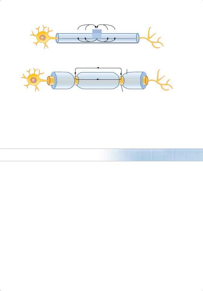
4. Propagation of action potentials (Figure 1.7)
■occurs by the spread of local currents to adjacent areas of membrane, which are then depolarized to threshold and generate action potentials.
12 |
brs Physiology |
|
|
|
|
|
|
|
|
|
+ |
+ |
+ |
+ |
– |
+ |
+ |
+ |
+ |
|
– |
– |
– |
– |
+ |
– |
– |
– |
– |
FIGure 1.7 Unmyelinated axon showing spread of depolarization by local current flow. Box shows active zone where action potential had reversed the polarity.
Myelin sheath
Node of Ranvier
FIGure 1.8 Myelinated axon. Action potentials can occur at nodes of Ranvier.
■ Conduction velocity is increased by:
a.↑ fiber size. Increasing the diameter of a nerve fiber results in decreased internal resistance; thus, conduction velocity down the nerve is faster.
b.Myelination. Myelin acts as an insulator around nerve axons and increases conduction velocity. Myelinated nerves exhibit saltatory conduction because action potentials can be generated only at the nodes of ranvier, where there are gaps in the myelin sheath (Figure 1.8).
V.neuroMusCular anD synaPTIC TransMIssIon
a.General characteristics of chemical synapses
1.an action potential in the presynaptic cell causes depolarization of the presynaptic terminal.
2.As a result of the depolarization, Ca2+ enters the presynaptic terminal, causing release of neurotransmitter into the synaptic cleft.
3.Neurotransmitter diffuses across the synaptic cleft and combines with receptors on the postsynaptic cell membrane, causing a change in its permeability to ions and, consequently, a change in its membrane potential.
4.Inhibitory neurotransmitters hyperpolarize the postsynaptic membrane: excitatory neurotransmitters depolarize the postsynaptic membrane.
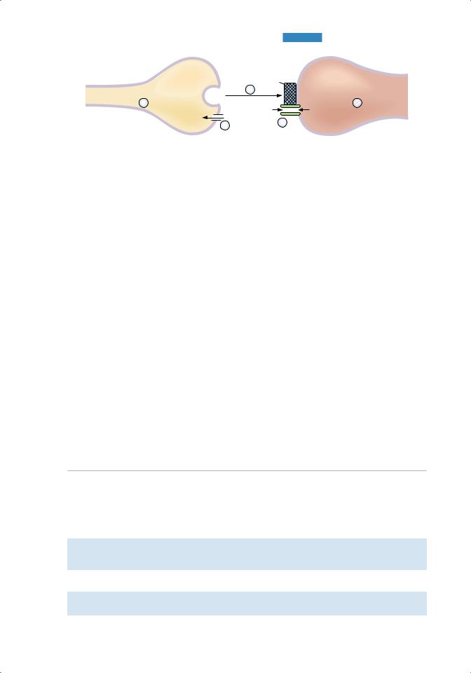
b.neuromuscular junction (Figure 1.9 and Table 1.2)
■is the synapse between axons of motoneurons and skeletal muscle.
■The neurotransmitter released from the presynaptic terminal is aCh, and the postsynaptic membrane contains a nicotinic receptor.
1.synthesis and storage of aCh in the presynaptic terminal
■Choline acetyltransferase catalyzes the formation of ACh from acetyl coenzyme A (CoA) and choline in the presynaptic terminal.
■ACh is stored in synaptic vesicles with ATP and proteoglycan for later release.
2.Depolarization of the presynaptic terminal and Ca2+ uptake
■Action potentials are conducted down the motoneuron. Depolarization of the presynaptic terminal opens Ca2+ channels.
|
|
|
Chapter 1 Cell Physiology |
13 |
|
|
|
AChR |
|
|
|
Action potential in nerve |
ACh |
3 |
Action potential in muscle |
|
|
|
|
||||
1 |
|
|
5 |
|
|
|
Na+ |
K+ |
|
||
|
|
|
|
||
|
|
Ca2+ |
4 |
|
|
|
|
2 |
|
|
|
|
|
|
|
|
|
Motoneuron |
|
|
|
Muscle |
|
Figure 1.9 Neuromuscular junction. ACh = acetylcholine; AChR = acetylcholine receptor.
■When Ca2+ permeability increases, Ca2+ rushes into the presynaptic terminal down its electrochemical gradient.
3. Ca2+ uptake causes release of ACh into the synaptic cleft
■The synaptic vesicles fuse with the plasma membrane and empty their contents into the cleft by exocytosis.
4. Diffusion of ACh to the postsynaptic membrane (muscle end plate) and binding of ACh to nicotinic receptors
■The nicotinic ACh receptor is also a Na+ and K+ ion channel.
■Binding of ACh to α subunits of the receptor causes a conformational change that opens the central core of the channel and increases its conductance to Na+ and K+. These are examples of ligand-gated channels.
5. End plate potential (EPP) in the postsynaptic membrane
■Because the channels opened by ACh conduct both Na+ and K+ ions, the postsynaptic membrane potential is depolarized to a value halfway between the Na+ and K+ equilibrium potentials (approximately 0 mV).
■The contents of one synaptic vesicle (one quantum) produce a miniature end plate potential (MEPP), the smallest possible EPP.
■MEPPs summate to produce a full-fledged EPP. The EPP is not an action potential, but simply a depolarization of the specialized muscle end plate.
6. Depolarization of adjacent muscle membrane to threshold
■Once the end plate region is depolarized, local currents cause depolarization and action potentials in the adjacent muscle tissue. Action potentials in the muscle are followed by contraction.
|
|
|
Agents Affecting Neuromuscular Transmission |
|
t a b l e |
|
1.2 |
||
|
|
|
|
|
|
|
|
|
Effect on Neuromuscular |
Example |
Action |
Transmission |
||
|
|
|
||
Botulinus toxin |
Blocks release of ACh from |
Total blockade |
||
|
|
presynaptic terminals |
|
|
Curare |
Competes with ACh for receptors |
|
on motor end plate |
Decreases size of EPP; maximal doses produce paralysis of respiratory muscles and death
Neostigmine |
Inhibits acetylcholinesterase |
Prolongs and enhances action of ACh at |
|
|
muscle end plate |
Hemicholinium |
Blocks reuptake of choline into |
Depletes ACh stores from presynaptic terminal |
|
presynaptic terminal |
|
ACh = acetylcholine; EPP = end plate potential.

14 |
BRS Physiology |
7. Degradation of Ach
■The EPP is transient because ACh is degraded to acetyl CoA and choline by acetylcholinesterase (AChE) on the muscle end plate.
■One-half of the choline is taken back into the presynaptic ending by Na+-choline cotransport and used to synthesize new ACh.
■AChE inhibitors (neostigmine) block the degradation of ACh, prolong its action at the muscle end plate, and increase the size of the EPP.
■Hemicholinium blocks choline reuptake and depletes the presynaptic endings of ACh stores.
8. Disease—myasthenia gravis
■is caused by the presence of antibodies to the ACh receptor.
■is characterized by skeletal muscle weakness and fatigability resulting from a reduced number of ACh receptors on the muscle end plate.
■The size of the EPP is reduced; therefore, it is more difficult to depolarize the muscle membrane to threshold and to produce action potentials.
■Treatment with AChE inhibitors (e.g., neostigmine) prevents the degradation of ACh and prolongs the action of ACh at the muscle end plate, partially compensating for the reduced number of receptors.
C.Synaptic transmission
1. Types of arrangements
a. One-to-one synapses (such as those found at the neuromuscular junction)
■An action potential in the presynaptic element (the motor nerve) produces an action potential in the postsynaptic element (the muscle).
b. Many-to-one synapses (such as those found on spinal motoneurons)
■An action potential in a single presynaptic cell is insufficient to produce an action potential in the postsynaptic cell. Instead, many cells synapse on the postsynaptic cell to depolarize it to threshold. The presynaptic input may be excitatory or inhibitory.
2. Input to synapses
■The postsynaptic cell integrates excitatory and inhibitory inputs.
■When the sum of the input brings the membrane potential of the postsynaptic cell to threshold, it fires an action potential.
a. Excitatory postsynaptic potentials (EPSPs)
■are inputs that depolarize the postsynaptic cell, bringing it closer to threshold and closer to firing an action potential.
■are caused by opening of channels that are permeable to Na+ and K+, similar to the ACh
channels. The membrane potential depolarizes to a value halfway between the equilibrium potentials for Na+ and K+ (approximately 0 mV).
■Excitatory neurotransmitters include ACh, norepinephrine, epinephrine, dopamine, glutamate, and serotonin.
b. Inhibitory postsynaptic potentials (IPSPs)
■are inputs that hyperpolarize the postsynaptic cell, moving it away from threshold and farther from firing an action potential.
■are caused by opening Cl− channels. The membrane potential is hyperpolarized toward the Cl− equilibrium potential (−90 mV).
■Inhibitory neurotransmitters are γ-aminobutyric acid (GABA) and glycine.
3. Summation at synapses
a. Spatial summation occurs when two excitatory inputs arrive at a postsynaptic neuron simultaneously. Together, they produce greater depolarization.

|
Cell Physiology |
15 |
Chapter 1 |
b. Temporal summation occurs when two excitatory inputs arrive at a postsynaptic neuron in rapid succession. Because the resulting postsynaptic depolarizations overlap in time, they add in stepwise fashion.
c. Facilitation, augmentation, and posttetanic potentiation occur after tetanic stimulation of the presynaptic neuron. In each of these, depolarization of the postsynaptic neuron is greater than expected because greater than normal amounts of neurotransmitter are released, possibly because of the accumulation of Ca2+ in the presynaptic terminal.
■ Long-term potentiation (memory) involves new protein synthesis.
4. Neurotransmitters a. ACh (see V B)
b. Norepinephrine, epinephrine, and dopamine (Figure 1.10)
(1) Norepinephrine
■is the primary transmitter released from postganglionic sympathetic neurons.
■is synthesized in the nerve terminal and released into the synapse to bind with a or b receptors on the postsynaptic membrane.
■is removed from the synapse by reuptake or is metabolized in the presynaptic terminal by monoamine oxidase (MAO) and catechol-O-methyltransferase (COMT).
The metabolites are:
(a)3,4-Dihydroxymandelic acid (DOMA)
(b)Normetanephrine (NMN)
(c)3-Methoxy-4-hydroxyphenylglycol (MOPEG)
(d)3-Methoxy-4-hydroxymandelic acid or vanillylmandelic acid (VMA)
■In pheochromocytoma, a tumor of the adrenal medulla that secretes catecholamines, urinary excretion of VMA is increased.
(2) Epinephrine
■is synthesized from norepinephrine by the action of phenylethanolamine- N-methyltransferase in the adrenal medulla
■a methyl group is transferred to norepinephrine from S-adenosylmethionine
Tyrosine
tyrosine hydroxylase
L-dopa
dopa decarboxylase
Dopamine
dopamine β-hydroxylase
Norepinephrine
phenylethanolamine-N-methyltransferase (adrenal medulla)
Epinephrine
Figure 1.10 Synthetic pathway for dopamine, norepinephrine, and epinephrine.

16brs Physiology
(3)Dopamine
■is prominent in midbrain neurons.
■is released from the hypothalamus and inhibits prolactin secretion; in this context, it is called prolactin-inhibiting factor (PIF).
■is metabolized by MAO and COMT.
(a)D1 receptors activate adenylate cyclase via a Gs protein.
(b)D2 receptors inhibit adenylate cyclase via a Gi protein.
(c)Parkinson disease involves degeneration of dopaminergic neurons that use the D2 receptors.
(d)schizophrenia involves increased levels of D2 receptors.
c.serotonin
■is present in high concentrations in the brain stem.
■is formed from tryptophan.
■is converted to melatonin in the pineal gland.
d.Histamine
■is formed from histidine.
■is present in the neurons of the hypothalamus.
e.Glutamate
■is the most prevalent excitatory neurotransmitter in the brain.
■There are four subtypes of glutamate receptors.
■Three subtypes are ionotropic receptors (ligand-gated ion channels) including the nMDa (N-methyl-d-aspartate) receptor.
■One subtype is a metabotropic receptor, which is coupled to ion channels via a heterotrimeric G protein.
f.Gaba
■is an inhibitory neurotransmitter.
■is synthesized from glutamate by glutamate decarboxylase.
■has two types of receptors:
(1)The Gabaa receptor increases Cl− conductance and is the site of action of benzodiazepines and barbiturates.
(2)The Gabab receptor increases K+ conductance.
g.Glycine
■ is an inhibitory neurotransmitter found primarily in the spinal cord and brain stem. ■ increases Cl− conductance.
h.nitric oxide (no)
■ is a short-acting inhibitory neurotransmitter in the gastrointestinal tract, blood ves-
sels, and the central nervous system.
■is synthesized in presynaptic nerve terminals, where no synthase converts arginine to citrulline and NO.
■is a permeant gas that diffuses from the presynaptic terminal to its target cell.
■also functions in signal transduction of guanylyl cyclase in a variety of tissues, including vascular smooth muscle.
VI. sKeleTal MusCle
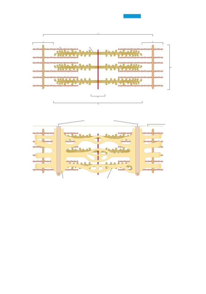
a.Muscle structure and filaments (Figure 1.11)
■Each muscle fiber is multinucleate and behaves as a single unit. It contains bundles of myofibrils, surrounded by sr and invaginated by transverse tubules (T tubules).
|
|
|
Chapter 1 Cell Physiology |
17 |
A |
Motoneuron |
|
Muscle |
|
|
Sarcomere |
|
||
|
|
|
|
|
|
I band |
|
I band |
|
|
Thin filament |
Thick filament |
|
|
Myofibril
Z line |
M line |
Z line |
|
H band |
|
|
A band |
|
B
Transverse tubules
Sarcolemmal membrane
Terminal cisternae Sarcoplasmic reticulum
Figure 1.11 Structure of the sarcomere in skeletal muscle. A: Arrangement of thick and thin filaments. B: Transverse tubules and sarcoplasmic reticulum.
■Each myofibril contains interdigitating thick and thin filaments arranged longitudinally in sarcomeres.
■Repeating units of sarcomeres account for the unique banding pattern in striated muscle. A sarcomere runs from Z line to Z line.
1. Thick filaments
■are present in the A band in the center of the sarcomere.
■contain myosin.
a. Myosin has six polypeptide chains, including one pair of heavy chains and two pairs of light chains.
b. Each myosin molecule has two “heads” attached to a single “tail.” The myosin heads bind ATP and actin and are involved in cross-bridge formation.
2. Thin filaments
■are anchored at the Z lines.
■are present in the I bands.

18BRS Physiology
■interdigitate with the thick filaments in a portion of the A band.
■contain actin, tropomyosin, and troponin.
a. Troponin is the regulatory protein that permits cross-bridge formation when it binds Ca2+.
b. Troponin is a complex of three globular proteins:
■Troponin T (“T” for tropomyosin) attaches the troponin complex to tropomyosin.
■Troponin I (“I” for inhibition) inhibits the interaction of actin and myosin.
■Troponin C (“C” for Ca2+) is the Ca2+-binding protein that, when bound to Ca2+, permits the interaction of actin and myosin.
3. T tubules
■are an extensive tubular network, open to the extracellular space, that carry the depolarization from the sarcolemmal membrane to the cell interior.
■are located at the junctions of A bands and I bands.
■contain a voltage-sensitive protein called the dihydropyridine receptor; depolarization causes a conformational change in the dihydropyridine receptor.
4. SR
■is the internal tubular structure that is the site of Ca2+ storage and release for excitation– contraction coupling.
■has terminal cisternae that make intimate contact with the T tubules in a triad arrangement.
■membrane contains Ca2+-ATPase (Ca2+ pump), which transports Ca2+ from intracellular fluid into the SR interior, keeping intracellular [Ca2+] low.
■contains Ca2+ bound loosely to calsequestrin.
■contains a Ca2+ release channel called the ryanodine receptor.
B.Steps in excitation–contraction coupling in skeletal muscle (Figures 1.12 and 1.13)
1. Action potentials in the muscle cell membrane initiate depolarization of the T tubules.
2. Depolarization of the T tubules causes a conformational change in its dihydropyridine receptor, which opens Ca2+ release channels (ryanodine receptors) in the nearby SR, caus-
ing release of Ca2+ from the SR into the intracellular fluid.
3. Intracellular [Ca2+] increases.
4. Ca2+ binds to troponin C on the thin filaments, causing a conformational change in troponin that moves tropomyosin out of the way. The cross-bridge cycle begins (see Figure 1.12):
a. At first, no ATP is bound to myosin (A) and myosin is tightly attached to actin. In rapidly
contracting muscle, this stage is brief. In the absence of ATP, this state is permanent (i.e., rigor).
b. ATP then binds to myosin (B) producing a conformational change in myosin that causes myosin to be released from actin.
c. Myosin is displaced toward the plus end of actin. There is hydrolysis of ATP to ADP and inorganic phosphate (Pi). ADP remains attached to myosin (C)
d. Myosin attaches to a new site on actin, which constitutes the power (force-generating) stroke (D) ADP is then released, returning myosin to its rigor state.
e. The cycle repeats as long as Ca2+ is bound to troponin C. Each cross-bridge cycle “walks” myosin further along the actin filament.
5. Relaxation occurs when Ca2+ is reaccumulated by the SR Ca2+-ATPase (SERCA). Intracellular Ca2+ concentration decreases, Ca2+ is released from troponin C, and tropomyosin again blocks the myosin-binding site on actin. As long as intracellular Ca2+ concentration is low, cross-bridge cycling cannot occur.
6. Mechanism of tetanus. A single action potential causes the release of a standard amount of Ca2+ from the SR and produces a single twitch. However, if the muscle is stimulated repeatedly, more Ca2+ is released from the SR and there is a cumulative increase in intracellular [Ca2+], extending the time for cross-bridge cycling. The muscle does not relax (tetanus).
|
|
|
Chapter 1 Cell Physiology |
19 |
|
|
Actin filament |
|
|
– |
|
|
+ |
|
|
|
Myosin |
Myosin |
|
|
|
head |
|
|
|
|
filament |
|
|
|
|
|
|
|
|
A |
|
|
|
– |
+ |
|
– |
+ |
|
|
|
||
ADP |
|
|
ATP |
|
D |
|
|
B |
|
– |
|
|
+ |
|
|
|
ADP Pi |
|
|
C
Figure 1.12 Cross-bridge cycle. Myosin “walks” toward the plus end of actin to produce shortening and force generation. ADP = adenosine diphosphate; ATP = adenosine triphosphate; Pi = inorganic phosphate.
C.Length–tension and force–velocity relationships in muscle
■Isometric contractions are measured when length is held constant. Muscle length (preload) is
fixed, the muscle is stimulated to contract, and the developed tension is measured. There is no shortening.
■Isotonic contractions are measured when load is held constant. The load against which the muscle contracts (afterload) is fixed, the muscle is stimulated to contract, and shortening is measured.
1. Length–tension relationship (Figure 1.14)
■measures tension developed during isometric contractions when the muscle is set to fixed lengths (preload).
Figure 1.13 Relationship of the action potential, the increase in intracellular [Ca2+], and muscle contraction in skeletal muscle.
Response
Action potential
Intracellular [Ca2+]
Twitch tension
Time

20 |
brs Physiology |
|
|
|
Total |
|
Tension |
Passive |
|
|
|
|
Length at maximum |
|
|
cross-bridge |
Active |
|
overlap |
|
|
|
|
|
Muscle length |
FIGure 1.14 Length–tension relation- |
|
|
ship in skeletal muscle. |
a.Passive tension is the tension developed by stretching the muscle to different lengths.
b.Total tension is the tension developed when the muscle is stimulated to contract at different lengths.
c.active tension is the difference between total tension and passive tension.
■Active tension represents the active force developed from contraction of the muscle. It can be explained by the cross-bridge cycle model.
■active tension is proportional to the number of cross-bridges formed. Tension will be maximum when there is maximum overlap of thick and thin filaments. When the muscle is stretched to greater lengths, the number of cross-bridges is reduced because there is less overlap. When muscle length is decreased, the thin filaments collide and tension is reduced.
2.Force–velocity relationship (Figure 1.15)
■ measures the velocity of shortening of isotonic contractions when the muscle is chal-
lenged with different afterloads (the load against which the muscle must contract).
■ The velocity of shortening decreases as the afterload increases.
VII. sMooTH MusCle
■has thick and thin filaments that are not arranged in sarcomeres; therefore, they appear homogeneous rather than striated.
a.Types of smooth muscle
1.Multiunit smooth muscle
■is present in the iris, ciliary muscle of the lens, and vas deferens.
■behaves as separate motor units.
Initial velocity of shortening
Afterload
FIGure 1.15 Force–velocity relationship in skeletal muscle.