


100 Muscles, synovial bursae and sheaths
|
1 |
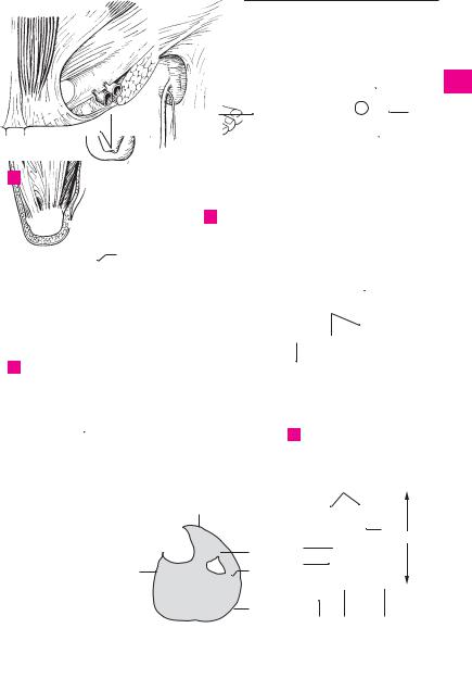
Femoral ring. Anulus femoralis. Entrance into |
15 |
Inferior (fibular) peroneal retinaculum. Reti- |
||||||||||||
|
|
the femoral canal bordered by the femoral vein, |
|
naculum mm. peroneorum (fibularium) in- |
||||||||||||
|
|
inguinal ligament, falx inguinalis and pectineal |
|
ferius. Lower band that holds the peroneal ten- |
||||||||||||
|
|
ligament. A |
|
|
|
|
|
dons in place. It passes from the extensor reti- |
||||||||
|
2 |
Femoral septum. Septum |
femorale. Fibrous |
|
naculum to the outer surface of the calcaneus. |
|||||||||||
|
|
A fibrous tract goes to the peroneal trochlea |
||||||||||||||
3 |
|
membrane that |
closes the |
entrance of the |
|
|||||||||||
|
|
and separates the upper lying peroneus brevis |
||||||||||||||
|
femoral canal. A |
|
|
|
|
|
||||||||||
|
|
|
|
|
|
|
from the peroneus longus muscle. C |
|
||||||||
|
3 Saphenous opening. Hiatus saphenus. Large |
|
|
|||||||||||||
4 |
16 |
Fascia dorsalis pedis. Thin fascia on the dor- |
||||||||||||||
|
opening in the fascia lata directly below the in- |
|||||||||||||||
|
|
guinal |
ligament |
for |
passage of |
the great |
|
sum of the foot connected above with the infe- |
||||||||
|
|
|
||||||||||||||
5 |
|
saphenous vein. B |
|
|
|
|
rior extensor retinaculum. C D |
|
|
|||||||
|
|
|
|
17 |
Plantar |
aponeurosis. Aponeurosis |
plantaris. |
|||||||||
|
4 |
Falciform margin. Margo falciformis. Curved, |
||||||||||||||
|
||||||||||||||||
6 |
|
principal lateral margin of the saphenous open- |
|
Tough, tendinous sheet on the sole of the foot |
||||||||||||
|
|
extending from the tuber calcanei to as far as |
||||||||||||||
|
ing. B |
|
|
|
|
|
|
|||||||||
|
5 |
Superior |
horn. Cornu superius. Upper, curved |
|
the middle phalanges. It braces the longitudinal |
|||||||||||
7 |
|
arch of the foot. E |
|
|
|
|
||||||||||
|
portion of the falciform margin. B |
|
18 |
Transverse |
fasciculi. |
Fasciculi |
transversi. |
|||||||||
|
|
|
||||||||||||||
|
6 |
Inferior horn. Cornu inferius. Lower, curved por- |
||||||||||||||
8 |
|
Transverse fibrous sheets in the distal plantar |
||||||||||||||
|
tion of the falciform magin. B |
|
|
aponeurosis. E |
|
|
|
|
||||||||
|
|
|
|
|
|
|
|
|||||||||
|
7 |
Cribriform fascia. Fascia cribrosa. Loose, per- |
19 |
Superficial |
transverse |
metatarsal |
ligament. |
|||||||||
9 |
||||||||||||||||
|
forated connective tissue lamina covering the |
|
Lig. metatarsale |
transversum |
superficiale. |
|||||||||||
|
|
saphenous opening. B |
|
|
|
|
Transverse fibrous tract in the vicinity of the |
|||||||||
10 |
8 |
Fascia of the leg (crural fascia). Fascia cruris. |
|
distal transverse fibers of the plantar |
||||||||||||
|
|
Superficial investing fascia of the leg which |
|
aponeurosis. E G |
|
|
|
|
||||||||
|
|
|
|
|
|
|
||||||||||
11 |
|
serves partially for muscle attachment and is |
19 a |
Synovial bursae (sacs) and sheaths. Bursae et |
||||||||||||
|
fused to the free bony margins of the tibia. C D F |
|
vaginae synoviales. |
|
|
|
||||||||||
|
|
|
|
|
|
|||||||||||
|
9 |
Anterior intermuscular septum of leg. Septum |
20 |
Synovial |
sheaths |
of the digits |
of |
the foot. |
||||||||
12 |
||||||||||||||||
|
intermusculare |
cruris |
anterius. |
Connective |
|
Vaginae synoviales tendinum digitorum pedis. |
||||||||||
|
|
tissue septum between the peroneal and exten- |
|
Synovial portion of the tendon sheaths for the |
||||||||||||
13 |
|
sor compartments. F |
|
|
|
|
flexors of the toes. G |
|
|
|
||||||
|
10 |
Posterior intermuscular septum of leg. Sep- |
21 |
Vincula |
tendinum. Connective |
tissue tract |
||||||||||
|
||||||||||||||||
14 |
|
tum intermusculare cruris posterius. Connec- |
|
passing obliquely through the tendon sheaths |
||||||||||||
|
tive tissue septum between the peroneal and |
|
||||||||||||||
|
|
|
bearing blood vessels. G |
|
|
|
||||||||||
|
|
flexor compartments. F |
|
|
|
22 |
Fibrous sheaths of the digits of the foot. |
|||||||||
15 |
|
|
|
|
||||||||||||
11 |
Superior extensor retinaculum. Retinaculum |
|||||||||||||||
|
Vaginae fibrosae tendinum digitorum pedis. |
|||||||||||||||
|
|
mm. |
extensorum |
superius. |
Transverse |
|
Tough fibrous sheath that reinforces the tendon |
|||||||||
16 |
|
thickened (about two finger’s breadth) of the |
|
sheaths on the flexor side of the toes. G |
||||||||||||
|
|
crural fascia that hold the extensor tendons in |
23 |
Annular part of fibrous sheath. Pars anularis |
||||||||||||
|
|
|||||||||||||||
|
|
place. C D |
|
|
|
|
||||||||||
|
|
|
|
|
|
|||||||||||
17 |
|
|
|
|
|
|
vaginae fibrosae. Circular tracts in the fibrous |
|||||||||
12 |
Flexor retinaculum. Retinaculum mm. flex- |
|
||||||||||||||
|
|
sheaths between the joints. G |
|
|
||||||||||||
|
|
orum. Fibrous band on the long flexor tendons |
24 |
Cruciate part of fibrous sheath. Pars cruci- |
||||||||||||
18 |
|
|||||||||||||||
|
that extends from the medial malleolus to the |
|||||||||||||||
|
|
calcaneus. It forms an osteofibrous compart- |
|
formis vaginae fibrosae. Crucitate connective |
||||||||||||
|
|
|
tissue tracts in the fibrous sheaths over the |
|||||||||||||
19 |
|
ment for the posterior tibial m., then divides |
|
|||||||||||||
|
|
joints. G |
|
|
|
|
|
|
||||||||
|
into two parts. The lower portion forms com- |
|
|
|
|
|
|
|
||||||||
|
|
|
|
|
|
|
|
|
|
|||||||
|
|
partments for the flexor digitorum longus and |
|
|
|
|
|
|
|
|
||||||
20flexor hallucis longus muscles. The tibial nerve and posterior tibial artery and vein lie between the two membranous parts. D
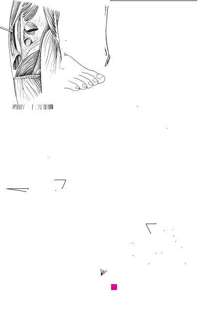
2113 Inferior extensor retinaculum. Retinaculum mm. extensorum inferius. Usually cruciate
22band that supports the extensor tendons, extending from both malleoli to the foot margins of the opposite side, primarily to the calcaneus.
23C D
24 |
14 Superior peroneal (fibular) retinaculum. Reti- |
naculum mm. peroneorum (fibularium) su- |
|
|
perius. Upper band that holds peroneal tendons |
25 |
in place; it extends from the lateral malleolus |
|
to the calcaneus. C |
Muscles, synovial bursae and sheaths 101
86.20
86.18
86.212
1
A Vascular lacuna from behind
811
13
14
15 16
C Foot, lateral view
 19
19
18
17
8
 5
5
7 |
3 |
4 |
|
 6
6
B Fascia lata in inguinal region
11 |
|
8 |
|
13
16
12
D Foot, medial view
|
|
24 |
|
8 |
|
|
|
|
|
|
23 |
|
|
|
22 |
9 |
20 |
|
|
|
|
|
|
10 |
21 |
|
24 |
|
|
||
|
|
|
|
8 |
|
|
|
|
20 |
19 |
19 |
E |
Plantar surface of foot |
F |
Cross-section of lower leg |
G |
Toes, plantar view |
1
2
3
4
5
6
7
8
9
10
11
12
13
14
15
16
17
18
19
20
21
22
23
24
25
a

102 Synovial bursae and sheaths
1 |
|
1 |
Tendon sheath of superior oblique muscle. |
16 |
Intratendinous bursa of olecranon. [B. in- |
|||||
|
|
Vagina tendinis m. obliqui superioris. Synovial |
|
tratendinea olecrani]. Synovial bursa within |
||||||
|
|
|
sheath of the superior oblique m. of the eyeball, |
|
the triceps tendon near the olecranon. F |
|||||
2 |
|
|
situated at the site where its tendon passes |
17 |
Subtendinous bursa of triceps brachii. B. sub- |
|||||
|
|
|
through the trochlea. See p. 364. 12 |
|
tendinea m. tricipitis brachii. Synovial bursa |
|||||
|
|
2 |
Synovial bursa of tensor veli palatini. Bursa m. |
|
||||||
3 |
|
|
between the triceps tendon and the olecranon. |
|||||||
|
|
tensoris veli palatini. Synovial bursa between |
|
F |
|
|
|
|
||
|
|
|
the pterygoid hamulus and the tendon of the |
18 |
Bicipitoradial bursa. B. bicipitoradialis. Syn- |
|||||
|
|
|
||||||||
4 |
|
|
||||||||
|
|
tensor veli palatini muscle. See pp. 116.20, 117. |
|
ovial bursa between the biceps tendon and the |
||||||
|
3 |
C |
|
anterior part of the radial tuberosity. F |
|
|||||
|
|
|
||||||||
5 |
Subcutaneous bursa of the laryngeal promi- |
19 |
[B. cubitalis |
interossea]. Synovial bursa be- |
||||||
|
|
|
nence. B. subcutanea prominentiae laryngealis. |
|
tween the biceps tendon and the ulna or ob- |
|||||
|
|
|
Synovial bursa between the skin and the laryn- |
|
||||||
|
|
|
|
lique cord. F |
|
|
|
|||
6 |
|
|
|
|
|
|
||||
|
|
geal prominence of the thyroid cartilage. A |
|
|
|
|
||||
|
|
20 Tendon sheath of abductor pollicis longus and |
||||||||
|
4 |
Retrohyoid bursa. B. retrohyoidea. Synovial |
||||||||
|
|
extensor pollicis brevis muscles. Vag. ten- |
||||||||
7 |
|
|
bursa between the body of the hyoid bone and |
|
||||||
|
|
|
dinum |
mm. abductoris longi et extensoris |
||||||
|
|
|
the median thyrohyoid ligament. A |
|
brevis pollicis. Common tendon sheath forming |
|||||
|
5 |
Infrahyoid bursa. B. infrahyoidea. Synovial |
|
|||||||
8 |
|
the first tendon compartment on the dorsum of |
||||||||
|
|
bursa between the upper end of the sternohy- |
|
the hand. G |
|
|
|
|||
|
|
|
oid muscle and the thyrohyoid membrane. A B |
21 Tendon sheath of extensor carpi radialis lon- |
||||||
9 |
|
|
||||||||
|
5 a Synovial bursae of upper limb. Bursae membri |
|||||||||
|
|
gus and brevis muscles. Vag. tendinum mm. |
||||||||
|
|
|
superioirs. |
|
extensorum carpi radialium. Common tendon |
|||||
|
|
|
|
|||||||
10 |
6 |
Subtendinous bursa of trapezius. B. subten- |
|
sheath forming the second tendon compart- |
||||||
|
ment on the dorsum of the hand. G |
|
||||||||
|
|
|
dinea m. trapezii. Synovial bursa between the |
|
|
|||||
|
|
|
|
|
|
|
|
|
||
|
|
|
trapezius muscle (ascending part) and the |
22 |
Tendon sheath of extensor pollicis longus |
|||||
11 |
|
|
||||||||
|
|
spine of the scapula. C |
|
muscle. Vag. tendinis m. extensoris pollicis |
||||||
|
7 |
[B. subcutanea acromialis]. Synovial bursa be- |
|
longi. Forms the third tendon compartment. G |
||||||
12 |
|
|||||||||
|
|
|
|
|
|
|||||
|
|
tween acromion and the skin. D |
23 |
Tendon sheath of extensor digitorum and ex- |
||||||
|
8 |
Subacromial bursa. B. subacromialis. Synovial |
|
tensor indicis muscles. Vag. tendinum mm. ex- |
||||||
|
|
|||||||||
13 |
|
|
bursa between the acromion, coracoacromial |
|
tensoris digitorum et extensoris indicis. Tendon |
|||||
|
|
ligament and supraspinous tendon. It and its |
|
sheath |
forming the fourth tendon compart- |
|||||
|
|
|
tendons lie on the joint capsule. D E |
|
ment on the dorsum of the hand. G |
|
||||
14 |
|
|
24 |
|
||||||
|
|
|
|
|
|
|
||||
9 |
Subdeltoid bursa. B. subdeltoidea. Synovial |
Tendon |
sheath of extensor |
digiti |
minimi |
|||||
|
|
muscle. Vag. tendinis m. extensoris digiti min- |
||||||||
|
|
|
bursa between the deltoid muscle and the |
|
||||||
15 |
|
|
greater tubercle of the humerus. It often com- |
|
imi. Forms the fifth tendon compartment on |
|||||
|
|
|
the dorsum of the hand. G |
|
|
|||||
|
|
|
municates with the subacromial bursa. D |
|
|
|
||||
|
|
|
25 |
Tendon sheath of extensor carpi ulnaris |
||||||
|
10 |
Coracobrachial bursa. [b. m. coracobrachialis]. |
||||||||
16 |
||||||||||
|
muscle. Vag. tendinis m. extensoris carpi ul- |
|||||||||
|
|
|
Synovial bursa between the tendons of the sub- |
|
naris. Forms the sixth tendon compartment on |
|||||
|
|
|
scapularis and coracobrachialis muscles below |
|
||||||
17 |
|
|
|
the dorsum of the hand. G |
|
|
||||
|
|
the apex of the coracoid process. D |
26 |
|
|
|||||
|
11 |
Subtendinous bursa of infraspinatus muscle. |
Sheath |
of |
extensor carpi |
radialis |
brevis |
|||
|
||||||||||
18 |
|
|
B. subtendinea m. infraspinati. Synovial bursa |
|
muscle. Vag. m. extensoris carpi radialis brevis. |
|||||
|
|
|
Synovial bursa at the attachment between the |
|||||||
|
|
between the tendon of the infraspinatus and |
|
|||||||
|
|
|
the capsule of the shoulder joint. E |
|
tendon and base of the 3rd metacarpal. |
|
||||
|
|
|
|
|
|
|
|
|
||
1912 Subtendinous bursa of subscapularis muscle.
B. subtendinea m. subscapularis. Synovial
20bursa between the tendon of the subscapularis and the capsule of the shoulder joint. It communicates with the joint cavity. D
2113 Subtendinous bursa of teres major muscle. B. subtendinea m. tertis majoris. Synovial bursa
22between the tendon of the teres major and the humerus. D
2314 Subtendinous bursa of latissimus dorsi muscle. B. subtendinea m. latissimi dorsi. Synovial bursa between the tendons of the teres
24major and latissimus dorsi. D
15 Subcutaneous bursa of olecranon. B. subcu-
25tanea olecrani. Synovial bursa between the olecranon and the skin. F
Synovial bursae and sheaths 103
1
4 |
|
|
|
|
|
|
|
|
5 |
|
|
|
|
|
|
|
|
|
|
|
|
|
2 |
||||||
|
|
|
|
|
|
|
|
|
|
|
|
|
|
|
|
|
|
|
|
|
|||||||||
5 |
|
|
|
|
|
|
|
|
|
|
|
|
|
|
|
|
|
|
|
|
|
|
|
|
|
|
|
||
|
|
|
|
|
|
|
|
|
|
|
|
|
|
|
|
|
|
6 |
|
|
|
|
|
|
|
|
|||
|
|
|
|
|
|
|
|
|
|
|
|
|
|
|
|
|
|
|
|
|
|
|
|
|
|
||||
|
|
|
|
|
|
|
|
|
|
|
|
|
|
|
|
|
|
|
|
|
|
|
|
|
|
|
3 |
||
|
|
|
|
|
|
|
|
|
|
|
|
|
|
|
|
|
|
|
|
|
|
|
|
|
|
|
|||
|
|
|
|
|
|
|
|
|
|
|
|
|
|
|
|
|
|
|
|
|
|
|
|
|
|
|
|
|
|
3 |
|
|
|
|
|
|
|
|
|
|
|
|
|
|
|
|
|
|
|
|
|
|
|
|
|
|
|
|
|
|
|
|
|
|
|
|
|
|
|
|
|
|
|
|
|
|
|
|
|
|
|
|
|
|
|
|
|
||
|
|
|
|
|
|
|
|
|
|
|
|
|
|
|
|
|
|
|
|
4 |
|||||||||
|
|
|
|
|
|
|
|
|
|
|
|
|
|
|
|
|
|
|
|
|
|
|
|
|
|
|
|||
|
|
|
|
|
|
|
|
|
|
|
|
|
|
|
|
|
|
|
|
|
|
|
|
|
|
|
|
|
|
|
|
|
|
|
|
|
|
|
|
|
|
|
|
|
|
|
|
|
|
|
|
|
|
|
|
|
|
|
|
|
|
|
|
|
|
|
|
|
|
|
|
|
|
|
|
|
|
|
|
|
|
|
|
|
|
|
|
|
|
|
|
|
|
|
|
|
|
|
|
|
|
|
|
|
|
|
|
|
|
|
|
|
|
|
|
|
|
|
5 |
|
|
|
|
|
|
|
|
|
|
|
|
|
|
|
|
|
|
|
|
|
|
|
|
|
|
|
|
|
|
|
|
|
|
|
|
|
|
|
|
|
|
|
|
|
|
|
|
|
|
|
|
|
|
|
|
|
|
|
|
|
|
|
|
|
|
|
|
|
|
|
|
|
|
|
|
|
|
|
|
|
|
|
|
|
|
|
|
|
6 |
|
A Sagittal section |
|
|
|
|
B Larynx, |
|
|
|
C Right shoulder, |
|
|
|||||||||||||||||
|
|
|
|
|
|
|
|
|
|
||||||||||||||||||||
|
|
|
|
7 |
|||||||||||||||||||||||||
|
|
|
of larynx |
|
|
|
|
|
|
|
lateral view |
|
|
|
posterior view |
|
|||||||||||||
|
|
|
|
|
|
|
|
|
|
|
|
|
|
|
|
|
|
|
|
|
|
|
|
|
|
|
|
|
|
7 |
|
|
|
|
|
|
|
|
|
|
|
|
|
|
|
|
|
|
|
|
|
|
|
|
|
8 |
|||
|
|
|
|
|
|
|
|
|
|
|
|
|
|
|
|
|
|
|
|
8 |
|
|
|||||||
|
|
|
|
|
|
|
|
|
|
|
|
|
|
|
|
|
|
|
|
|
|
||||||||
8 |
|
|
|
|
|
|
|
|
|
|
|
|
|
9 |
|||||||||||||||
|
|
|
|
|
|
|
|
|
|
|
|
|
|
|
|
|
|
|
|
|
|
||||||||
|
|
|
|
|
|
|
|
|
|
|
|
|
|
|
|
|
|
|
|
|
|||||||||
10 |
|
|
|
|
|
|
|
|
|
|
|
|
|
|
|
|
|
|
|
|
|
|
|||||||
|
|
|
|
|
|
|
|
|
|
|
|
|
|
|
|
|
|
|
|
|
10 |
||||||||
|
|
|
|
|
|
|
|
|
|
|
|
|
|
|
|
|
|
|
|
|
|
|
|
|
|
|
|
|
|
|
|
|
|
|
|
|
|
|
|
|
|
|
|
|
|
|
|
|
|
|
|
|
|
|
|
|
|
|
|
|
|
|
|
|
|
|
|
|
|
|
|
|
|
|
|
|
|
|
|
|
|
|
|
|
|
|
|
|
|
9 |
|
|
|
|
|
|
|
|
12 |
|
|
|
|
|
|
|
11 |
|
|
|
11 |
||||||||
|
|
|
|
|
|
|
|
|
|
|
|
|
|
|
|
|
|
|
|
|
|||||||||
|
|
|
|
|
|
|
|
|
|
|
|
|
|
|
|
|
|
|
|
||||||||||
|
|
|
|
|
|
|
|
|
|
|
|
|
|
|
|
|
|
|
|
|
|
|
|
|
|
|
|
|
|
|
|
|
|
|
|
|
|
|
|
|
|
|
|
|
|
|
|
|
|
|
|
|
|
|
|
|
|
|
|
|
|
|
|
|
|
|
|
|
|
|
|
|
|
|
|
|
|
|
|
|
|
|
|
|
|
|
|
|
12 |
|
|
|
|
|
|
|
|
|
|
|
|
|
|
|
|
|
|
|
|
|
|
|
|
|
|
|
|
|
|
|
|
|
|
|
|
|
|
|
|
|
|
|
|
|
|
|
|
|
|
|
|
|
|
|
|
|
|
|
|
|
|
|
|
|
|
|
|
|
|
|
|
|
|
|
|
|
|
|
|
|
|
|
|
|
|
|
|
|
13 |
14 |
|
|
|
|
|
|
|
|
|
|
|
|
|
|
|
|
|
|
|
|
|
|
|||||||
|
|
|
|
|
|
|
|
|
|
|
|
|
|
|
|
|
|
|
|
|
|
||||||||
|
|
|
|
|
|
|
|
|
|
|
|
|
|
|
|
|
|
|
|
14 |
|||||||||
|
|
|
|
|
|
|
|
|
|
|
|
|
|
|
|
|
|
|
|
|
|
|
|
|
|
|
|
|
|
|
|
|
|
|
|
|
|
|
|
|
|
|
|
|
|
|
|
|
|
|
|
|
|
|
|
|
|
|
|
|
|
|
|
|
|
|
|
|
|
|
|
|
|
|
|
|
|
|
|
|
|
|
|
|
|
|
|
|
|
13 |
|
D |
Shoulder joint, anterior view |
E |
Shoulder joint, posterior view |
|
15 |
||||||||||||||||||||||
|
|
|
|
|
|
|
|
|
|
|
|
|
|
|
|
|
|
|
|
|
|
|
|
|
|
|
|
|
|
|
|
|
|
|
|
|
|
|
|
|
|
|
|
|
|
|
|
|
|
|
|
|
|
|
|
|
|
|
16 |
|
|
|
|
|
|
|
|
|
|
|
|
|
|
|
|
|
|
|
|
|
|
|
|
|
|
|
|
|
|
|
|
|
|
|
|
|
|
|
|
|
|
|
|
|
|
|
|
|
|
|
|
|
|
|
|
|
|
|
|
|
|
|
|
|
|
|
|
|
|
|
|
|
|
|
|
|
|
|
|
|
|
|
|
|
|
|
|
|
17 |
|
|
|
|
|
|
|
|
|
|
16 |
|
|
|
|
|
|
|
|
|
|
|
|
|
||||||
|
|
|
|
|
|
|
|
|
|
|
|
|
|
|
|
|
|
|
|
|
|
|
|||||||
|
|
|
|
|
|
|
|
|
|
|
|
|
|
|
|
|
|
|
|
|
18 |
||||||||
|
|
|
|
|
|
|
|
|
|
|
|
|
|
|
17 |
|
|
|
|
|
|
|
|
|
|
|
|
||
|
|
|
|
|
|
|
|
|
|
|
|
|
|
|
|
|
|
|
|
|
|
|
|
|
|
|
|
||
|
|
|
|
|
|
|
|
|
|
|
|
|
|
15 |
|
|
|
|
|
|
|
|
|
|
|
|
|
||
|
|
|
|
|
|
|
|
|
|
|
|
|
|
|
|
|
|
|
|
|
|
|
|
|
19 |
||||
|
|
|
|
|
|
|
|
|
|
|
|
|
|
|
|
|
|
|
|
|
|
||||||||
|
|
|
|
|
|
|
|
|
|
|
|
|
|
|
|
|
|
|
|
|
|
|
|
|
|
|
|
|
|
|
|
|
|
|
|
|
|
|
|
|
|
|
|
|
|
|
|
|
|
|
|
|
|
|
|
|
|
|
|
|
|
|
|
|
|
|
|
|
|
|
|
|
|
|
|
|
|
|
|
|
|
|
|
|
|
|
|
|
20 |
|
|
|
|
|
|
|
|
|
|
|
|
|
|
|
|
|
|
|
|
|
|
|
|
|
|
|
|
24 |
|
|
|
|
|
|
|
|
|
|
|
|
|
|
|
|
|
|
|
|
|
|
|
|
|
|
|
|
|
|
|
|
|
|
|
|
|
|
|
|
|
|
|
|
|
|
|
|
|
|
|
|
|
|
|
|
|
|
|
|
|
|
|
|
|
|
|
|
|
|
|
|
|
|
|
|
|
|
|
22 |
|
|
|
|
|
|
|
|
|
21 |
|
|
|
|
|
|
|
|
|
|
|
|
|
|
|
|
|
|
|
|
|
|
|
|
|
|
|
|
|
||
|
|
|
|
|
|
|
|
|
|
|
|
|
|
|
|
|
|
|
|
|
|
|
|
|
|
|
|
|
|
19 |
|
|
|
|
|
|
|
|
|
|
|
|
|
|
21 |
|
|
23 |
|
|
|
|
|
|
|
||||
|
|
|
|
|
|
|
|
|
|
|
|
|
|
|
|
|
|
|
|
22 |
|||||||||
|
|
|
|
|
|
|
|
|
|
|
|
|
|
|
|
|
|
|
|
|
|
||||||||
|
|
|
|
|
|
|
|
|
|
|
|
|
|
20 |
|
|
|
|
|
|
|
|
|
|
|||||
|
|
|
|
|
|
|
|
|
|
|
|
|
|
|
|
|
|
|
|||||||||||
18 |
|
|
|
|
|
|
|
|
|
|
|
|
|
|
|
|
|
|
|
|
|
|
|
|
|||||
|
|
|
|
|
|
|
|
|
|
|
|
|
|
|
|
|
|
|
|
|
|
|
|
|
|
||||
|
|
|
|
|
|
|
|
|
|
|
|
|
|
|
|
|
|
|
|
|
|
|
|
|
|
||||
|
|
|
|
|
|
|
|
|
|
|
|
|
|
|
|
|
|
|
|
|
|
|
|
|
|
|
|
25 |
23 |
|
|
|
|
|
|
|
|
|
|
|
|
|
|
|
|
|
|
|
|
|
|
|
|
|
|
|
|
|
|
|
|
|
|
|
|
|
|
|
|
|
|
|
|
|
|
|
|
|
|
|
|
|
|
|
|
|
|
|
|
|
|
|
|
|
|
|
|
|
|
|
|
|
|
|
|
|
|
|
|
|
|
|
|
|
|
|
|
|
24 |
|
F Section of elbow joint sawed open |
G |
Wrist and hand, dorsal view |
|
|
||||||||||||||||||||||||
|
|
||||||||||||||||||||||||||||
25

104 Synovial bursae and sheaths
1 |
|
1 |
Tendon sheath of flexor carpi radialis muscle. |
15 |
Subtendinous iliac bursa. B. subtendinea iliaca. |
|
|
Vag. tendinis m. flexoris carpi radialis. In- |
|
Synovial bursa between the lesser trochanter |
|
|
|
|
dividual tendon sheath for the flexor carpi |
|
and the iliopsoas tendon. C |
2 |
|
|
radialis at the insertion of the tendon to the |
16 |
Superior bursa of biceps femoris muscle. B. m. |
|
|
|
base of the 2nd metacarpal bone. A |
|
bicipitis femoris superior. Synovial bursa be- |
|
|
|
|
|
|
3 |
|
2 Common sheath of flexor muscles. Vag. com- |
|
tween the origins of the biceps femoris and |
|
|
|
munis mm. flexorum. Common tendon sheath |
|
semimembranosus muscles. B |
|
|
|
|
|
||
|
|
|
for the two long flexors of the fingers. A |
17 |
Subcutaneous prepatellar bursa. B. subcu- |
|
|
|
|||
4 |
|
|
|||
|
|
|
|||
3 |
Tendon sheath of flexor pollicis longus |
|
tanea prepatellaris. Synovial bursa directly be- |
||
|
|
|
muscle. Vag. tendinis m. flexoris pollicis longi. |
|
tween the skin and the fascia in front of the |
5 |
|
|
Separate synovial sheath for the long flexor of |
|
knee. D |
|
|
|
the thumb. A |
18 |
Subfascial prepatellar bursa. [B. subfascialis |
|
|
|
|
||
6 |
|
4 Tendon sheaths for flexors in region of fin- |
|
prepatellaris]. Synovial bursa between the in- |
|
|
|
gers. Vag. tendinum digitorum manus. A |
|
vesting fascia of the knee and the tendon of the |
|
|
|
|
|
||
|
|
4 a Synovial bursae of lower limb. Bursae membri |
|
quadratus femoris muscle. D |
|
7 |
|
|
|||
|
19 |
Subtendinous prepatellar bursa. [B. subten- |
|||
|
|
inferioris. |
|||
|
|
5 Subcutaneous trochanteric bursa. Bursa sub- |
|
dinea prepatellaris]. Synovial bursa directly on |
|
8 |
|
|
the knee joint below the tendon of the quadra- |
||
|
|
cutanea trochanterica. Synovial bursa on the |
|
||
|
|
|
tendon of the gluteus maximus between the |
|
tus femoris. D |
|
|
|
20 |
Suprapatellar bursa. B. suprapatellaris. Syn- |
|
9 |
|
|
skin and greater trochanter. B |
||
6 |
Trochanteric bursa of gluteus maximus. B. |
|
ovial bursa between the quadriceps tendon and |
||
|
|
||||
|
|
the femur. It almost always communicates with |
|||
|
|
|
trochanterica m. glutei maximi. Synovial bursa |
|
|
10 |
|
|
|
the joint cavity. D |
|
|
|
between the tendon of the gluteus maximus |
|
||
|
|
|
and the greater trochanter. B |
21 |
Subcutaneous infrapatellar bursa. B. subcu- |
11 |
7 |
Trochanteric bursae of gluteus medius. Bb. |
|
tanea infrapatellaris. Synovial bursa between |
|
|
the ligamentum patellae and the skin. D |
||||
|
|
|
trochantericae m. glutei medii. This designa- |
|
|
|
|
|
22 |
Deep infrapatellar bursa. B. infrapatellaris pro- |
|
12 |
|
|
tion comprises two synovial bursae, an anterior |
||
|
|
one between the tendon of insertion of the glu- |
|
funda. Synovial bursa between the ligamentum |
|
|
|
|
|
||
|
|
|
teus medius and the greater trochanter and a |
|
patellae and the tibia. D |
13 |
|
|
|
||
|
|
posterior one between this tendon and the piri- |
23 |
Subcutaneous bursa of tibial tuberosity. B. |
|
|
|
|
formis muscle. B C |
|
subcutanea tuberositas tibiae. Synovial bursa |
|
|
|
|
||
14 |
8 |
Trochanteric bursa of gluteus minimus. B. tro- |
|
between the tibial tuberosity and the skin. It is |
|
|
|
|
chantericae m. glutei minimi. Synovial bursa |
|
mostly involved in kneeling. D |
|
|
|
|
||
|
|
|
between the tendon of insertion of the gluteus |
24 |
Subtendinous bursae of sartorius muscle. Bb. |
15 |
|
|
|||
|
|
minimus and the greater trochanter. B C |
|
subtendineae m. sartorii. Synovial bursae be- |
|
|
|
9 Bursa of piriformis muscle. B. m. piriformis. |
|
tween the sartorius tendon and the tendons of |
|
|
|
||||
16 |
|
|
Synovial bursa between the piriformis tendon, |
|
the gracilis and semitendinosus situated below |
|
|
|
femur and superior gemellus muscle. B |
|
it. E |
|
|
|
|
||
1710 Ischial bursa of obturator internus muscle. B. ischiadica (sciatica) m. obturatoris interni. Synovial bursa between the cartilage-covered sur-
18face of the lesser sciatic notch and the tendon of the obturator internus. B
1911 Subtendinous bursa of obturator internus muscle. B. subtendinea m. obturatoris interni. Synovial bursa below the insertion of the obtu-
20rator internus. B
12 Intermuscular bursae of gluteal muscles. Bb.
21intermusculares mm. gluteorum. 2−3 synovial bursae that extend inferiorly from the gluteus
22maximus to the linea aspera. B
13 Ischial bursa of gluteus maximus muscle. B.
23ischiadica (sciatica) m. glutei maximi. Synovial bursa between the ischial tuberosity and the inferior surface of the gluteus maximus. B
2414 Iliopectineal bursa. [B. iliopectinea]. Synovial bursa between the iliopsoas muscle and the
25pelvic bone. It lies above and often communicates with the hip joint. C
Synovial bursae and sheaths 105
4
|
4 |
|
|
|||
|
|
|
|
|
3 |
|
|
|
|||||
|
|
|
|
|
|
|
|
2 |
|
|
|
|
|
2 |
|
|
1 |
|
|
|
|
|
|
||||
|
|
|
|
|
|
|
|
3 |
|
|
|||
A Palmar view of hand
20
7 
8
 18
18
 17
17  19
19
22
21
23
9
8
7
10 
 11
11
5
|
6 |
|||
13 |
|
|
|
|
|
|
|
||
16 |
|
|
|
|
12 |
||||
|
||||
BDeep hip region, dorsal view
14
C Hip joint, anterior view
 15
15
24
D |
Knee, sagittal section sawed open |
E |
Knee, anterior view |
1
2
3
4
5
6
7
8
9
10
11
12
13
14
15
16
17
18
19
20
21
22
23
24
25

106
1 |
|
1 Anserine bursa. B. anserina. Synovial bursa on |
15 Common tendon sheath for peroneal muscles. |
|
|
|
the tibial collateral ligament below the tendons |
Vag. tendinum mm. peroneorum (fibularium) |
|
|
|
|
of the semitendinosus, gracilis and sartorius |
communis. It lies below the peroneal reti- |
2 |
|
|
muscles. It occasionally communicates with |
naculum and extends to the cuboid bone. C |
|
|
|
the subtendinous bursa of the sartorius. A |
16 Subtendinous bursa of tibialis anterior |
3 |
|
2 Inferior subtendinous bursa of biceps femoris |
muscle. B. subtendinea m. tibialis anterioris. |
|
|
|
muscle. B. subtendinea m. bicipitis femoris in- |
Synovial bursa between the tibialis anterior |
|
|
|
|
ferior. Synovial bursa located partially on the |
tendon and the medial cuneiform bone. D |
|
|
|
||
4 |
|
|
fibular collateral ligament below the tendon of |
17 Subcutaneous calcaneal bursa. B. sucutanea |
|
|
|
insertion of the biceps femoris. B |
calcanea. Synovial bursa between the skin and |
|
|
|
||
5 |
3 |
Subpopliteal recess. Recessus subpopliteus |
the posterior surface of the calcaneus. D |
|
|
|
[bursa m. poplitei]. Synovial bursa on the |
18 Bursa of calcaneal [[Achilles]] tendon. B. ten- |
|
|
|
|
||
|
|
|
lateral femoral condyle below the tendon of |
dinis calcanei [Achilles]. Synovial bursa be- |
6 |
|
|
||
|
|
origin of the popliteal muscle. It always com- |
tween the calcaneus and the Achilles tendon. D |
|
|
|
|
municates with the knee joint cavity, more |
19 Tendon sheath of peroneus longus muscle at |
|
|
|
||
7 |
|
|
rarely with the tibiofibular joint. B |
the sole of the foot. Vag. tendinis m. peronei |
|
4 Lateral subtendinous bursa of gastrocnemius |
|||
|
|
(fibularis) longi plantaris. D |
||
|
|
|
muscle. B. subtendinea m. gastrocnemii later- |
|
|
|
|
20 Tendon sheaths for the flexors of the toes. |
|
8 |
|
|
||
|
|
alis. Synovial bursa between the lateral condyle |
||
|
|
Vagg. tendinum digitorum pedis. D |
||
|
|
|
of the femur and the lateral gastrocnemius ten- |
|
|
|
|
|
|
9 |
|
|
don. B |
|
|
5 Medial subtendinous bursa of gastrocnemius |
|
||
|
|
|
||
|
|
|
muscle. B. subtendinea m. gastrocnemii medi- |
|
10alis. Synovial bursa between the medial condyle of the femur and the medial gastrocne-
11mius tendon. A B
6Bursa of semimembranosus muscle. B. m. semimembranosi. Synovial bursa between the
12semimembranosus tendon and the upper margin of the tibia. A
137 Subcutaneous bursa of lateral malleolus. B. subcutanea malleoli lateralis. Synovial bursa between the skin and the lateral malleolus. C
148 Subcutaneous bursa of medial malleolus. B. subcutanea malleoli medialis. Synovial bursa
15between the skin and the medial malleolus. D
9Tendon sheath of tibialis anterior muscle. Vag.
16tendinis m. tibialis anterioris. It begins just below the extensor retinaculum. D
17 |
10 Tendon sheath of extensor hallucis longus |
||
|
muscle. Vag. tendinis m. extensoris hallucis |
||
|
|
longi. Sheath extending below the extensor ret- |
|
18 |
|
inaculum and further distal. C D |
|
|
11 Tendon sheath of extensor digitorum longus |
||
|
|||
19 |
|
muscle. Vag. tendinum m. extensoris digitorum |
|
|
pedis longi. Sheath extending below the exten- |
||
|
|
||
|
|
sor retinaculum and further distal. C |
|
20 |
12 |
||
Tendon sheath of flexor digitorum longus |
|||
|
|
muscle. Vag. tendinum m. flexoris digitorum |
|
|
|
||
21 |
|
pedis longi. It lies behind and below the medial |
|
|
malleolus covered by the flexor retinaculum. D |
||
|
|
||
|
13 Tendon sheath of tibialis posterior muscle. |
||
22 |
|||
|
Vag. tendinis m. tibialis posterioris. It resides |
||
|
|
below the flexor retinaculum and begins at the |
|
23 |
|
point where it is crossed over by the flexor digi- |
|
|
|
torum longus. D |
|
|
14 |
Tendon sheath of flexor hallucis longus |
|
24 |
|||
|
muscle. Vag. tendinis m. flexoris hallucis longi. |
||
|
|
It extends up to the proximal end of the sole, |
|
25 |
|
where it crosses under the tendon of the flexor |
|
|
|
digitorum longus. D |
|
Synovial bursae and sheaths 107
|
|
|
|
|
|
|
|
|
|
|
|
|
|
|
|
|
|
|
|
|
|
|
|
|
|
|
|
|
|
|
|
|
|
|
1 |
|
|
|
|
|
|
|
|
|
|
|
|
|
|
|
|
|
|
|
|
|
|
|
|
|
|
|
|
|
|
|
|
|
|
|
2 |
|
|
|
|
|
|
|
|
|
|
|
|
|
|
5 |
|
|
|
|
|
|
|
|
|
|
|
|
|
|
|
|
|
|
|
|
3 |
|
|
|
|
|
|
|
|
|
|
|
|
|
|
|
|
|
|
|
|
|
|
|
|
|
|
|
|
|
|||||||
|
|
|
|
|
|
|
|
|
|
|
|
|
|
|
|
|
|
|
|
|
|
|
|
|
|
|
|
|
|
|
|
|
|
|
|
|
|
|
|
|
|
|
6 |
|
|
|
|
|
|
|
|
|
|
|
|
|
|
|
|
|
|
|
|
4 |
|||||||
|
|
|
|
|
|
|
|
|
|
|
|
|
|
|
|
|
|
|
|
|
|
||||||||||||||
|
|
|
|
|
|
|
|
|
|
|
|
|
|
|
|
|
|
|
|
|
|
|
|
|
|
|
|
|
|
|
|
|
|
|
|
|
|
|
|
|
|
|
|
|
|
|
|
|
|
|
|
|
|
|
|
|
|
|
|
|
|
|
|
|
|
|
|
|
|
|
|
|
|
|
|
|
|
|
|
|
|
|
|
|
|
|
|
|
|
|
|
|
|
|
|
|
|
|
|
|
|
|
|
|
|
|
5 |
|
1 |
|
|
|
|
|
|
|
|
|
|
|
|
|
|
|
|
|
|
|
|
|
|
|
|
|
|
|
|
|
|
|
|||
|
|
|
|
|
|
|
|
|
|
|
|
|
|
|
|
|
|
|
|
|
|
|
|
|
|
|
|
|
|
|
|
||||
|
|
|
|
|
|
|
|
|
|
|
|
|
|
|
|
|
6 |
||||||||||||||||||
|
|
|
|
|
|
|
|
|
|
|
|
|
|
|
|
|
|
|
|
|
|
|
|
|
|
|
|
|
|
|
|||||
|
|
|
|
|
|
|
|
|
|
|
|
|
|
|
|
|
|
|
|
|
|
|
|
|
|
|
|
|
|
|
|
|
|
|
|
|
|
|
|
|
|
|
|
|
|
|
|
|
|
|
|
|
|
|
|
|
|
|
|
|
|
|
|
|
|
|
|
|
|
|
|
|
|
|
|
|
|
|
|
|
|
|
|
|
|
|
|
|
|
|
|
|
|
|
|
|
|
|
|
|
|
|
|
|
|
|
|
|
|
|
|
|
|
|
|
|
|
|
|
|
|
|
|
|
|
|
|
|
|
|
|
|
|
|
|
|
|
|
|
|
|
|
7 |
|
|
|
|
|
|
|
|
|
|
|
|
|
|
|
|
|
|
|
|
|
|
|
|
|
|
|
|
|
|
|
|
|
|
|
|
|
|
|
|
|
|
|
|
|
|
|
|
|
|
|
|
|
|
|
|
|
|
|
|
|
|
|
|
|
|
|
|
|
|
|
|
|
|
|
|
|
|
|
|
|
|
|
|
|
|
|
|
|
|
|
|
|
|
|
|
|
|
|
|
|
|
|
|
|
|
|
8 |
|
5 |
|
|
|
|
|
|
|
|
|
|
|
|
|
|
|
4 |
|
|
|
|
||||||||||||||
|
|
|
|
|
|
|
|
|
|
|
|
|
|
|
|
|
|
|
|
||||||||||||||||
|
|
|
|
|
|
|
|
|
|
|
|
|
|
|
9 |
||||||||||||||||||||
|
|
|
|
|
|
|
|
|
|
|
|
|
|
|
|
|
|
|
|||||||||||||||||
|
|
|
|
|
|
|
|
|
|
|
|
|
|
|
|
|
|
|
|
|
|
|
|
|
|
|
|
||||||||
|
|
|
|
|
|
|
|
|
|
|
|
|
|
|
|
|
|
|
|
|
|
|
|
|
|
|
|
|
|
|
|
|
|
|
|
|
|
|
|
|
|
|
|
|
|
|
|
|
|
|
|
|
|
|
|
|
|
|
|
|
|
|
|
|
|
3 |
|
|
|
|
|
|
|
|
|
|
|
|
|
|
|
|
|
|
|
|
|
|
|
|
|
|
|
|
|
|
|
|
|
||||||||
|
|
|
|
|
A |
Right knee joint, posterior view |
|
|
|
|
|
|
|
|
|
|
|
|
|
10 |
|||||||||||||||
|
|
|
|
|
|
|
|
|
|
|
|
||||||||||||||||||||||||
|
|
|
|
|
|
|
|
|
|
|
|
|
|
|
|
|
|
|
|
|
|||||||||||||||
|
|
|
|
|
|
|
|
|
|
|
|
|
|
|
|
|
|
|
|
|
|
|
|
|
|
|
|
|
|
2 |
|
|
|
||
|
|
|
|
|
|
|
|
|
|
|
|
|
|
|
|
|
|
|
|
|
|
|
|
|
|
|
|||||||||
|
|
|
|
|
|
|
|
|
|
|
|
|
|
|
|
|
|
|
|
|
|
||||||||||||||
|
|
|
|
|
|
|
|
|
|
|
|
|
|
|
|
|
|
|
|
|
|
|
|
|
11 |
||||||||||
|
|
|
|
|
|
|
|
|
|
|
|
|
|
|
|
|
|
|
|
|
|
|
|
|
|
|
|
|
|
|
|
|
|
|
|
|
|
|
|
|
|
|
|
|
|
|
|
|
|
|
|
|
|
|
|
|
|
|
|
|
|
|
|
|
|
|
|
|
|
|
|
|
|
|
|
|
|
|
|
|
|
|
|
|
|
|
|
|
|
|
|
|
|
|
|
|
|
|
|
|
|
|
|
|
|
|
|
|
|
|
|
|
|
|
|
|
|
|
|
|
|
|
|
|
|
|
|
|
|
|
|
|
|
|
|
|
|
|
|
|
|
|
12 |
|
|
|
|
|
|
|
|
|
10 |
|
|
|
|
|
|
|
|
|
|
|
|
|
|
|
|
|
|
|
|
|
|
|
|
|
|
|
|
|
|
|
|
|
|
|
|
|
|
|
|
|
|
|
|
|
|
|
|
|
|
|
|
|
|
|
|
|
|
|
|
||
|
|
|
|
|
|
|
|
|
|
|
|
|
|
|
|
|
|
|
|
|
|
|
|
|
|
|
|
|
|
|
|
13 |
|||
|
|
|
|
|
|
|
|
|
|
|
|
|
|
|
|
|
|
|
|
||||||||||||||||
|
|
|
|
|
|
|
|
|
|
|
|
|
|
|
|
|
|
|
|
|
|
|
|
|
|
|
|
|
|
|
|
|
|
|
|
|
|
|
|
|
|
|
|
|
|
|
|
|
|
|
|
|
|
|
|
|
|
|
|
|
|
|
|
|
|
|
|
|
|
|
|
|
|
|
|
|
|
|
|
|
|
|
|
|
|
|
|
|
|
|
|
|
|
|
|
|
|
|
|
|
|
|
|
|
|
|
|
|
|
|
|
|
|
|
|
|
|
|
|
|
|
|
|
|
|
|
|
|
|
|
|
|
|
|
|
|
|
|
|
|
|
|
14 |
|
|
|
|
|
|
|
|
|
|
|
|
|
|
|
|
B |
Right knee joint, posterior view |
||||||||||||||||||
|
|
|
|
|
|
|
|
|
|
|
|
|
|
|
|
|
|||||||||||||||||||
7 |
10 |
|
|
|
|
|
|
|
|
|
|
|
|
|
|
|
|
|
|
|
|
|
|
|
|||||||||||
|
|
|
|
|
|
|
|
|
|
|
|
|
|
|
|
15 |
|||||||||||||||||||
|
|
|
|
|
|
|
|
|
|
|
|
|
|
|
|
|
|
|
|
|
|
|
|
|
|
|
|
|
|
|
|
|
|
||
15 |
|
|
|
|
|
|
|
|
|
|
|
|
11 |
|
|
|
|
|
|
|
|
|
|
|
|
|
|
|
|
|
|
|
|
|
|
|
|
|
|
|
|
|
|
|
|
|
|
|
|
|
|
|
|
|
|
|
|
|
|
|
|
|
|
|
|
|
|
|
|
||
|
|
|
|
|
|
|
|
|
|
|
|
|
|
|
|
|
|
|
|
|
|
|
|
|
|
|
|
|
|
|
16 |
||||
|
|
|
|
|
|
|
|
|
|
|
|
|
|
|
|
|
|
|
|
|
|
|
|
|
|
|
|
|
|
|
|
|
|
|
|
|
|
|
|
|
|
|
|
|
|
|
|
|
|
|
|
|
|
|
|
|
|
|
|
|
|
|
|
|
|
|
|
|
|
|
|
|
|
|
|
|
|
|
|
|
|
|
|
|
|
|
|
|
|
|
|
|
|
|
|
|
|
|
|
|
|
|
|
|
|
|
|
|
|
|
|
|
|
|
|
|
|
|
|
|
|
|
|
|
|
|
|
|
|
|
|
|
|
|
|
|
|
|
|
|
|
|
17 |
|
|
|
|
|
|
|
|
|
|
|
|
|
|
|
|
|
|
|
|
|
|
|
|
|
|
|
|
|
|
|
|
|
|
|
|
|
|
|
|
|
|
|
|
|
|
|
|
|
|
|
|
|
|
|
|
|
|
|
|
|
|
|
|
|
|
|
|
|
|
|
|
|
|
|
|
|
|
|
|
|
|
|
|
|
|
|
|
|
|
|
9 |
|
8 |
|
|
|
|
|
|
|
|
|
18 |
||||
|
|
|
Foot, lateral view |
|
|
|
|
|
|
|
|
|
|
|
|||||||||||||||||||||
|
|
C |
|
|
|
|
|
|
|
|
|
|
|
|
|
|
|
|
|
||||||||||||||||
|
|
|
10 |
|
|
|
|
|
|
13 |
|
19 |
|||||||||||||||||||||||
|
|
|
|
|
|
|
|
|
|
|
|
|
|
|
|
|
|
|
|
|
|
|
|
|
|
|
|
|
|
|
|
12 |
|
|
|
|
|
|
|
|
|
|
|
|
|
|
|
|
|
|
|
|
|
|
|
|
|
|
|
|
|
|
|
|
|
|
14 |
|
20 |
||
|
|
|
|
|
|
|
|
|
|
|
|
|
|
|
|
|
|
|
|
|
|
|
|
|
|
||||||||||
|
|
|
|
|
|
|
|
|
|
|
|
|
|
|
|
|
|
|
|
|
|
|
|
|
|
|
|
|
|
|
|
|
18 |
|
|
|
|
|
|
|
|
|
|
|
|
|
|
|
|
|
|
|
|
|
|
|
|
|
|
|
|
|
|
|
|
|
|
||||
|
|
|
|
|
|
|
|
|
|
|
|
|
|
|
|
|
|
|
|
|
|
|
|
|
|
|
|
|
|
|
|
|
|
|
|
|
|
|
|
|
|
|
|
|
|
|
|
|
|
|
|
|
|
|
|
|
|
|
|
|
|
|
|
|
|
|
|
|
17 |
|
21 |
|
|
|
|
|
|
|
|
|
|
|
|
|
|
|
|
|
|
|
|
|
|
|
|
|
|
|
|
|
|
|
|
|
|
||
|
|
|
|
|
|
|
|
|
|
|
|
|
|
|
|
|
|
|
|
|
|
|
|
|
|
|
|
|
|
|
|
|
|
||
|
|
|
|
|
|
|
|
|
|
|
|
|
|
|
|
|
|
|
|
|
|
|
|
|
|
|
|
|
|
|
|||||
|
|
|
|
|
|
|
|
|
|
|
|
|
|
|
|
|
|
|
|
|
|
||||||||||||||
|
|
|
|
|
|
|
|
|
|
|
|
|
|
|
|
|
|
|
|
|
22 |
||||||||||||||
|
|
|
|
|
|
|
|
|
|
|
|
|
|
|
|
|
|
|
|
|
|
|
|||||||||||||
|
|
|
|
|
|
|
|
|
|
|
|
|
|
|
|
|
|
|
|
|
|
|
|
|
|
|
|
|
|
|
|
|
|
|
|
|
16 |
19 |
12 14 |
|
|
|
|
|
|
|
|
|
|
|
|||||||||||||||||||||
|
20 |
|
|
|
|
|
|
|
|
|
|
|
|
|
|
|
|
|
|
|
|
23 |
|||||||||||||
|
|
|
|
|
|
|
|
|
|
|
|
|
|
|
|
|
|
|
|
|
|
|
|
|
|
|
|
|
|
|
|
|
|
|
|
D Foot, medial view
24
25