


|
74 |
|
Muscles |
|
|
|
|
|
|
|
|
|||
|
|
|
|
|
|
|
13 |
M. longissimus cervicis. o: Transverse proces- |
||||||
|
|
|
|
|
|
|
||||||||
1 |
|
|
|
|
MUSCULAR SYSTEM |
|||||||||
|
|
1 DORSAL MUSCLES. Musculi dorsi. The muscles |
|
ses T1−6. i: Transverse processes of vertebrae |
||||||||||
|
|
|
|
of the back. True back muscles are innervated |
|
C2−7. It lies between the iliocostalis cervicis |
||||||||
2 |
|
|
|
by the dorsal rami of spinal nerves, whereas the |
|
and longissimus capitis muscles. C |
|
|
||||||
|
|
|
|
muscles of the shoulder girdle are not. A B C |
14 |
M. longissimus capitis. o: Transverse |
||||||||
|
|
|
2 M. trapezius. o: Spinous processes of vertebrae |
|
processes |
of |
vertebrae |
C3−T3. |
i: |
Mastoid |
||||
3 |
|
|
|
|||||||||||
|
|
|
T1-T12 and C1, the nuchal ligament, occipital |
|
process. It lies between the longissimus cervi- |
|||||||||
|
|
|
|
protuberance, and superior nuchal line. i: Spine |
|
cis and semispinalis capitis muscles. A: Lateral |
||||||||
4 |
|
|
|
of scapula, acrimonion, and clavicle. A: Rotates, |
|
and backward flexion of the head. It rotates the |
||||||||
|
|
|
|
raises, lowers, and adducts the scapula; rotates |
|
face toward the ipsilateral side. C |
|
|
||||||
|
|
|
|
|
|
|
||||||||
5 |
|
|
|
the head. I: Accessory nerve, cervical plexus. A |
15 |
M. iliocostalis. Iliocostal muscle, which con- |
||||||||
|
|
3 M. transversus nuchae. (Rare, 25%). Platysmal |
|
sists of the following three segments. |
|
|||||||||
|
|
|
|
|
||||||||||
|
|
|
|
muscle situated between the insertions of the |
16 |
M. iliocostalis lumborum. o: Iliac crest. i: |
||||||||
6 |
|
|
|
|||||||||||
|
|
|
trapezius and sternocleidomastoid. It passes |
|
Angle of ribs 5−12. A: Extenson and lateral flex- |
|||||||||
|
|
|
|
transversely, either superficial or deep, to the |
|
ion of lower vertebral column. B C |
|
|
||||||
|
|
|
|
|
|
|
||||||||
7 |
|
|
|
trapezius. A |
17 |
M. iliocostalis thoracis. o: Medial sides of 6 |
||||||||
|
|
4 M. latissimus dorsi. o: Thoracolumbar fascia, |
||||||||||||
|
|
|
|
lower rib angles. i: Six uppermost rib angles. A: |
||||||||||
|
|
|
|
spinous processes of vertebrae T7−L5, sacrum, |
|
Flattening of thoracic kyphosis, lateral flexion. |
||||||||
8 |
|
|
|
|
||||||||||
|
|
|
iliac crest, and four lower ribs. i: Crest of lesser |
|
B C |
|
|
|
|
|
|
|||
|
|
|
|
tubercle of humerus. A: Adduction and medial |
18 |
M. iliocostalis cervicis. o: Upper and middle |
||||||||
|
|
|
|
|||||||||||
9 |
|
|
|
rotation of the arm. I: Thoracodorsal nerve. A |
|
ribs. i: Transverse processes of middle cervical |
||||||||
|
|
5 M. rhomboideus major. o: Spinous processes |
|
|||||||||||
|
|
|
|
vertebrae. C |
|
|
|
|
||||||
|
|
|
|
of vertebrae T1−4. i: Medial margin of scapula. |
19 |
M. splenius cervicis. o: Spinous processes of |
||||||||
10 |
|
|
|
|||||||||||
|
|
|
A: Medial and upward movement of scapula. I: |
|
T3−5. |
i: |
Posterior tubercle of |
transverse |
||||||
|
|
|
|
Dorsal scapular nerve. A |
|
processes of C1−2. A: Backward flexion and ro- |
||||||||
|
|
|
|
|
||||||||||
11 |
6 |
M. rhomboideus minor. o: Spinous processes |
|
tation of head. B |
|
|
|
|||||||
|
|
|
|
of cervical vertebrae 6−7. i: Medial margin of |
20 M. splenius capitis. o: Spinous processes of C4− |
|||||||||
|
|
|
|
scapula above the spine. A: Medial and upward |
||||||||||
12 |
|
|
|
|
T3. i: External half of superior nuchal line and |
|||||||||
|
|
|
movement of scapula. I: Dorsal scapular nerve. |
|
||||||||||
|
|
|
|
mastoid process. A: Backward flexion and rota- |
||||||||||
|
|
|
|
A |
|
|||||||||
|
|
|
|
|
tion of head. A B |
|
|
|
||||||
13 |
|
|
7 M. levator scapulae. o: Posterior tubercle of |
|
|
|
|
|||||||
|
|
21 |
MM. INTERTRANSVERSARII. Muscular connec- |
|||||||||||
|
|
|
transverse processes of cervical vertebrae 1−4. |
|||||||||||
|
|
|
|
|
tion of adjacent transverse processes. A: Lateral |
|||||||||
|
|
|
|
i: Superior angle of scapula. A: Elevates super- |
|
|||||||||
14 |
|
|
|
|
flexion. See p. 77 C D E |
|
|
|
||||||
|
|
|
ior angle of scapula and rotates neck. I: Dorsal |
|
|
|
|
|||||||
|
|
|
22 Mm. intertransversarii laterales lumborum. |
|||||||||||
|
|
|
|
scapular nerve and cervical plexus. A |
||||||||||
|
|
|
|
|
Muscles between adjacent costal processes. I: |
|||||||||
15 |
8 |
M. serratus posterior inferior. o: Spinous |
|
|||||||||||
|
Ventral rami of spinal nerves. See p. 77 C |
|||||||||||||
|
|
|
|
processes of vertebrae T11−L2. i: Four lower |
23 Mm. intertransversarii mediales lumborum. |
|||||||||
|
|
|
|
ribs. A: Retroversion of four lower ribs. I: Inter- |
||||||||||
16 |
|
|
|
|
Muscles between the mamillary processes. See |
|||||||||
|
|
|
costal nerves. A |
|
||||||||||
|
9 |
M. serratus posterior superior. o: Spinous |
|
p. 77 C |
|
|
|
|
|
|||||
|
24 Mm. intertransversarii thoracis. Usually ab- |
|||||||||||||
17 |
|
|
|
processes of vertebrae C6−T2. i: Second to fifth |
||||||||||
|
|
|
|
sent. See p. 77 C |
|
|
|
|||||||
|
|
|
ribs. A: Raises ribs in inspiration. I: Intercostal |
|
|
|
|
|||||||
|
|
|
|
nerves. A B |
25 |
Mm. intertransversarii |
posteriores |
cervicis. |
||||||
18 |
|
|
|
|||||||||||
10 |
M. ERECTOR SPINAE. Collective term for the |
|
Muscles between posterior tubercles of trans- |
|||||||||||
|
|
|
|
muscles of the lateral and medial tracts of the |
|
verse processes of cervical vertebrae. See p. 77 |
||||||||
|
|
|
|
|
||||||||||
19 |
|
|
|
back. I: Posterior rami of spinal nerves. o: Iliac |
26 |
Pars medialis. Medial part of 25. |
|
|
||||||
|
|
|
|
crest, spinous processes of L1−S4, mamillary |
27 |
Pars lateralis. Lateral part of 25. I: Ventral |
||||||||
|
|
|
|
processes of L1−2, transverse processes of T7− |
|
ramus of spinal nerves. |
|
|
|
|||||
20 |
|
|
|
|
|
|
|
|||||||
|
|
|
12. i: Costal and accessory processes of lumbar |
28 |
Mm. |
intertransversarii |
anteriores |
cervicis. |
||||||
|
|
|
|
vertebrae, angle of 11 lower ribs, all thoracic |
||||||||||
|
|
|
|
|
Muscles connecting the anterior tubercles of |
|||||||||
21 |
|
|
|
transverse processes. A: Lateral and backward |
|
|||||||||
|
|
|
|
the cervical |
transverse |
processes. I: Ventral |
||||||||
|
|
|
flexion of vertebral column. B C |
|
||||||||||
|
|
|
|
|
ramus of spinal nerves. See p. 77 E |
|
|
|||||||
|
|
|
|
|
|
|
|
|
|
|||||
10 a [[Lateral tract]]. [Tractus laterale].
2211 M. longissimus. It consists of the following three parts. B
2312 M. longissimus thoracis. o: Iliac crest, spinous processes of L1−S4, mamillary processes of L1− 2, transverse processes of T7−I2. i: Costal and
24accessory processes of lumbar vertebrae, angle
of lower 11 ribs all thoracic transverse
25processes, A: Lateral and backward flexion of vertebral column. B C

Muscles 75
3 |
|
|
|
|
|
|
80.26 |
|
|
|
|
|
|
|
|
|
||||
|
|
|
|
|
|
|
|
|
|
|
|
|
|
|
||||||
20 |
|
|
|
|
|
|
|
|
|
|
|
|||||||||
|
|
|
|
|
|
|
|
|||||||||||||
|
|
|
|
|
|
|
|
|
|
|
|
|
||||||||
|
|
|
|
|
|
|
|
9 |
|
|
|
|
|
|
|
|
||||
|
|
|
|
|
|
|
|
2 |
|
|
|
|
|
|
|
|
|
|||
2 |
|
|
|
|
|
|
|
|
|
|||||||||||
|
|
|
|
|
|
|
|
|
|
|||||||||||
|
|
|
|
|
|
|
|
6 |
|
|
|
|
|
|
|
|
|
|
|
|
|
|
|
|
|
|
|
|
5 |
|
|
|
88.10 |
|
|
|
|||||
|
|
|
|
|
|
|
|
|
|
|
|
|
|
|||||||
|
|
|
|
|
|
|
|
4 |
|
|
|
|||||||||
|
|
|
|
|
|
|
|
|
|
|
|
|
|
|
|
|||||
4 |
|
|
|
|
|
|
|
|
|
|
|
|
|
|
|
|
|
|||
|
|
|
|
|
|
|
|
8 |
|
|
|
|
|
|
|
|
|
|
||
4 |
|
|
|
|
|
|
|
|
|
|
|
|
|
|||||||
|
|
|
|
|
|
|
|
20 |
|
|
|
|||||||||
86.9 |
|
|
|
|
|
|
|
|
|
|
|
|
|
|
|
|
|
19 |
76.4 |
|
|
|
|
|
|
|
|
|
|
|
|
|
|
|
|
|
|
|
|
||
|
|
|
|
|
|
|
|
86.18 |
|
|
|
|
|
|
|
|
|
|||
|
|
|
|
|
|
|
|
9 |
|
|
|
|
||||||||
|
Superficial dorsal muscles |
|
|
|
|
|
|
|
|
19 |
|
|||||||||
|
|
|
||||||||||||||||||
|
|
|
|
|
|
|
|
|
||||||||||||
A |
17 |
|
|
|
|
|
|
|
||||||||||||
|
|
|
|
|
|
|||||||||||||||
76.3 
 76.2
76.2
 11 12
11 12
16
86.9 
14
13
 18
18
12
17
 16
16
12
B Deep dorsal muscles C Autochthonous
dorsal muscles, schematic view
a
3
4
5
6
7
8
9
10
11
12
13
14
15
16
17
18
19
20
21
22
23
24
25

|
76 |
Muscles |
|
|
|
|
||||
|
|
|
[[Medial tract]]. [Tractus mediale]. |
|
20 |
Thoracolumbar fascia. Fascia thoracolumbalis. |
||||
1 |
|
1 |
|
|||||||
|
2 |
M. spinalis. Muscular system attached to the |
|
Encasing fascia of the erector spinae muscle. It |
||||||
|
|
|
is attached to the spinous processes with a su- |
|||||||
|
|
|
|
spinous processes and consisting of the follow- |
|
|||||
2 |
|
|
|
|
perficial layer, to the costal processes with a |
|||||
|
|
|
ing three segments: See p. 75 B |
|
|
|||||
|
|
3 |
M. spinalis thoracis. o: Transverse processes |
|
deep layer, both layers being united laterally. |
|||||
|
|
|
Associated muscles: transversus abdominis, |
|||||||
|
|
|
||||||||
3 |
|
|||||||||
|
|
|
of T11−L2. i: Spinous processes of T2−11. A: |
|
serrati posteriores, latissimus dorsi and, in |
|||||
|
|
|
|
Back flexion. See p. 75 C |
|
|
some cases, the internal oblique muscle of the |
|||
|
|
|
|
|
|
|||||
4 |
4 |
M. spinalis cervicis. o: Transverse processes of |
|
abdomen. F |
||||||
|
|
|||||||||
|
|
|
|
C6−T2. i: Spinous processes of C2−4. A: Back |
21 |
Nuchal fascia. Fascia nuchae (nuchalis). Dorsal |
||||
|
|
|
|
flexion. See p. 75 C |
|
|
|
continuation of the superficial layer of the cer- |
||
5 |
|
|
|
|
|
|
||||
5 |
M. spinalis capitis. Inconstant part of semispi- |
|
vical fascia. (Investing fascia of the true neck |
|||||||
|
|
|
|
nalis capitis with additional origins from the |
|
musculature.) |
||||
6 |
|
|
|
|
||||||
|
|
|
|
|
||||||
|
|
|
upper thoracic and lower cervical spinous |
22 |
HEAD MUSCLES. Musculi capitis. |
|||||
|
|
|
|
processes. |
|
|
23 |
MM. SUBOCCIPITALES. The following seven |
||
|
|
|
|
|
|
|||||
7 |
6 |
MM. INTERSPINALES. Unlike the spinal muscles, |
||||||||
|
muscles: |
|||||||||
|
|
|
|
these muscles extend only between the |
24 |
M. rectus capitis anterior. o: Lateral mass of |
||||
|
|
|
|
|||||||
|
|
|
|
spinous processes of continuous vertebrae. A: |
||||||
8 |
|
|
|
|||||||
|
|
|
Dorsiflexion. C D |
|
|
|
the atlas. i: Basilar part of occipital bone. A: For- |
|||
|
7 |
Mm. interspinales cervicis. These are paired |
|
ward flexion of head. I: Anterior rami of spinal |
||||||
|
|
|||||||||
|
|
nerves. E |
||||||||
9 |
|
|||||||||
|
|
|
because of the bifid cervical spinous processes. |
25 M. rectus capitis posterior major. o: Spinous |
||||||
|
|
|
|
D |
|
|
|
|||
|
|
|
|
|
|
|
|
process of axis. i: Middle of inferior nuchal line. |
||
10 |
8 |
Mm. interspinales thoracis. Usually absent. C |
|
|||||||
|
A: Outward rotation and dorsiflexion of head. I: |
|||||||||
|
9 |
Mm. |
interspinales |
lumborum. |
Especially |
|
Suboccipital nerve. D. See also p. 79 A |
|||
|
|
|||||||||
11 |
|
|
|
strong muscle bands. C |
|
26 |
M. rectus capitis posterior minor. o: Posterior |
|||
|
10 |
MM. TRANSVERSOSPINALES. Collective term for |
|
tubercle of atlas. i: Inner third of inferior nuchal |
||||||
|
|
|||||||||
12 |
|
|
|
the following nine muscles. A B C |
|
|
line. A: Mainly dorsiflexion of head. I: Sub- |
|||
|
|
|
|
|
occipital nerve. D. See also p. 79 A |
|||||
|
11 M. semispinalis. The longest superficial por- |
|
||||||||
|
|
|
|
|||||||
13 |
|
|
|
tion of the transversospinales. It spans four or |
|
|
||||
|
|
|
more vertebrae and comprises the following |
|
|
|||||
|
|
|
|
|
|
|||||
|
|
|
|
three segments: A B |
|
|
|
|
||
14 |
|
|
|
|
|
|
|
|||
12 |
M. semispinalis thoracis. o: Transverse |
|
|
|||||||
|
|
|
|
processes of T7−I2. i: Spinous processes of C6− |
|
|
||||
15 |
|
|
|
T6. A: Primarily dorsiflexion. A |
|
|
|
|||
|
13 |
M. |
semispinalis |
cervicis. o: |
Transverse |
|
|
|||
|
|
|
||||||||
16processes of T1−6. i: Spinous processes of C2−5. A: Mainly dorsiflexion. A
17 |
14 |
M. semispinalis capitis. o: Transverse |
|
|
processes of C4−T6. i: Occipital bone between |
||
|
|
superior and inferior nuchal lines. A: Dorsiflex- |
|
18 |
|
ion of head and rotation, depending on starting |
|
|
|
position. A |
|
19 |
15 |
Mm. multifidi. Portion of the transversospinal |
|
|
|
system spanning 2−4 vertebrae. A: Dor- |
|
|
|
solateral flexion and slight rotation. B |
|
20 |
|
||
16 |
Mm. rotatores. Deepest layer of the transver- |
||
|
|
sospinal system with short fibers taking an |
|
21 |
|
especially transverse course, thereby providing |
|
|
|
stronger rotation. They attach to an adjacent or |
|
|
|
||
|
|
superior vertebra. A B C |
|
22 |
|
||
17 |
Mm. rotatores cervicis. o: Inferior articular |
||
|
|
process. i: Arch or root of spinous process of |
|
23 |
|
||
|
cervical vertebrae. A |
||
|
18 |
Mm. rotatores thoracis. o: Transverse process |
|
24 |
|||
|
of thoracic vertebrae. i: Spinous process. A C |
19 Mm. rotatores lumborum. o: Mamillary
25process. i: Roots of spinous process of lumbar vertebrae. A C

Muscles 77
18
14 |
|
|
|
74.24 |
|
|
|
|
|
|
||
|
|
|
|
|
|
|
|
|
|
|||
|
|
|
|
|
|
|
|
8 |
||||
|
|
|
|
17 |
|
|
|
|
|
|
|
|
|
|
|
|
|
13 |
|
|
|
|
|
|
|
|
|
|
|
|
|
|
|
|
|
|
|
|
|
|
|
|
|
|
|
|
|
|
|
|
|
|
|
|
|
74.22 |
|
|
|
|
|
|
||
|
|
|
|
|
|
|
|
|
||||
|
|
|
|
74.23 |
|
|
|
|
|
9 |
||
|
|
|
|
|
|
|
|
|
||||
|
|
|
|
20 |
|
|
|
|||||
|
|
|
|
|
|
|||||||
|
|
|
|
|
|
|
|
|
|
|||
18 |
|
|
|
11 |
|
|
|
|
|
|
|
|
|
|
|
|
|
12;11 |
|
|
|
|
|
|
|
|
|
|
|
|
|
|
|
|
|
|
|
|
|
|
|
|
74.21 |
|
6 |
||||||
|
|
|
|
15 |
|
|
||||||
|
|
|
|
|
|
|
|
|
|
|
||
19 |
|
|
|
|
|
|
|
19 |
|
|||
16 |
|
|
|
20 |
|
|
|
|
|
|
||
|
|
|
16 |
|
|
|
|
|
|
|
||
|
|
|
|
|
|
|
|
|
|
|||
|
|
|
|
|
|
|
|
|
|
|
||
A |
Systems of mm. |
B |
Transversospinal |
C |
Short muscles of the |
|
transversospinales |
|
system |
|
vertebral column |
24
|
78.1 |
26 |
74.28 |
25
78.2
E Deep neck muscles, anterior view
78.3
|
|
|
|
94.4 |
|
|
|
|||||
74.25 |
|
|
7;6 |
|
|
|
|
86.32 |
||||
|
|
|
|
|
|
|||||||
|
|
|
|
|
||||||||
|
Short neck muscles |
74.10 |
|
|
|
|
|
|
|
|
||
|
|
|
|
|
|
|
|
|
||||
|
|
|
|
|
|
|
|
|
||||
D |
|
|
20 |
|
||||||||
F Thoracolumbar fascia
a
3
4
5
6
7
8
9
10
11
12
13
14
15
16
17
18
19
20
21
22
23
24
25

|
78 |
Muscles |
|
|
|||
|
|
|
M. rectus capitis lateralis. o: Transverse |
|
of the eye. Comprises three segments. It closes |
||
1 |
|
1 |
|
||||
|
|
|
process of atlas. i: Jugular process of occipital |
|
the eyelids and assists the flow of tears into the |
||
|
|
|
|
bone. A: Lateral flexion of head. I: Anterior rami |
|
lacrimal sac and into the nose. i: Facial nerve. C |
|
2 |
|
|
|
of spinal nerves C1−2. A B, see also p. 77 E |
|
D |
|
|
|
2 |
M. obliquus capitis superior. o: Transverse |
18 |
Palpebral part. Pars palpebralis. Fibers sit- |
||
3 |
|
|
|
process of atlas. i: Field above attachment of |
|
uated in the eyelids passing from the medial |
|
|
|
|
rectus capitis posterior major. A: Backward and |
|
palpebral ligament and the adjacent bones to |
||
|
|
|
|
lateral flexion of head. I: Posterior rami of spi- |
|
the lateral palpebral ligament. C |
|
4 |
|
|
|
|
|||
|
|
|
nal nerves C1−2. A, see also p. 77 D |
19 |
Orbital part. Pars orbitalis. Arises from the |
||
|
3 |
M. obliquus inferior. o: Spinous process of axis. |
|||||
|
|
medial palpebral ligament and adjacent bones, |
|||||
5 |
|
|
|
i: Transverse process of atlas. A: Lateral rotation |
|
thus encircling the eye. C |
|
|
|
|
of atlas and face toward the same side. I: Poste- |
|
|||
|
|
|
20 |
Lacrimal part. Pars lacrimalis. o: Posterior |
|||
|
|
|
|
rior rami of spinal nerves C1−2. A, see also p. 77 |
|||
6 |
|
|
|
D |
|
lacrimal crest. It curves around the lacrimal |
|
|
|
|
|
canaliculus, extends partially behind the lacri- |
|||
|
|
|
4 M. longus capitis. o: Anterior tubercle of trans- |
|
|||
|
|
|
|
mal sac and radiates into the palpebral part of |
|||
7 |
|
|
|
verse processes of C3−6. i: Basal part of occipi- |
|
||
|
|
|
|
the orbicularis oculi muscle below the medial |
|||
|
|
|
tal bone. A: Forward and lateral flexion of head |
|
|||
|
|
|
|
and cervical vertebral column. I: Anterior rami |
|
palpebral ligament. D |
|
|
|
|
|
|
|||
8 |
|
|
|
of spinal nerves C1−2. B |
21 |
M. corrugator supercilii. o: Nasal part of fron- |
|
|
|
|
5 FACIAL AND MASTICATORY MUSCLES. Musculi |
|
tal bone. i: Skin over the middle of the eyebrow. |
||
|
|
|
|
||||
|
|
|
|
faciales et masticatorii. |
|
Located below the orbicularis oculi muscle. I: |
|
9 |
|
|
|
|
|||
|
|
6 M. epicranius. Collective term for the muscles |
|
Facial nerve. D |
|||
|
|
|
|
attaching to the galea aponeurotica. I: Facial |
22 |
M. depressor supercilii. Muscle medial to the |
|
10 |
|
|
|
||||
|
|
|
nerve. C |
|
corrugator supercilii that radiates from the |
||
|
7 |
M. occipitofrontalis. Muscle extending ante- |
|
orbicular oculi into the skin of the medial part |
|||
|
|
||||||
11 |
|
|
|
riorly and posteriorly into the galea |
|
of the eyebrow. I: Facial nerve. D |
|
|
|
|
|
|
|||
|
|
|
|
aponeurotica. C |
23 |
M. auricularis anterior. Muscle in front of the |
|
|
|
|
|
||||
|
|
|
8 Frontal belly. Venter frontalis. The portion of the |
|
ear. o: Temporal fascia. i: Spine of helix. I: Facial |
||
12 |
|
|
|
||||
|
|
|
occipitofrontalis which passes from the galea |
|
nerve. C |
||
|
|
|
|
aponeurotica to the eyebrows. A: Moves scalp |
24 M. auricularis superior. o: Galea aponeurotica. |
||
13 |
|
|
|
forward and raises the eyebrows. C |
|||
|
|
|
|
i: Root of pinna. I: Facial nerve. C |
|||
|
|
|
9 Occipital belly. Venter occipitalis. The portion of |
25 M. auricularis posterior. o: Mastoid process. i: |
|||
|
|
|
|||||
14 |
|
|
|
the occipitofrontalis which passes from the su- |
|||
|
|
|
|
Root of pinna. I: Facial nerve. C |
|||
|
|
|
preme nuchal line to the galea aponeurotica. A: |
|
|||
|
|
|
|
Moves galea aponeurotica backward. C |
26 |
M. orbicularis oris. Encircles the mouth open- |
|
15 |
10 |
M. temporoparietalis. o. Region of superior |
|
ing and consists of two parts (see nos. 27, 28). It |
|||
|
|
|
|
auricular muscle. i: Galea aponeurotica. C |
|
assists in closing the lips and helps to empty |
|
|
|
|
|
|
|||
|
11 |
Galea aponeurotica (aponeurosis epicrania- |
|
the vestibule of the mouth. I: Facial nerve. C D E |
|||
16 |
|
||||||
27 |
Marginal part. Pars marginalis. The peripheral |
||||||
|
|
|
|
lis). Displaceable, helmet-like, superficial ten- |
|||
|
|
|
|
don for the two parts of the epicranius. It lies |
|
margin radiating into the neighboring muscles. |
|
17 |
|
|
|
|
|||
|
|
|
against the periosteum and is attached to the |
|
D |
||
|
|
|
|
supreme nuchal line and to the external occipi- |
28 |
Labial part. Pars labialis. The main part of the |
|
|
|
|
|
||||
18 |
|
|
|
tal protuberance. C |
|
orbicularis oris including the portion which |
|
12 |
M. procerus. o: Dorsum of nose. i: Skin above |
|
|||||
|
takes a hook-like bend externally below the red |
||||||
|
|
|
|
the nose. A: Depression of frontal skin. I: Facial |
|
portion of the lips. C D E |
|
19 |
|
|
|
|
|||
|
|
|
nerve. C |
|
|
||
|
|
13 M. nasalis. Common term for the following two |
|
|
|||
|
|
|
|||||
20 |
|
|
|
nasal muscles: I: Facial nerve. D |
|
|
|
|
|
14 Transverse part of nasalis. Pars transversa |
|
|
|||
|
|
|
|||||
|
|
|
|
[[compressor naris]]. o: Field over root of canine |
|
|
|
21 |
|
|
|
|
|
||
|
|
|
tooth. i: Superficial tendon on dorsum of nose. |
|
|
||
|
|
|
|
D |
|
|
|
22 |
|
|
|
|
|
||
15 |
Alar part of nasalis. Pars alaris [[dilatator |
|
|
||||
|
|
|
|
naris]]. o: Above the lateral incisor tooth. i: |
|
|
|
|
|
|
|
|
|
||
23 |
|
|
|
Margins of the nasal openings and the adjacent |
|
|
|
|
|
|
region. D |
|
|
||
|
|
|
|
|
|
|
|
16 M. depressor septi. o: Above the medial incisor
24tooth. i: Cartilaginous nasal septum. A: Depresses tip of nose. I: Facial nerve. D
25
17 M. orbicularis oculi. Circular sphincter muscle

Muscles 79
2
76.26 |
1 |
|
76.25
3
A Deep suboccipital muscles, lateral view
|
11 |
|
|
|
|
|
|
|
8 |
|
|
|
10 |
19 |
|||
|
|
|
|||
24 |
|
|
|
|
12 |
|
|
|
|||
9 |
18 |
|
|
|
|
|
|
|
19 |
|
|
|
|
|
|
|
|
|
23 |
|
|
|
|
 28 25
28 25 
C Superficial muscles of head
28 

28
ESagittal section through the lips
|
|
|
|
|
|
|
|
|
|
|
76.24 |
|
|
|
|
|
|
|
|
1 |
|
|
|
|
|
|
|
||
|
|
|
|
|
|
|
|
|
|
|
|
|||
|
|
|
|
|
|
|
|
|
|
|
|
|||
|
|
|
|
|
|
|
|
|
|
3 |
||||
|
|
|
|
|
|
|
|
|
|
|
|
|
|
|
|
|
|
|
|
|
|
|
|
|
|
|
|
|
|
|
|
|
|
|
|
|
|
|
|
|
|
|
|
|
|
|
|
|
|
|
|
|
|
|
|
|
|
|
4 |
|
|
|
|
|
4 |
|
|
|
|
|
|
|||
|
|
|
|
|
|
|
|
|
|
|
||||
|
|
|
|
|
|
|
|
|
|
5 |
||||
|
|
|
|
|
|
|
|
|
|
|
|
|
|
|
|
|
|
|
|
|
|
|
|
|
|
|
|
|
|
|
|
|
|
|
|
|
|
|
|
|
|
|
|
|
|
|
|
|
|
|
|
|
|
|
|
|
|
|
6 |
|
|
|
|
|
|
|
|
|
|
|
|
|
|
|
|
|
|
|
|
|
|
|
|
|
|
|
|
|
|
|
|
|
|
|
|
|
|
|
|
|
|
|
|
7 |
|
|
|
|
|
|
|
|
|
|
|
|
|
|
|
|
|
|
|
|
|
|
|
|
|
|
|
|
|
|
|
|
|
|
|
|
|
|
|
|
|
|
|
|
8 |
|
|
|
|
|
|
|
|
|
|
|
|
|
82.1 |
|
|
|
|
|
|
|
|
|
|
|
|
|
|
|
|
|
|
|
|
|
|
|
|
|
|
|
9 |
|||
|
|
|
|
|
|
|
|
|
|
|
|
|||
|
|
|
|
|
|
|
|
|
|
|
|
|
|
|
|
|
|
|
|
|
|
|
|
|
|
|
|
|
|
|
|
|
|
|
|
|
|
|
|
|
|
|
|
|
|
|
|
|
|
|
|
|
|
|
|
|
|
|
10 |
|
|
|
|
|
|
|
|
|
|
|
|
|
|
|
|
|
|
|
|
|
|
|
|
|
|
|
|
|
|
|
|
|
|
|
|
|
|
|
|
|
|
|
|
11 |
|
|
|
|
|
|
|
|
|
|
|
|
|
|
|
|
|
|
|
|
|
|
|
|
|
|
|
|
|
|
|
|
|
|
|
|
|
|
|
|
|
|
|
|
12 |
|
|
|
|
Neck, anterior view |
|
|
|
|
||||||
|
|
B |
|
|
|
|
||||||||
|
|
|
13 |
|||||||||||
|
|
|
|
|
|
|
|
|
|
|
|
|
|
|
|
|
|
|
|
|
|
|
|
|
|
|
|
|
|
|
|
|
|
|
|
|
|
|
|
|
|
|
|
|
|
|
|
|
|
|
|
|
|
|
|
|
|
|
14 |
|
|
|
|
|
|
|
|
|
|
|
|
|
|
|
|
|
|
|
|
|
|
|
|
|
|
|
|
|
|
|
|
|
|
|
22 |
21 |
|
|
|
15 |
||||
|
|
|
|
|
|
|
|
|
||||||
|
|
|
|
|
|
|
|
|
|
|
|
|
|
|
|
|
|
|
|
|
|
|
|
|
|
|
|
|
16 |
|
|
|
|
|
|
|
|
|
|
|
|
|
|
|
|
|
|
|
|
|
|
|
|
|
|
|
|
|
|
|
|
|
|
|
|
|
|
|
|
|
|
|
|
|
|
|
|
|
|
|
|
|
|
|
|
|
|
|
|
|
|
|
|
|
|
|
|
|
|
|
|
|
17 |
|
13 |
|
|
|
|
20 |
|
|
|
|
|||||
|
|
|
|
|
|
|
|
|
|
|
||||
|
|
|
|
|
|
|
|
|
|
|
|
|
|
|
|
|
|
|
|
|
|
|
|
|
|
|
|
|
|
|
|
|
|
|
|
|
|
|
|
|
|
|
|
|
15 |
|
|
|
|
|
|
|
|
|
|
|
|
|
18 |
|
|
|
|
27 |
|
|
|
|
|
|
|
|||
16 |
|
|
|
|
|
|
|
|
|
|
|
|||
|
|
|
|
|
|
|
|
|
19 |
|||||
|
|
|
|
|
|
|
|
|
|
|
|
|
||
28 |
|
|
|
|
|
|
|
|
|
|
|
|
|
|
|
|
|
|
|
|
|
|
|
|
|
|
|
|
|
|
|
|
|
|
|
|
|
|
|
|
|
|
20 |
|
|
|
|
|
|
|
|
|
|
|
|
|
|
||
|
|
|
|
|
|
|
|
|
|
|
|
|
|
|
|
|
|
|
|
|
|
|
|
|
|
|
|
|
|
|
|
|
|
|
|
|
|
|
|
|
|
|
|
21 |
|
|
|
|
|
|
|
|
|
|
|
|
|
|
|
22
D Deep mimic muscles
23
24
25
a

|
80 |
Muscles |
|
|
|||
|
|
|
M. depressor anguli oris. [[Triangularis]]. o: |
16 |
Lateral pterygoid muscle. M. pterygoideus |
||
1 |
|
1 |
|||||
|
|
|
Anterior and lateral part of mandible. i: Angle |
|
lateralis. o: Lateral surface of lateral pterygoid |
||
|
|
|
|
of mouth. I: Facial nerve. A |
|
plate and lower surface of greater wing of sphe- |
|
2 |
|
2 |
M. transversus menti. Transverse muscular |
|
noid. Arises by two heads (variant: three heads), |
||
|
|
|
|
connection between the right and left depres- |
|
one from the discocapsular system, the other |
|
|
|
|
|
|
from the pterygoid fossa. The inferior head pulls |
||
|
|
|
|
sor anguli oris muscles below the chin. I: Facial |
|
||
3 |
|
|
|
|
|||
|
|
|
|
the mandible and discocapsular system forward. |
|||
|
|
|
nerve. A |
|
|||
|
|
|
|
|
The superior head determines the velocity at |
||
|
|
|
3 M. risorius. o: Parotid fascia and buccal skin. i: |
|
|||
4 |
|
|
|
which the discocapsular system is pulled back |
|||
|
|
|
Angle of mouth. I: Facial nerve. A |
|
|||
|
|
|
|
into place. I: Mandibular nerve. B |
|||
|
4 |
M. zygomaticus major. o: Lateral side of zygo- |
|
||||
|
17 |
Medial pterygoid muscle. M. pterygoideus me- |
|||||
5 |
|||||||
|
|
|
matic bone. i: Angle of mouth. I: Facial nerve. A |
|
dialis. o: Pterygoid fossa and tuber of maxilla. i: |
||
|
|
|
5 M. zygomaticus minor. o: Anterior side of zy- |
|
Pterygoid tuberosity and inner aspect of angle of |
||
|
|
|
|
||||
6 |
|
|
|
gomatic bone. i: Upper lip. I: Facial nerve. A |
|
mandible. It courses obliquely downward and |
|
6 |
M. levator labii superioris [[quadratus labii |
|
backward and is a synergist to the temporalis |
||||
|
|
||||||
|
|
and masseter muscles. I: Mandibular nerve. B |
|||||
|
|
|
|
sup., M. levator nasi et labii maxillaris lat.]]. o: |
|
||
7 |
|
|
|
|
|||
|
|
|
18 |
Buccopharyngeal fascia. Fascia buccopharyn- |
|||
|
|
|
Above the infra-orbital foramen. Radiates into |
||||
|
|
|
|
orbicularis oris. I: Facial nerve. A |
|
gea. It lies on the buccinator muscle and extends |
|
8 |
7 |
M. levator labii superioris alaeque nasi [[quad- |
|
from the angle of the mouth deeply as far as the |
|||
|
pharyngeal constrictor muscle. C |
||||||
|
|
|
|
ratus labii sup., M. lev. nasi et labii maxillaris |
|
||
|
|
|
|
19 |
Masseteric fascia. Fascia masseterica. Fascia |
||
9 |
|
|
|
med]]. o: Medial to orbit. i: Nasal ala and upper |
|||
|
|
|
|
covering the masseter muscle, part of which at- |
|||
|
|
|
lip. I: Facial nerve. A |
|
|||
|
|
|
|
|
taches below the parotid at the upper end of the |
||
|
8 |
M. depressor labii inferioris [[quadr. labii inf.]]. |
|
||||
10 |
|
zygomatic arch. D |
|||||
|
|
|
Located below the depressor anguli oris. o: Pla- |
|
|||
|
|
|
20 Parotid fascia. Fascia parotidea. Fascial covering |
||||
|
|
|
|
tysmaandmandible.i:Lowerlip.I:Facialnerve.A |
|||
|
|
|
|
|
of the parotid, partly identical with the masse- |
||
11 |
9 |
M. levator anguli oris [[Caninus]]. o: Canine |
|
||||
|
teric fascia. D |
||||||
|
|
|
|
fossa. i: Angle of mouth. I: Facial nerve. A |
21 |
Temporal fascia. Fascia temporalis. External |
|
|
|
|
|
||||
12 |
|
|
9 a Modiolus. Palpable muscular mass lateral to the |
|
connective tissue investment of the temporalis |
||
|
|
|
angle of the mouth. Point of convergence of adja- |
|
|||
|
|
|
|
|
muscle between the superior temporal line and |
||
|
|
|
|
cent muscles radiating into the orbicularis oris. |
|
the zygomatic arch. It consists of the following |
|
13 |
|
|
|
|
|||
10 |
M. buccinator. Cheek muscle. o: Pterygoman- |
|
two layers. D |
||||
|
|
|
|
dibular raphe and adjoining parts of upper and |
22 |
Superficial layer. Lamina superficialis. Layer of |
|
14 |
|
|
|
lower jaw. i: Angle of mouth and orbicularis oris. |
|
the temporal fascia attached to the outer margin |
|
|
|
|
|
I: Facial nerve. A B |
|
of the zygomatic arch. D |
|
|
|
|
|
|
|||
15 |
11 |
M. mentalis. Arises over the roots of the lower |
23 |
Deep layer. Lamina profunda. Layer of the tem- |
|||
|
|
|
|
incisors. i: Skin of chin (chin dimple). I: Facial |
|
poral fascia attached to the inner margin of the |
|
|
|
|
|
nerve. A |
|
zygomatic arch. D |
|
16 |
|
|
|
|
|||
12 |
M. masseter. Most prominent masticatory |
24 |
NECK MUSCLES. Musculi colli (cervicis). A C |
||||
|
|
|
|
muscle. Closes jaw and, together with the tem- |
25 |
Platysma. Cutaneous muscle occupying an ex- |
|
|
|
|
|
||||
17 |
|
|
|
poral and medial pterygoid muscles, determines |
|
tensive area of the neck. It extends from the |
|
|
|
|
the degree of masticatory power. It is comprised |
|
lower part of the face to the upper thorax. I: Fa- |
||
|
|
|
|
|
|||
|
|
|
|
of a superficial and deep part. I: Mandibular |
|
cial nerve. A D |
|
18 |
|
|
|
|
|||
|
|
|
nerve. A C |
26 |
Sternocleidomastoid muscle. M. sternoclei- |
||
|
13 |
Superficial part. Pars superficialis. o: Anterior |
|
domastoideus. o: Sternum und clavicle. i: Mas- |
|||
19 |
|
|
|
two-thirds of zygomatic arch. i: Angle of |
|
toid process and superior nuchal line. It elevates |
|
|
|
|
|
mandible. It courses obliquely backward and |
|
the chin and rotates it to the opposite side. I: Ac- |
|
20 |
|
|
|
downward. It also draws the mandible some- |
|
cesory nerve, cervical plexus. C |
|
|
|
|
what forward. C |
|
|
||
14 Deep part. Pars profunda. Size varies. Arises
21from the zygomatic arch proximal to the mandibular joint and discocapsular system. i: Mandible. Action: Together with fibers of the
22temporal muscles, it ensures lateral stabilization of the discocapsular system during laterotrusal
23movement. C
15M. temporalis. o: Temporal fossa. i: Coronoid process that extends downward to the occlusal
24plane and to the region of the pterygomandibular raphe. Action: Elevation and retraction of the
25mandible, fixation of the pharynx when swallowing; cf. pp. 12, 14. I: Mandibular nerve. B

Muscles 81
|
|
|
|
|
|
|
|
|
|
|
|
|
|
|
|
7 |
|
3 |
|
6 |
9 |
|
|
4 |
|||
5 |
|||
|
410
3 |
|
|
|
|
12 |
|
|
|
|
|
5 |
||
|
|
|
|
|
|
|
|
|
|
|
|||
|
|
|
|
|
|
|
|
|
|
|
|
|
6 |
1 |
8 |
11 |
|
|
|
|
|
|
|
|
|||
|
|
|
|
|
|
|
|
||||||
|
|
|
|
|
|
|
7 |
||||||
|
|
|
|
|
|
|
|
|
|
|
|
|
|
|
|
|
|
|
|
|
|
|
|
|
|
|
|
|
|
|
|
|
|
|
|
|
|
|
|
|
|
|
|
|
|
|
|
|
|
|
|
|
|
|
8 |
|
|
25 |
2 |
|
|
|
|
15 |
|
|
|
||
|
|
|
|
|
|
|
|
|
|
|
|||
|
|
|
|
|
|
|
|
|
|
|
|
|
|
|
|
|
|
|
|
|
|
|
|
|
|
9 |
|
|
|
|
|
|
|
|
|
|
|
|
|
|
|
|
|
|
|
|
|
|
|
|
|
|
|
|
|
|
|
|
|
|
|
|
|
|
|
|
|
|
|
|
|
|
|
|
|
|
|
|
|
|
|
|
10 |
A |
Facial muscles, anterior view |
16 |
|
|
|
||||||||
|
|
|
|||||||||||
|
|
11 |
|||||||||||
|
|
|
|
|
|
|
|
||||||
|
|
|
|
|
|
12 |
|
|
|
|
|
|
|
|
|
|
|
|
|
|
|
|
|
|
|
|
|
|
|
|
|
|
|
|
12 |
||||||
|
|
|
|
|
|
17 |
10 |
||||||
|
|
|
|
|
|
|
|
Masticatory |
|
||||
|
|
|
|
|
|
|
|
|
|||||
14 |
|
|
|
|
|
B |
|
13 |
|||||
|
|
|
|
|
|||||||||
|
|
|
|
|
|
muscles |
|
||||||
|
|
|
|
|
|
|
|
|
|
||||
13 |
|
|
|
|
14 |
|
|
|
|
|
|
||
|
|
18 |
|
|
|
|
26 |
|
|
|
15 |
||
|
|
|
|
|||
|
|
|
|
|
||
|
|
|
|
|
||
|
|
|
|
16 |
||
|
|
|
|
|
|
|
|
|
|
|
|
|
|
|
|
|
|
|
|
|
|
|
|
|
|
|
17 |
|
|
22 |
|
|
|
|
|
|
|
|
|
|
|
|
|
|
|
|
18 |
|
|
|
|
|
|
|
|
|
|
|
|
|
|
|
|
|
|
|
|
|
|
|
|
|
|
|
|
19 |
|
|
|
23 |
|
||
|
|
|
|
|||
|
|
|
20 |
|||
|
|
|
|
|
|
|
|
Muscles of head and neck, |
|
|
|
|
|
|
|
|
|
|
|
|
C |
20 |
|
|
|
21 |
|
|
right inferior view |
|
|
|
||
|
|
|
|
|
|
|
|
|
19 |
|
|
|
|
|
|
|
|
|
22 |
|
|
|
|
|
|
|
|
|
|
|
|
|
|
|
|
|
|
|
|
|
23 |
|
|
|
|
25 |
|
|
|
|
|
|
|
|
|
24
D Fasciae of the head
25
a

|
82 |
Muscles |
|
|
|
|
|
|||
|
|
|
M. longus colli. Arches to connect the 2nd to 5th |
15 |
M. omohyoideus. o: Upper margin of scapula |
|||||
1 |
|
1 |
||||||||
|
|
|
cervical vertebrae with the lower cervical and |
|
medial to scapular notch. i: Body of hyoid bone. |
|||||
|
|
|
|
upper thoracic vertebrae. Its fibers also extend |
|
An intermediate tendon situated above the |
||||
2 |
|
|
|
from the vertebral bodies to the transverse |
|
jugular vein divides it into two bellies. A: Draws |
||||
|
|
|
|
processes with the C6 transverse process as the |
|
the hyoid downward and tenses the cervical fas- |
||||
|
|
|
|
central point. A: Lateral and forward flexion of |
|
cia. I: see p. 13. A C |
|
|
||
|
|
|
|
|
|
|
||||
3 |
|
|
|
|
|
|
||||
|
|
|
the neck. I: Ventral ramus of spinal nerve. D |
16 |
Superior belly. Venter superior. Upper segment |
|||||
|
|
|
|
|
||||||
|
2 |
M. scalenus anterior. o: Transverse processes of |
|
of omohyoid between the hyoid and interme- |
||||||
4 |
|
|||||||||
|
|
|
C3−6. i: Scalene tubercle on 1st rib. A: Elevation of |
|
diate tendon. A |
|
|
|
||
|
|
|
|
1st rib, lateral flexion and rotation of neck. It sep- |
17 |
Inferior belly. Venter inferior. Lower half of |
||||
|
|
|
|
|||||||
5 |
|
|
|
arates the anterior and posterior or scalenus |
||||||
|
|
|
gaps. I: see p. 1. D |
|
omohyoid from the intermediate tendon to the |
|||||
|
|
|
|
|
scapular notch. A |
|
|
|||
|
|
|
3 M. scalenus medius. o: Transverse processes of |
|
|
|
||||
6 |
|
|
18 |
M. sternothyroideus. o: Posterior surface of |
||||||
|
|
|
C2−7. i: 1st rib behind groove for subclavian |
|||||||
|
|
|
|
artery. A: Elevation of 1st rib and lateral flexion of |
|
manubrium and 1st rib. i: Oblique line of thyroid |
||||
7 |
|
|
|
|
cartilage. A: Draws larynx downward. I: see p. 13. |
|||||
|
|
|
neck. I: see p. 1. D |
|
||||||
|
|
|
|
A |
|
|
|
|||
|
|
|
|
|
|
|
|
|
||
|
4 |
M. scalenus posterior. o: Transverse processes |
19 |
M. thyrohyoideus. o: Oblique line of thyroid |
||||||
8 |
||||||||||
|
|
|
of C4−6. i: Upper margin of 2nd rib. A: Elevates the |
|
cartilage. i: Greater horn of hyoid bone. A: Brings |
|||||
|
|
|
|
rib, laterally flexes the neck. I: see p. 1. D |
|
hoyid and thyroid cartilage closer together. I: C1 |
||||
|
|
|
|
|
||||||
9 |
5 |
[M. scalenus minimus]. Extra muscle occasion- |
|
via hypoglossal nerve. A |
|
|
||||
|
|
|
|
ally present between scalenus anterior and me- |
20 [M. levator glandulae thyroideae]. Part of the |
|||||
|
|
|
|
|||||||
10 |
|
|
|
dius. o: Transverse processes of C6 or 7. i: 1st rib |
|
thyrohyoid muscle that extends to the thyroid |
||||
|
|
|
and pleural cupola. |
|
||||||
|
|
|
|
gland. |
|
|
|
|||
|
6 |
SUPRAHYOID MUSCLES M. suprahyoidei. The |
21 CERVICAL FASCIA. Fascia cervicalis. Collective |
|||||||
11 |
||||||||||
|
|
|
following muscles above the hyoid bone. A |
|||||||
|
|
|
|
term for the connective tissue layers of the neck. |
||||||
|
|
|
7 M. digastricus. o: Notch medial to the mastoid |
22 Superficial (investing) layer. Lamina superfi- |
||||||
12 |
|
|
||||||||
|
|
|
process. i: Inner side of mandible. It has an inter- |
|||||||
|
|
|
|
cialis. Superficial layer of cervical fascia that sur- |
||||||
|
|
|
|
mediate tendon which acts on the lesser horn of |
|
rounds the sternocleidomastoid and trapezius |
||||
13 |
|
|
|
the hyoid bone by means of a connective tissue |
|
|||||
|
|
|
|
muscles. It is attached to the anterior margin of |
||||||
|
|
|
|
sling. A: Elevation of hyoid. A |
|
the manubrium, the clavicle and the mandible. C |
||||
|
8 |
Anterior belly. Venter anterior. It extends from |
|
|||||||
14 |
23 |
Pretracheal layer. Lamina pretrachealis. Layer |
||||||||
|
|
|
the mandible to the intermediate tendon. A: |
|||||||
|
|
|
|
Pulls mandible forward and depresses it. I: Mylo- |
|
that spreads |
between |
the |
two omohyoid |
|
15 |
|
|
|
|
muscles and is attached to the posterior margin |
|||||
|
|
|
hyoid nerve. A E |
|
||||||
|
|
|
|
of the manubrium and clavicle. It surrounds the |
||||||
|
|
|
|
|
|
|||||
|
9 |
Posterior belly. Venter posterior. It passes from |
|
infrahyoid muscles. C |
|
|
||||
16 |
|
|
|
|||||||
|
|
|
the mastoid process to the intermediate tendon. |
24 |
Prevertebral |
layer. Lamina |
prevertebralis. |
|||
|
|
|
|
A: It draws back the hyoid bone. I: Facial nerve. A |
||||||
|
|
|
|
|
Layer that lies between the vertebral column and |
|||||
17 |
|
|
|
E |
|
pharyngeal constrictors as well as the |
||||
|
|
|
|
|
||||||
|
10 |
M. stylohyoideus. o: Styloid process. i: Lesser |
|
esophagus, covers the scaleni muscles and con- |
||||||
|
|
|||||||||
18 |
|
|
|
horn of hyoid bone. It accompanies the posterior |
|
tains the sympathetic trunk and phrenic nerve. C |
||||
|
|
|
|
|
|
|
|
|||
|
|
|
belly of the digastric muscle and can pass |
25 |
Carotid sheath. Vagina |
carotica. Connective |
||||
|
|
|
|
|||||||
|
|
|
|
through it via a fissure. A: It pulls the hyoid back- |
||||||
|
|
|
|
|
tissue investing the neurovascular bundle |
|||||
19 |
|
|
|
|
||||||
|
|
|
ward and upward. I: Facial nerve. A E |
|
||||||
|
|
|
|
(carotid artery, jugular vein and vagus nerve) |
||||||
|
11 |
M. mylohyoideus. Muscle of the floor of the |
|
and continuous with the pretracheal layer. C |
||||||
|
|
|
|
|
|
|||||
20mouth. o: Mylohyoid line of mandible. i: Body of hyoid bone. A: Draws the hyoid forward and upward and forms the diaphragma oris. I: Mylohy-
21oid nerve. A B
22 |
12 |
M. geniohyoideus. o: Mental spine. i: Body of |
|
hyoid bone. A: Draws the hyoid forward and up- |
|
|
|
ward. I: C1 via the hypoglossal nerve. B |
23 |
|
|
13 |
Infrahyoid muscles. Mm. infrahyoidei. The |
|
|
|
muscles below the hyoid bone (infrahyoid m.). I: |
24 |
|
Ansa cervicalis. A |
14 M. sternohyoideus. o: Posterior surface of ma-
25nubrium sterni. i: Body of hyoid bone. A: Draws the hyoid downward. I: see p. 13. A
Muscles 83
|
|
|
|
|
|
|
|
|
|
|
|
|
10 |
|
8 |
|||||
|
|
|
|
|
|
|||||||||||||||
|
|
|
|
|
|
|
|
|
|
|
|
|
|
|
9 |
|
||||
9 |
10 |
|
|
|
|
|
|
|
|
|
|
|
|
|
||||||
|
|
|
|
|
|
|
|
|
|
|
|
|
|
|
|
|||||
|
|
|
|
|
|
|
|
|
|
|
|
|
|
|
|
|||||
|
|
|
8 |
|
|
|
|
|
|
|
|
|
|
|
|
|
||||
|
|
|
|
|
|
|
|
|
|
|
|
|
Segment of A |
|||||||
|
|
|
|
|
|
|
|
|
|
|
|
E |
||||||||
|
|
|
11 |
|
|
|
|
|
|
|
|
|
|
|
|
|
|
|||
|
|
|
19 |
|
|
|
16 |
|
|
|
|
|
|
|
|
|
|
|
|
|
|
|
|
14 |
|
|
|
|
|
|
|
|
|
|
|
|
|
|
|
||
|
|
|
|
|
|
|
|
|
|
|
|
|
|
|
|
|
||||
|
|
|
|
|
|
|
|
|
|
|
|
|
|
|
|
|
|
|
||
|
|
|
|
18 |
|
|
|
|
|
|
|
|
|
|
|
11 |
|
|
||
|
|
|
|
|
|
|
|
|
|
|
|
|
|
|
|
|
||||
|
|
|
11 |
12 |
|
|
|
|||||||||||||
|
|
|
|
|
|
|
|
|
|
|
|
|
|
|
|
|
|
|
|
|
|
|
|
|
|
|
|
|
|
|
|
|
|
|
|
|
|
|
|
|
|
17 |
|
|
|
|
|
|
|
|
|
|
|
|
|
|
|
|
|
|
||
|
Muscles of hyoid bone |
|
|
|
|
|
|
Muscles of floor of mouth |
||||||||||||
A |
|
|
|
|
B |
|||||||||||||||
23 |
22 |
|
|
|
|
|
|
|
from above and behind |
|||||||||||
|
|
|
|
|
|
|
|
|
|
|
|
|
|
|
|
|
||||
80.26 
25
15
23
22
24
3
2
|
|
5 |
C |
Fasciae of the neck |
4 |
|
|
1 1
DDeep neck muscles, anterior view
a
3
4
5
6
7
8
9
10
11
12
13
14
15
16
17
18
19
20
21
22
23
24
25

|
84 |
Muscles |
|
|
|||
|
|
|
MUSCLES OF THORAX. Musculi thoracis. A−F |
18 |
Internal intercostal membrane. Membrana in- |
||
1 |
|
1 |
|||||
|
2 |
[M. sternalis]. Muscle that occasionally (4%) |
|
tercostalis interna. Continuation of the internal |
|||
|
|
|
intercostal muscles from the rib angle to the |
||||
|
|
|
|
crosses the pectoralis major muscle parallel and |
|
||
2 |
|
|
|
|
vertebrae. E |
||
|
|
|
proximal to the sternum. A |
|
|||
|
|
3 |
M. pectoralis major. Consists of the three parts |
19 |
Innermost intercostal muscles. Mm. inter- |
||
|
|
||||||
3 |
|
costales intimi. Internal portion of the internal |
|||||
|
|
|
listed below (nos. 4−6). o: Clavicle, sternum, first |
|
|||
|
|
|
|
intercostal muscles separated by the intercostal |
|||
|
|
|
|
4−6 costal cartilages and rectus sheath. i: Crest of |
|
||
|
|
|
|
|
vessels. F |
||
4 |
|
|
|
greater tubercle and humerus. A: Adduction and |
|
||
|
|
|
20 |
Subcostal muscles. Mm. subcostales. Internal |
|||
|
|
|
medial rotation of arm. I: Medial and lateral pec- |
||||
|
|
|
|
toral nerves. A |
|
intercostal muscles that pass over 1−2 ribs. I: |
|
5 |
4 |
Clavicular part. Pars clavicularis. The portion |
|
see p. 17. E |
|||
21 |
M. transversus thoracis. Situated on the inner |
||||||
|
|
|
|
originating from the clavicle. A |
|||
|
|
|
|
||||
6 |
5 |
Sternocostal part. Pars stenocostalis. The por- |
|
surface of the anterior thoracic wall, it radiates |
|||
|
obliquely upward from the sternum to costal |
||||||
|
|
|
|
tion arising from the sternum and ribs. A |
|
cartilages 2−6. I: see p. 17. C |
|
|
|
|
|
|
|||
7 |
6 |
Abdominal part. Pars abdominalis. The portion |
|
||||
22 |
Thoracic fascia. Fascia thoracica. Epimysium of |
||||||
|
|
|
|
arising from the rectus sheath. A |
|
the inner thoracic musculature. |
|
|
|
|
|
|
|||
87 M. pectoralis minor. It lies beneath the pec- 23 Diaphragm. Diaphragma [thoraco-abdomi- toralis major. o: Ribs 3−5. i: Coracoid process. A: nale]. Dome-shaped, muscular partition be-
|
|
Draws scapula forward and downward and ribs |
|
tween the thoracic and abdominal cavities. I: |
||
9 |
|
|
||||
|
upward. Accessory respiratory muscle. I: see |
|
Phrenic nerve. C D |
|||
|
|
p. 3. A |
24 Lumbar part of diaphragm. Pars lumbalis dia- |
|||
|
|
|||||
10 |
8 |
M. subclavius. o: 1st costal cartilage. i: Lower |
||||
|
phragmatis. Medial part of the diaphragm aris- |
|||||
|
|
surface of clavicle. A: Stabilizes sternoclavicular |
|
ing from the lumbar vertebral bodies, inter- |
||
|
|
|
||||
|
|
joint against tension. I: Subclavian nerve. A |
|
vertebral discs and fibrous arches. D |
||
11 |
|
|
||||
9 |
Pectoral fascia. Fascia pectoralis. It covers the |
25 |
Right crus of lumbar part. Crus dextrum. o: |
|||
|
||||||
|
|
pectoralis major muscle, is attached to the |
|
L1−3(4). D |
|
|
12 |
|
|
|
|||
|
clavicle and sternum and is continuous with the |
26 |
Left crus of |
lumbar part. Crus sinistrum. o: |
||
|
|
axillary fascia. |
||||
|
|
|
L1−2(3). D |
|
||
13 |
10 |
Clavipectoral fascia. Fascia clavipectoralis. Fas- |
|
|
||
27 |
Costal part of diaphragm. Pars costalis dia- |
|||||
|
|
cia attached to the coracoid process and the |
|
phragmatis. The part of the diaphragm originat- |
||
|
|
|
||||
14 |
|
clavicle. It covers the pectoralis minor and sub- |
|
ing from ribs 7−12. C D |
||
|
clavius muscles. A |
28 Sternal part of diaphragm. Pars sternalis dia- |
||||
|
11 |
M. serratus anterior. o: Ribs 1−9. i: Medial mar- |
||||
|
|
phragmatis. The part of the diaphragm arising |
||||
15 |
|
|||||
|
gin of scapula. A: Supports, lowers and rotates |
|
from the sternum. C D |
|||
|
|
the scapula and draws it forward. It assists in |
29 Aortic (opening) hiatus. Hiatus aorticus. Pas- |
|||
16 |
|
raising the arm high above the horizontal plane. |
||||
|
|
sageway for the aorta between the right and left |
||||
|
I: Long thoracic nerve. A |
|
||||
|
|
|
crus of the lumbar part. D |
|||
|
12 Mm. levatores costarum. Muscles behind and |
|
||||
17 |
30 |
Esophageal |
(opening) hiatus. Hiatus oe- |
|||
|
below the long back muscles. o: Thoracic trans- |
|||||
|
|
|
sophageus. Passageway for the esophagus and |
|||
|
|
verse processes. i: Ribs. I: Posterior ramus of spi- |
|
the vagus nerves above the aortic opening. D |
||
18 |
|
nal nerve. B |
|
|||
|
31 Central tendon. Centrum tendineum. Clover- |
|||||
|
13 |
Mm. levatores costarum longi. o: Transverse |
||||
|
|
leaf-shaped, tendinous central area of the dia- |
||||
19 |
|
processes. i: Passes over a rib to insert on the next |
|
|||
|
|
phragm. D |
|
|||
|
|
lower rib. A: Elevates the ribs. B |
32 Foramen for the inferior vena cava. Foramen |
|||
|
14 |
Mm. levatores costarum breves. o: Transverse |
||||
20 |
|
venae cavae. Opening in the central tendon for |
||||
|
|
process. i: Next lower rib. B |
|
the inferior vena cava. D |
||
|
15 |
External intercostal muscles. Mm. inter33 |
Medial arcuate ligament. Lig. arcuatum medi- |
|||
21 |
||||||
|
costales externi. They extend obliquely forward |
|
ale. Tendinous arch between the body and trans- |
|||
|
|
and downward between the ribs. A: Inspiration, |
|
verse process of L1 or L2 forming the passageway |
||
22 |
|
bracing of the ribs. I: Intercostal nerves. A E F |
|
for the psoas muscle. D |
||
|
16 |
External intercostal membrane. Membrana in- |
34 |
Lateral arcuate ligament. Lig. arcuatum |
||
|
||||||
23 |
|
tercostalis externa. Membrane that replaces the |
|
laterale. Tendinous arch over the quadratus lum- |
||
|
external intercostal muscles anteriorly between |
|
borum muscle between the transverse process |
|||
|
|
|
||||
|
|
the costal cartilages. A |
|
of L1 and the 12th rib. D |
||
24 |
|
|
||||
17 |
Internal intercostal muscles. Mm. intercostales |
35 |
Median arcuate ligament. Lig. arcuatum medi- |
|||
|
|
interni. They pass obliquely backward and |
|
anum. Tendinous arch over the aortic hiatus. D |
||
25downward between the ribs. A: Partially expiratory, bracing of the ribs. I: Intercostal nerves. E F

Muscles 85
|
|
|
|
10 |
|
|
8 |
|
|
|
|
|
|
|
|
|
|
|
|
|
|
|
||
|
|
|
|
|
|
4 |
|
|
|
|
|
|
|
|
|
|
|
|
|
|
|
|
14 |
|
|
|
|
|
|
|
|
|
|
|
|
|
|
|
|
|
|
|
|
|
|
|
|||
|
|
|
|
|
|
|
|
|
|
|
|
|
|
|
|
|
|
|
|
|
|
|||
|
|
|
|
|
|
|
|
|
|
|
|
|
|
|
|
|
|
|||||||
|
|
|
|
|
|
|
|
|
|
|
|
|
|
|
|
|
|
12 |
||||||
|
|
|
|
|
|
|
|
7 |
|
|
|
|
|
|
|
|
|
|
|
|
13 |
|||
|
|
|
|
|
|
|
|
|
|
|
|
|
|
|
|
|
|
|||||||
|
|
|
|
|
|
|
|
|
|
|
|
|
|
|
|
|
|
|
|
|
|
|
|
|
|
|
|
|
|
|
3 |
|
|
|
|
|
|
|
|
|
|
|
|
|
|
|
|
|
|
|
|
|
|
|
|
5 |
|
|
|
|
|
|
|
|
|
|
|
|
|
|
|
|
|
|
|
|
|
|
|
|
2 |
11 |
|
|
|
|
|
|
|
|
|
|
|
|
|
|
|||
|
|
|
|
|
|
|
|
|
|
|
|
|
|
|
|
|
|
|
|
|
|
|||
|
|
|
|
11 |
|
6 |
15 |
|
|
|
|
|
|
|
|
|
|
|
|
|||||
|
|
|
|
|
|
|
|
16 |
|
|
B |
Levator muscles of ribs |
||||||||||||
|
|
|
|
|
|
|
|
|
|
|
|
|
28 |
|
||||||||||
|
|
|
|
|
|
|
|
|
|
|
|
|
|
|
|
|
|
|
||||||
|
|
|
|
Muscles of thorax, anterior view |
|
|
|
|
|
|
|
|
|
|
|
|
|
|
|
|
||||
|
A |
|
|
|
|
|
|
|
|
31 |
|
|
|
|
|
|
||||||||
|
|
|
|
|
|
|
|
|
|
|
|
|
32 |
|
30 |
|
|
|
|
|||||
|
|
|
|
|
|
|
|
|
|
|
|
|
|
|
|
|
|
|
|
|
|
|
||
|
|
|
|
|
|
|
|
|
|
|
|
|
|
|
|
|
|
|
|
|
|
|
||
|
|
|
|
|
|
|
|
|
|
|
|
|
|
|
|
|
|
27 |
||||||
|
|
|
|
|
|
|
|
35 |
|
|
|
|
|
|
|
|
|
|
|
|
|
|
|
|
|
|
|
|
|
|
|
|
29 |
|
|
|
|
|
|
|
|
|
|
|
|
|
|
|
|
|
|
|
|
|
|
|
|
|
|
|
|
|
|
|
|
|
|
|
|
|
|
|
|
|
|
|
|
|
|
|
|
|
33 |
|
|
|
|
|
|
|
|
26 |
|
|
|
|
|
||
|
|
|
|
|
|
|
|
|
|
|
|
|
|
|
|
|
|
|
|
|
||||
|
|
|
|
21 |
|
|
|
34 |
|
|
|
|
|
25 |
|
|
|
|
|
|
|
|||
|
|
|
|
|
|
|
|
|
|
|
|
|
|
|
|
|
|
|
||||||
|
|
|
|
|
|
|
|
|
|
|
|
|
|
|
|
|
|
|
|
|
|
|||
|
|
|
|
|
|
|
|
|
|
|
|
|
|
|
|
|
|
|
|
|
|
|
|
|
|
|
|
|
|
|
|
|
|
|
|
|
|
|
|
|
24 |
|
|
|
|
|
|
|
|
|
|
|
|
|
|
|
|
|
|
|
|
|
|
|
|
|
|
|
|
|
|
|
||
|
|
|
|
|
|
|
|
|
|
|
|
|
Diaphragm, inferior view |
|||||||||||
|
|
|
|
|
|
|
|
|
|
|
|
D |
||||||||||||
|
|
|
|
27 |
|
28 |
|
|
|
|||||||||||||||
|
|
|
|
|
|
|
|
|
|
|
|
|
|
|
|
|
|
|
|
|
|
|||
|
|
|
|
Anterior thoracic wall, |
|
|
|
|
|
|
|
|
|
|
|
|
|
|
|
|
|
|||
|
C |
|
|
|
|
|
|
|
|
|
|
|
|
|
|
|
|
|
||||||
|
|
|
|
posterior view |
15 |
|
|
|
|
|
|
|
|
|
|
|
|
|
|
|||||
|
|
|
|
|
|
|
|
15 |
|
|
|
|
|
|
|
|
|
|
|
|
|
|
||
|
|
|
|
|
|
17 |
|
|
|
|
|
18 |
|
|
|
|
|
|
|
|
|
|
|
|
|
|
|
|
|
|
20 |
|
|
|
|
|
|
|
|
15 |
|
|
|
|
|
|
|
|
|
|
|
|
|
|
|
|
|
|
|
|
|
|
|
|
|
|
|
|
|
|
|
|
|
|
|
|
|
|
|
|
20 |
|
|
|
|
|
|
|
|
17 |
|
|
|
|
|
|
|
|
|
|
|
|
|
|
|
|
|
|
|
|
|
|
|
|
|
|
|
|
|
|
||||
|
|
|
|
|
|
|
|
|
|
|
|
|
|
19 |
|
|
|
|
|
|
|
|
||
|
|
|
|
|
|
|
|
|
|
|
|
|
|
|
|
|
|
|
|
|
||||
|
|
|
Posterior thoracic wall, |
|
|
|
|
|
|
|
|
|
|
|
Intercostal muscles |
|||||||||
|
E |
|
|
|
|
|
|
|
|
|
|
|||||||||||||
|
|
|
|
|
|
|
|
|
|
|
|
F |
||||||||||||
|
|
|
|
anterior view |
|
|
|
|
|
|
|
|
|
|
|
|
|
|
|
|
|
|
|
|
|
|
|
|
|
|
|
|
|
|
|
|
|
|
|
|
|
|
|
|
|
|
|
|
|
a
3
4
5
6
7
8
9
10
11
12
13
14
15
16
17
18
19
20
21
22
23
24
25
|
86 |
Muscles |
|
|
|
|||
|
|
|
ABDOMINAL MUSCLES. Musculi abdominis. |
|
19 |
M. cremaster. Muscle fibers derived primarily |
||
1 |
|
1 |
|
|||||
|
||||||||
|
2 |
M. rectus abdominis. o: Costal cartilages 5−7. i: |
|
|
from the internal oblique abdominal muscle. It |
|||
|
|
|
|
invests the spermatic cord and elevates the |
||||
|
|
|
|
Pubis and symphysis. A: Forward flexion of |
|
|
||
2 |
|
|
|
|
|
testis. A |
||
|
|
|
trunk, lowering of thorax, elevation of pelvis. I: |
|
|
|||
|
|
|
|
Intercostal nerves 7−12. A E |
|
20 |
M. transversus abdominis. o: Inner surface of |
|
|
3 |
Tendinous intersections. Intersectiones ten- |
|
|
ribs 7− 12, thoracolumbar fascia, iliac crest, in- |
|||
3 |
|
|
||||||
|
|
guinal ligament. i: Rectus sheath. I: Intercostal |
||||||
|
|
|
dineae. The 3−4 intermediate tendons of the rec- |
|
|
|||
|
|
|
|
tus abdominis muscle. They fuse with the ante- |
|
|
nerves 7−12, iliohypogastric, ilio-inguinal, geni- |
|
|
|
|
|
|
|
|||
4 |
|
|
|
|
|
tofemoral nerves. A |
||
|
|
|
rior wall of the rectus sheath. A |
|
|
|||
|
|
|
4 |
Rectus |
sheath. Vagina m. recti abdominis. |
|
21 |
Conjoined tendon. Falx inguinalis (tendo con- |
5 |
|
|
|
junctivus). Fibers arching from the aponeurosis |
||||
|
|
|
Covering of the rectus abdominis muscle formed |
|
|
|||
|
|
|
|
|
of the transversus muscle into the pectineal liga- |
|||
|
|
|
|
by the |
aponeuroses of the flat abdominal |
|
|
|
|
|
|
|
|
|
ment. A E |
||
6 |
|
|
|
muscles. A |
|
|
||
|
|
|
|
22 |
Linea alba. The white tendinous line (raphe) |
|||
|
|
5 |
Anterior layer of rectus sheath. Lamina anterior. A |
|
||||
|
|
|
|
formed by the abdominal aponeuroses. It is sit- |
||||
|
|
|
6 |
Posterior layer of rectus sheath. Lamina posterior. |
|
|
||
7 |
|
|
|
uated between the right and left rectus abdom- |
||||
|
|
|
A |
|
|
|
inis muscles. A E |
|
|
|
|
|
|
|
|
||
8 |
|
7 |
Arcuate line. Linea arcuata. Caudal end of the |
|
23 |
Umbilical ring. Anulus umbilicalis. Fibrous ring |
||
|
|
|
posterior layer of the rectus sheath. A |
|
|
around the umbilicus in the linea alba. A |
||
|
|
8 |
M. pyramidalis. Muscle enclosed within the |
|
24 |
Adminiculum lineae albae. Inferior attach- |
||
9 |
|
|
|
split anterior layer of the rectus sheath. It passes |
|
|
ment of the linea alba at the symphysis. A E |
|
|
|
|
|
from the pubis and symphysis to the linea alba. I: |
|
25 |
Suspensory ligament of penis (clitoris). Lig. |
|
10 |
|
|
|
Subcostal nerve. A |
|
|
suspensorium penis/clitoridis. It radiates from |
|
|
|
|
|
|
|
|
||
|
|
9 |
External abdominal oblique muscle. M. ob- |
|
|
the pubis into the deep fascia of the penis (cli- |
||
|
|
|
|
|||||
11 |
|
|
|
liquus externus abdominis. o: Outer surface of |
|
|
toris). A |
|
|
|
|
ribs 5−12. i: Iliac crest, rectus sheath, linea alba. |
|
26 |
Fundiform ligament of penis. Lig. fundiforme |
||
|
|
|
|
A: Lowering of thorax, rotation of trunk, lateral |
|
|
penis. Elastic band arising from the abdominal |
|
12 |
|
|
|
flexion. I: Intercostal nerves 5−12. A B D |
|
|
fascia and linea alba and extending sling-like |
|
|
|
|
|
|
|
|
||
|
|
10 |
Inguinal ligament. Lig. inguinale (Arcus ingui- |
|
|
onto the penis. A |
||
13 |
|
|
|
nalis). The lower edge of the external oblique |
|
27 |
Lumbar triangle. Trigonum lumbale. Weak area |
|
|
|
|
aponeurosis. It passes from anterior superior |
|
|
of the back bordered by the latissimus dorsi, ex- |
||
14 |
|
|
|
iliac spine to the pubic tubercle. D E |
|
|
ternal abdominal oblique m. and iliac crest. B |
|
|
11 |
Lacunar ligament. Lig. lacunare. Connective |
|
28 |
Transversalis fascia. Fascia transversalis. Fascia |
|||
|
|
|
|
tissue fibers arching downward to the pubis at |
|
|
between the peritoneum and abdominal |
|
15 |
|
|
|
the medial attachment of the inguinal ligament. |
|
|
musculature. A E |
|
|
|
|
D |
|
|
29 |
Deep inguinal ring. Anulus inguinalis profun- |
|
|
|
|
12 |
Pectineal ligament. Lig. pectineale. Continua- |
|
|||
16 |
|
|
|
dus. Inner inguinal ring at the transition of the |
||||
|
|
|
tion of the lacunar ligament along the pecten |
|
|
transversalis fascia into the internal spermatic |
||
|
|
|
|
pubis. D |
|
|
|
fascia. A E |
17 |
|
13 |
Reflected ligament. Lig. reflexum. Curved band of |
|
30 |
Inguinal canal. Canalis inguinalis. Walls: Ingui- |
||
|
|
|
|
fibers passing upward from the medial attach- |
|
|
nal ligament, external oblique aponeurosis, in- |
|
18 |
|
|
|
ment of the inguinal ligament and forming the |
|
|
ternal oblique and transversus abdominis |
|
|
|
|
medial lining of the superficial inguinal ring. D |
|
|
muscles, interfoveolar ligament. It contains the |
||
19 |
|
|
14 |
Superficial inguinal ring. Anulus inguinalis su- |
|
|
spermatic cord. E |
|
|
|
31 |
Interfoveolar ligament. Lig. interfoveolare. |
|||||
|
|
|
perficialis. External opening of the inguinal |
|
||||
|
|
|
|
canal. A D |
|
|
Thickened portion of the transversalis fascia be- |
|
20 |
|
|
15 |
Medial crus. Crus mediale. Group of fibers of the |
|
|
hind the inguinal canal. A E |
|
|
|
32 |
M. quadratus lumborum. o: Iliac crest and |
|||||
|
|
|
|
external oblique aponeurosis ascending ob- |
|
|||
21 |
|
|
|
liquely medial to the superficial inguinal ring. D |
|
|
processes of lumbar vertebrae. i: 12th rib, trans- |
|
|
16 |
Lateral crus. Crus laterale. Group of fibers of the |
|
|
verse processes of lumbar vertebrae. A: Lowers |
|||
|
|
|
|
external oblique aponeurosis ascending lateral |
|
|
ribs, lateral flexion. I: 12th intercostal nerve, lum- |
|
|
|
|
|
|
|
bar plexus. C |
||
22 |
|
|
|
to the superficial inguinal ring. D |
|
|
||
|
|
|
|
33 Linea semilunaris. Arched musculotendinous |
||||
|
|
|
17 |
Intercrural fibers. Fibrae intercrurales. Curved |
|
|||
|
|
|
|
margin of the transversus abdominis muscle. |
||||
23 |
|
|
|
fibers between the medial and lateral crura. D |
|
|
||
|
|
|
|
|
|
|||
|
|
18 |
Internal |
abdominal oblique muscle. M. ob- |
|
|
|
|
|
|
|
|
|
||||
24 |
|
|
|
liquus internus abdominis. o: Thoracolumbar |
|
|
|
|
|
|
|
fascia, iliac crest, inguinal ligament. i: Ribs 10−12 |
|
|
|
||
|
|
|
|
and rectus sheath. A: Lowering of ribs, forward |
|
|
|
|
25 |
|
|
|
and lateral flexion. I: Intercostal nerves 8−12, il- |
|
|
|
|
|
|
|
|
iohypogastric and ilio-inguinal nerves. A B |
|
|
|
|
|
|
|
|
|
|
|
|
|

Muscles 87
|
|
|
|
|
|
|
|
2 |
|
|
|
|
|
2 |
|
4 |
|
4 |
|
|
|
9 |
|
5 |
|
|
|
|
|
9 |
||
|
|
|
|
|
|
|
||||
|
|
|
|
3 |
|
|
|
|
|
|
|
|
|
|
|
|
|
|
|
|
|
|
|
|
|
|
|
|
|
|
|
|
|
|
|
|
|
|
|
|
|
|
|
|
|
|
|
|
|
|
|
|
|
|
|
|
|
|
|
22 |
|
|
|
||
|
|
|
|
3 |
|
|
|
23 |
|
|
|
|
|
|
3 |
|
|
|
|
||
|
|
|
|
|
|
|
|
|
|
|
|
18 |
|
|
|
|
|
|
5 |
|
|
|
|
|
|
|
|
|
|
|
||
|
|
|
|
|
|
|
6 |
20 |
||
|
|
|
|
|
|
|
|
|||
|
|
|
|
|
|
|
|
|
|
|
27
|
9 |
|
28 |
8 |
18 |
|
|
24 |
|
14 |
21 |
19 25 26 31 29
A Abdominal muscles, anterior view
9
15
16
17
 14
14
13
12
11
D Inguinal region, anterior view
a
9
18
27
B Lumbar triangle
XII
I
II
III 32
IV
V
CMusculus quadratus lumborum, anterior view
228
22 |
|
29 |
|
21 |
|
||
31 |
10 |
||
|
|||
|
24 |
30 |
E Inguinal region, posterior view
3
4
5
6
7
8
9
10
11
12
13
14
15
16
17
18
19
20
21
22
23
24
25

|
88 |
Muscles |
|
|
|
|||
|
|
|
COCCYGEAL MUSCLES. Musculi coccygei. |
17 |
M. coracobrachialis. o: Coracoid process. i: |
|||
1 |
|
1 |
||||||
|
2 |
M. coccygeus. o: Ischial spine. i: Coccyx and ad- |
|
Middle third of humerus anteriorly. A: Forward |
||||
|
|
|
flexion, weak adduction and medial rotation of |
|||||
|
|
|
|
joining sacrum. It lies medial to the sacrospinous |
|
|||
2 |
|
|
|
|
the arm. I: Musculocutaneous nerve. C D E |
|||
|
|
|
ligament. I: Spinal nerve. A. See also p. 174. 13. |
|
||||
|
|
3 |
[[Ventral (anterior) sacrococcygeus muscle]]. |
18 |
M. brachialis. o: Lower 2/3 of anterior surface of |
|||
|
|
|||||||
3 |
|
|
|
M. sacrococcygeus ventralis. Occasional rudi- |
|
humerus. i: Ulnar tuberosity. A: Flexion of the |
||
|
|
|
|
elbow joint. I: Musculocutaneous nerve. D E F |
||||
|
|
|
ment of the primitive coccygeal musculature |
|
||||
|
|
|
|
passing anteriorly from the sacrum to the coc- |
19 |
M. triceps brachii. Three-headed upper arm |
||
4 |
|
|
|
|||||
|
|
|
cyx. A |
|
|
muscle with common attachment to the |
||
|
4 |
[[Dorsal (posterior) sacrococcygeus muscle]]. |
|
olecranon. G |
||||
|
|
|||||||
5 |
|
|
|
M. sacrococcygeus dorsalis. Rudimentary rem- |
20 |
Long head of triceps. Caput longum. o: In- |
||
|
|
|
|
nant of coccygeal musculature passing posteri- |
|
fraglenoid tubercle. i: Olecranon. A: Extension of |
||
|
|
|
|
|
||||
|
|
|
|
orly from the sacrum to the coccyx. A |
|
the elbow joint, adduction of the shoulder joint. |
||
6 |
|
|
|
|
||||
5 |
MUSCLES OF THE UPPER LIMB. Musculi mem- |
|
It separates the medial (triangular) from the |
|||||
|
|
|
|
bri superioris. |
|
lateral (quadrangular) space. B C D G |
||
7 |
|
|
|
|
||||
|
|
|
21 |
Lateral head of triceps. Caput laterale. o: Post- |
||||
6 |
M. deltoideus. o: Spine of scapula, acromion |
|||||||
|
|
|
|
clavicle. i: Deltoid tuberosity of humerus. A: |
|
erior surface of humerus lateral and proximal to |
||
|
|
|
|
|
||||
8 |
|
|
|
Lateral and medial rotation, abduction, adduc- |
|
groove for radial nerve. i: Olecranon. A: Exten- |
||
|
|
|
|
sion of the elbow joint. F G |
||||
|
|
|
tion, forward and backward flexion. I: Axillary |
|
||||
|
|
|
|
nerve. B E F G |
22 |
Medial head of triceps. Caput mediale. o: Me- |
||
9 |
|
|
|
|||||
7 |
M. supraspinatus. o: Supraspinous fossa. i: |
|
dial and distal to groove for radial nerve. i: |
|||||
|
|
Olecranon. A: Extension of the elbow joint. F G |
||||||
|
|
|
|
Greater tubercle of humerus. A: Abduction, me- |
|
|||
10 |
|
|
|
dial and lateral rotation. I: Suprascapular nerve. |
23 |
Superior and inferior apertures. [[Aperturae |
||
|
|
|
|
B E F G |
|
|
superior et inferior]]. Medial and lateral ends of |
|
|
|
|
|
|
|
|||
|
8 |
M. infraspinatus. o: Infraspinous fossa. i: |
|
the fascial cone for the large neurovascular |
||||
11 |
|
|||||||
|
bundles in the axilla. |
|||||||
|
|
|
Greater tubercle of humerus. A: Lateral rotation. |
|
||||
|
|
|
|
I: Suprascapular nerve. B F G |
24 |
M. anconaeus. o: Lateral humeral epicondyle. |
||
|
|
|
|
|||||
12 |
9 |
M. teres minor. o: Beside the infraspinatus |
|
Continuation of medial head of triceps. i: Lateral |
||||
|
|
|
|
muscle. i: Greater tubercle. A: Lateral rotation |
|
margin of olecranon and posterior surface of |
||
|
|
|
|
|
||||
13 |
|
|
|
and weak adduction. I: Axillary nerve. B F G |
|
ulna. A: Extension of the elbow joint. I: Radial |
||
|
|
|
|
nerve. G |
||||
10 |
M. teres major. o: Lateral margin of scapula. i: |
|
||||||
|
25 |
M. articularis cubiti. Articular muscle of elbow. |
||||||
14 |
|
|
|
Crest of lesser tubercle. A: Medial rotation and |
||||
|
|
|
|
Its fibers pass from the triceps brachii to the ar- |
||||
|
|
|
adduction with dorsiflexion of arm. I: Subscapu- |
|
||||
|
|
|
|
|
ticular capsule. A: Tenses the capsule. I: Radial |
|||
|
|
|
|
lar nerve. B D E G |
|
|||
15 |
|
|
|
|
nerve. |
|||
11 |
M. subscapularis. o: Subscapular fossa. i: Lesser |
|
||||||
26 M. pronator teres. o: Medial epicondyle of |
||||||||
|
|
|
|
tubercle. Medial rotation of arm. I: Subscapular |
||||
|
|
|
|
|
humerus and coronoid process of ulna. i: Middle |
|||
16 |
|
|
|
nerve. C D E |
|
|
||
|
|
|
|
|
of lateral surface of radius. A: Pronation and flex- |
|||
12 |
M. biceps |
brachii. Two-headed upper arm |
|
|||||
|
|
ion of the elbow joint. I: Median nerve. D |
||||||
|
|
|||||||
17 |
|
|
|
muscle with the three parts described below |
27 |
Humeral head of pronator teres. Caput |
||
|
|
|
(nos. 13−15). i: Radial tuberosity and ulna via the |
|||||
|
|
|
|
bicipital aponeurosis. A: Flexion and supination |
|
humerale. The portion arising from the medial |
||
|
|
|
|
|
||||
|
|
|
|
|
epicondyle. D E |
|||
18 |
|
|
|
of the elbow joint. I: Musculocutaneous nerve. D |
|
|||
|
|
|
28 |
Ulnar head of pronator teres. Caput ulnare. |
||||
|
13 |
Long head |
of biceps. Caput longum. o: Su- |
|||||
|
|
The part arising from the coronoid process. D |
||||||
|
|
|
|
praglenoid |
tubercle. i: Radial tuberosity and |
|
||
19ulna. A: Flexion and supination of the elbow joint, weak abduction of the shoulder joint. C D
2014 Intertubercular tendon sheath. Vagina tendinis intertubercularis. Synovial sheath for the tendon of the long head of the biceps in the inter-
21tubercular groove. D
22 |
15 |
Short head of biceps. Caput breve. o: Coracoid |
|
process. i: Radial tuberosity of ulna. A: Flexion of |
|
|
|
the elbow joint and supination, forward flexion |
23 |
|
of the arm at the shoulder joint. C D |
|
16 |
Bicipital aponeurosis. Aponeurosis m. bici- |
|
||
24 |
|
pitis brachii (aponeurosis bicipitalis) [[lacertus |
|
fibrosus]]. Medial expansion of the biceps ten- |
|
|
|
don that attaches to the forearm fascia. A: Trans- |
25 |
|
mits the pull of the biceps to the ulna when the |
|
|
arm is supinated. D |
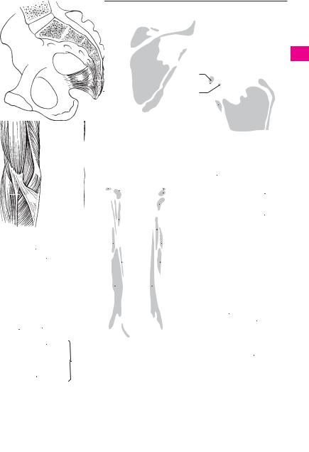
Muscles 89
|
|
|
|
|
|
|
|
|
|
|
|
|
|
|
|
|
|
|
|
|
|
7 |
|
|
|
|
|
|
|
|
|
|
6 |
|
|
|
|
|
|
|
|
|
|
|
|
|
|
|
|
|
|
|
|
|
|
|
|
|
|
|
|
|
|
|
|
|
|
|
|
|
|
|
|
|
|
|
|
|
|
|
|
|
|
|
|
|
|
|
|
|
|
|
|
|
|
|
|
|
|
|
|
|
|
|
|
|
|
|
|
|
|
|
|
|
|
|
|
|
|
|
|
|
|
|
|
|
|
|
|
|
20 |
|
|
|
|
|
|
|
|
|
|
|
|
|
|
|
|
|
|
|
|
|
|
|
|
|
|
|
|
|
|
|
|
|
|
|
|
|
8 |
|
|
|
|
|
|
|
|
|
|
|
|
|
|
|
|
|
|
|
|
|
|
|
|
||
|
|
|
|
|
|
|
|
|
|
|
|
|
|
|
|
|
3 |
|
|
|
|
|
|
9 |
|
|
|
|
|
|
|
|
|
|
|
|
|
|
|
|
||||||||
|
|
|
|
|
|
|
|
|
|
2 |
|
|
|
|
|
|
|
|
|
|
|
|
|
|
|
|
|
|
|
|
|
|
|
|
|
|
|
|
|
|
|
|
|
|
|
|||
|
|
|
|
|
|
|
|
|
|
|
|
|
|
|
4 |
|
|
|
|
|
|
|
|
|
|
|
|
|
|
|
|
|
|
|
|
|
|
|
|
|
|
|
|
|
|
|||
|
|
|
|
|
|
|
|
|
|
|
|
|
|
|
|
|
|
|
|
|
|
|
10 |
|
|
|
|
|
|
|
|
|
|
|
|
|
|
11 |
|
|
|
|
|
|
||||
|
|
|
|
|
|
|
|
|
|
|
|
|
|
|
|
|
|
|
|
|
|
|
|
|
|
|
|
|
|
|
|
|
|
|
|
|
|
|
|
|
|
|
|
|
||||
|
|
|
|
|
|
|
|
|
|
|
|
|
|
|
|
|
|
|
|
|
|
|
|
|
|
|
|
|
|
|
|
|
|
|
|
|
|
|
|
|
|
|
|
|
||||
|
|
|
|
|
|
|
|
|
|
|
|
|
|
|
|
|
|
|
|
|
|
|
|
|
|
|
|
|
|
|
|
|
|
|
|
|
|
|
|
|
|
|
|
|
|
|
||
|
|
|
|
|
|
|
|
|
|
|
|
|
|
|
|
|
|
|
|
Scapula, posterior view |
|
|
|
|
|
|
|
|
||||||||||||||||||||
|
|
|
|
|
|
|
|
|
|
|
|
|
|
|
|
|
|
|
B |
|
|
|
|
|
|
|
|
|||||||||||||||||||||
|
|
|
Right half of pelvis, medial view |
|
|
|
|
|
|
|
|
|
|
|
|
|
|
|
|
|
|
|
|
|
|
Scapula, anterior view |
||||||||||||||||||||||
|
A |
|
|
|
|
|
|
|
|
|
|
|
|
|
|
|
|
|
|
|
|
C |
||||||||||||||||||||||||||
14 |
|
|
|
|
|
|
|
|
|
11 |
|
|
|
|
7 |
11 |
|
|
|
|
|
7 |
|
8 |
7 |
|
|
|
|
|
|
|
|
|
|
|
|
|
|
|
||||||||
|
|
|
|
|
|
|
|
|
|
|
|
|
|
|
|
|
|
|
|
|
|
|
|
|
|
|
|
|
|
|
||||||||||||||||||
|
|
|
|
|
|
|
|
|
|
|
|
|
|
|
|
|
|
|
|
|
|
|
|
|
|
|
8 |
|
|
|
|
|
|
|
|
|||||||||||||
|
|
|
|
|
|
|
|
|
|
|
|
|
|
|
|
|
|
|
|
|
|
|
|
|
|
|
|
|
|
|
|
|
|
|
||||||||||||||
|
|
|
|
|
|
|
|
|
|
|
|
|
|
|
|
|
|
|
|
|
|
|
|
|
|
|
|
|
|
|
|
|
|
|
|
|
|
|
|
|||||||||
|
|
|
|
|
|
|
|
|
|
|
|
|
|
|
|
|
|
|
10 |
|
|
|
|
|
|
|
|
|
|
9 |
9 |
|
|
6 |
|
|
||||||||||||
|
|
|
|
|
|
|
|
|
|
10 |
|
|
|
|
|
|
|
|
|
|
21 |
|
|
|
|
|
|
|
|
|
||||||||||||||||||
|
|
|
|
|
|
|
|
|
|
|
|
|
|
|
|
|
|
|
|
|
|
|
|
|
|
|
||||||||||||||||||||||
|
|
|
|
|
|
|
|
|
|
|
|
|
|
|
|
|
|
|
|
|
|
|
|
|
|
|
|
|
10 |
|
|
|
|
|
|
|
|
|
|
|
||||||||
|
|
|
|
|
|
|
|
|
|
|
6 |
|
|
|
|
|
|
|
|
|
|
|
|
6 |
|
|
|
|
|
20 |
|
|
|
|
|
|
||||||||||||
|
|
|
|
|
|
|
|
|
|
|
|
|
|
|
|
|
|
|
|
|
|
|
|
|
|
|
|
|
|
|
|
|
||||||||||||||||
|
|
|
|
|
|
|
|
|
|
|
|
|
|
|
|
|
|
|
|
|
|
|
|
|
|
|
|
|
|
|
|
|
|
|
|
|||||||||||||
|
|
|
|
|
|
|
|
|
|
|
|
|
|
|
|
|
|
|
|
|
|
|
|
|
|
|
|
|
|
|
|
|
|
|
||||||||||||||
|
|
|
|
|
|
|
|
|
|
|
|
|
|
|
|
|
|
|
|
|
|
|
|
|
|
|
|
|
|
|
|
|
|
|
|
|
|
|
||||||||||
|
|
|
|
|
|
|
|
|
|
|
|
|
|
|
|
|
|
|
|
|
|
|
|
|
|
|
|
|
|
|
|
|
|
|
|
|
|
|
|
|
|
|||||||
|
|
|
|
|
|
|
|
|
|
|
17 |
|
|
|
|
|
|
|
|
|
|
|
|
|
|
|
|
|
|
|
|
|
|
|
|
21 |
|
|
|
|
|
|
||||||
|
|
|
|
|
|
|
|
|
|
|
|
|
|
|
|
|
|
|
|
|
|
|
|
|
|
|
|
|
|
|
|
|
|
|
|
|
|
|
|
|
||||||||
|
|
|
|
|
|
|
|
|
|
|
|
|
|
|
|
|
|
|
|
|
|
|
|
|
|
|
|
|
|
|
|
|
|
|
|
|
|
|
|
|
|
|
|
|
|
|||
|
|
|
|
|
|
|
|
|
|
|
|
|
|
|
|
|
|
|
|
|
|
|
|
|
|
|
|
|
|
|
|
|
|
|
|
|
|
|
|
|
|
|
|
|
|
|||
|
|
|
|
|
|
|
|
|
|
|
20 |
|
|
|
|
|
|
|
|
|
|
|
|
|
|
|
|
|
|
|
|
|
|
|
|
|
|
|
|
|
|
|
|
|
|
|
|
|
13 |
12 |
15 |
|
|
18 |
|
|
|
|
|
|
17 |
22 |
|
|
|
|
|
|
|
18 |
|
|
|
|
|
|
|
|
|
|
|
|
|
|
|
|
|||||||||||
|
|
|
|
|
|
|
|
|
|
|
|
|
|
|
|
|
|
|
|
|
|
|
|
|
|
|
|
|
|
|
|
|
|
|
||||||||||||||
|
|
|
|
|
|
|
|
|
|
|
|
|
|
|
|
|
|
|
|
|
|
|
|
|
|
|
|
|
|
|
|
|
|
|
|
|
||||||||||||
|
|
|
|
|
|
|
|
|
|
|
|
|
|
|
|
|
|
|
|
|
|
|
|
|
|
|
|
|
|
|
|
|
|
|
|
|
|
|
|
|
||||||||
|
|
|
|
|
|
|
|
|
|
|
|
|
|
|
|
|
|
|
|
|
|
|
|
|
|
|
|
|
|
|
19 |
|
|
|
|
|
|
|||||||||||
|
|
|
|
|
|
|
|
|
|
|
|
|
|
|
|
|
|
|
|
|
|
|
|
|
|
|
|
|
|
|
|
|
|
|
|
|
|
|
|
|
|
|||||||
|
|
|
|
|
|
|
|
|
|
|
|
|
|
|
|
|
|
|
|
|
|
|
|
|
|
|
|
|
|
|
|
|
|
|
|
|
|
|
|
|
|
|||||||
|
|
|
|
|
|
|
|
|
|
|
|
|
|
|
|
|
|
|
|
|
|
|
|
|
|
|
|
|
|
|
|
|
|
|
|
|
|
|
|
|
|
|
|
|
|
|
||
18 |
|
|
|
|
|
|
|
|
|
|
18 |
|
|
|
|
|
27 |
|
|
|
|
|
|
|
|
|
|
|
22 |
|
|
|
|
|
|
|
|
22 |
||||||||||
|
|
|
|
|
|
|
|
|
|
|
|
|
|
|
|
|
|
|
|
|
|
|
|
|
|
|
|
|
|
|
|
|
|
|||||||||||||||
|
|
|
|
|
|
|
|
|
|
|
|
|
|
|
|
|
|
|
|
|
|
|
|
|
|
|
|
|
|
|
|
|
|
|
|
|
|
|||||||||||
|
|
|
|
|
|
|
|
|
|
|
|
|
|
|
|
|
|
|
|
|
|
|
|
|
|
|
|
|
|
|
|
|
|
|
||||||||||||||
|
|
|
|
|
|
|
|
|
|
|
|
|
|
|
|
|
|
|
|
|
|
|
|
|
|
|
|
|
|
|
|
|
|
|
|
|
|
|
|
|
|
|||||||
|
|
|
|
|
|
|
|
|
|
|
|
|
|
|
|
|
|
|
|
|
|
|
|
|
|
|
|
|
|
|
|
|
|
|
|
|
|
|
|
|
|
|||||||
|
|
|
|
|
|
|
|
|
|
|
|
|
|
|
|
|
|
|
|
|
|
|
|
|
|
|
|
|
|
|
|
|
|
|
|
|
|
|
|
|
||||||||
|
|
|
|
|
|
|
|
|
|
|
27 |
|
|
|
|
|
|
|
|
|
|
|
|
|
|
|
|
|
|
|
|
|
|
|
|
|
|
|
|
|
|
|
|
|
|
|
24 |
|
|
|
|
|
|
|
|
|
|
|
|
|
|
|
|
|
|
|
|
|
|
|
|
|
|
|
|
|
|
|
|
|
|
|
|
|
|
|
|
|
|
|
|
|
|
|
|||
|
|
|
|
|
|
|
16 |
26 |
|
|
|
|
|
|
|
|
|
|
|
|
|
|
|
|
|
|
|
|
|
|
|
|
|
|
|
|
|
|
|
|
||||||||
|
|
|
|
|
|
|
|
|
|
|
|
|
|
|
|
|
|
|
|
|
|
|
|
|
|
|
|
|
|
|
|
|
|
|
|
|
||||||||||||
|
|
|
|
|
|
|
|
|
|
|
|
|
|
|
|
|
|
|
|
|
|
|
|
|
|
|
|
|
|
|
|
|
|
|
|
|
|
|
|
|
|
|
|
|||||
|
|
|
|
|
|
|
|
|
|
|
|
|
E |
|
Humerus, |
|
F |
|
Humerus, |
|
|
|
|
|
|
|
|
|
|
|
|
|
|
|
|
|||||||||||||
|
|
|
|
|
|
|
|
|
|
|
28 |
|
|
|
anterior view |
|
|
posterior view |
|
|
|
|
|
|
|
|
||||||||||||||||||||||
|
|
|
|
|
|
|
|
|
|
|
|
|
|
|
|
|
|
|
|
|
|
|
||||||||||||||||||||||||||
|
|
Upper arm, anterior view |
|
|
|
|
|
|
|
|
|
|
|
|
|
|
|
|
|
|
|
|
|
|
|
|
|
Upper arm, posterior view |
||||||||||||||||||||
|
D |
|
|
|
|
|
|
|
|
|
|
|
|
|
|
|
|
|
|
|
|
|
|
|
|
G |
||||||||||||||||||||||
a
3
4
5
6
7
8
9
10
11
12
13
14
15
16
17
18
19
20
21
22
23
24
25

|
90 |
Muscles |
|
|
|||
|
|
|
M. flexor carpi radialis. o: Medial epicondyle of |
15 |
M. extensor digitorum. o: Lateral epicondyle of |
||
1 |
|
1 |
|||||
|
|
|
humerus. i: Base of 2nd metacarpal bone. A: Pro- |
|
humerus. i: Distal phalanges 2−5 via their dorsal |
||
|
|
|
|
nation, flexion and radial abduction of the wrist |
|
aponeurosis. A: Extension of fingers and dor- |
|
2 |
|
|
|
joint. I. Median nerve. A |
|
siflexion of the wrist joint. I: Radial nerve. C |
|
|
|
2 |
M. palmaris longus. o: Medial epicondyle of |
16 |
Intertendinous connections. Connexus inter- |
||
|
|
|
|
humerus. i: Palmar aponeurosis. A: Tenses |
|
tendineus [[juncturae tendineum]]. Oblique ten- |
|
3 |
|
|
|
|
|||
|
|
|
aponeurosis and flexes the wrist joint and fin- |
|
dinous connections between the extensor ten- |
||
|
|
|
|
gers 2−5 at the metacarpophalangeal joints. Its |
|
dons of the fingers on the dorsum of the hand. C |
|
4 |
|
|
|
tendon lies above the flexor retinaculum. I: Me- |
17 M. extensor digiti minimi. o: Lateral epicon- |
||
|
|
|
|
dian nerve. A |
|
dyle of humerus. i: Dorsal aponeurosis of 5th fin- |
|
|
|
|
|
|
|||
5 |
3 |
M. flexor carpi ulnaris. o: Medial epicondyle of |
|
ger. A: Extension of little finger, abduction and |
|||
|
|
|
humerus, olecranon and ulna. i: Pisiform bone as |
|
dorsiflexion of the wrist joint. I: Radial nerve. C |
||
|
|
|
|
well as the hamate and 5th metacarpal bones via |
18 |
M. extensor carpi ulnaris. o: Lateral epicondyle |
|
6 |
|
|
|
the pisohamate and pisometacarpal ligaments. |
|
of humerus, radial collateral ligament, posterior |
|
|
|
|
|
A: Ulnar abduction and flexion of the wrist joint. |
|
surface of ulna. i: Base of 5th metacarpal. A: Dor- |
|
|
|
|
|
I: Ulnar nerve. A |
|
siflexion, ulnar abduction. I. Radial nerve. C D |
|
7 |
|
|
|
|
|||
4 |
Humeral head of flexor carpi ulnaris. Caput |
19 |
Humeral head of extensor carpi ulnaris. |
||||
|
|||||||
|
|
|
|
humerale. The part arising from the epicondyle |
|
Caput humerale. The part arising from the |
|
8 |
|
|
|
of the humerus. |
|
humerus. |
|
|
5 |
Ulnar head of flexor carpi ulnaris. Caput ul- |
20 Ulnar head of extensor carpi ulnaris. Caput |
||||
|
|||||||
9 |
|
|
|
nare. The portion originating from the ulna. E |
|
ulnare. The portion arising from the ulna. F |
|
6 |
M. flexor digitorum superficialis. o: Medial |
21 |
M. supinator. o: Lateral epicondyle, radial col- |
||||
|
|||||||
|
|
|
|
epicondyle of humerus, coronoid process of ulna |
|
lateral ligament, supinator crest of ulna. i: Ante- |
|
10 |
|
|
|
|
|||
|
|
|
and anterior surface of radius. i: Middle phalanx |
|
rior surface of radius. A: Supination. I: Radial |
||
|
|
|
|
of fingers 2−5. A: Flexion of all joints it crosses. |
|
nerve. D E F |
|
|
|
|
|
|
|||
11 |
|
|
|
Each tendon is perforated by the corresponding |
22 |
M. abductor pollicis longus. o: Dorsal side of |
|
|
|
|
tendon from the flexor digitorum profundus. I. |
||||
|
|
|
|
|
interosseous membrane and adjacent surfaces |
||
|
|
|
|
Median nerve. A B |
|
||
|
|
|
|
|
of radius and ulna. i: Base of 1st metacarpal. A: |
||
12 |
|
|
|
|
|||
7 |
Humeroulnar head of flexor digitorum su- |
|
Radial abduction and dorsiflexion of the meta- |
||||
|
|
|
|
perficialis. Caput humeroulnare. The portion |
|
carpophalangeal joint of the thumb. Supination. |
|
|
|
|
|
|
|||
13 |
|
|
|
arising from the humerus and ulna. A E F |
|
I: Radial nerve. C D F |
|
8 |
Radial head of flexor digitorum superfi- |
23 |
M. extensor pollicis brevis. o: Radius (extensor |
||||
|
|||||||
|
|
|
|
cialis. Caput radiale. The portion arising from |
|
side) and interosseous membrane. i: Base of pro- |
|
14 |
|
|
|
|
|||
|
|
|
the radius. A E |
|
ximal phalanx of thumb. A: Abduction and ex- |
||
|
9 |
M. flexor digitorum profundus. o: Upper half |
|
tension of thumb at the metacarpophalangeal |
|||
15 |
|
|
|
of ulna. i: Base of distal phalanges of fingers 2−5. |
|
joint. I: Radial nerve. C D F |
|
|
|
|
A: Flexion of all joints crossed. The tendon per- |
24 M. extensor pollicis longus. o: Interosseous |
|||
|
|
|
|
||||
|
|
|
|
forates the corresponding superficial flexor ten- |
|
membrane and dorsal surface of ulna. i: Distal |
|
16 |
|
|
|
|
|||
|
|
|
don. I: Median and ulnar nerves. B E F |
|
phalanx of thumb. A: Adducts and extends the |
||
|
10 |
M. flexor pollicis longus. o: Middle of anterior |
|
thumb. Supinator. I: Radial nerve. C D F |
|||
17 |
|
|
|
surface of radius and usually the medial epicon- |
25 |
M. extensor indicis. o: Interosseous membrane |
|
|
|
|
|
dyle. i: Distal phalanx of thumb. A: Flexion of |
|
and dorsal surface of ulna. i: Dorsal aponeurosis |
|
|
|
|
|
joints crossed. I: Median nerve. B E |
|
of index finger. A: Extends the index finger and |
|
18 |
|
|
|
|
|||
11 |
M. pronator quadratus. o: Lower fourth of |
|
wrist joint. I: Radial nerve. D F |
||||
|
|
|
|
anterior surface of ulna. i: Lower fourth of ante- |
26 M. palmaris brevis. o: Ulnar side of palmar |
||
19 |
|
|
|
rior surface of radius. A: Pronation. I: Median |
|
aponeurosis. i: Skin of ulnar aspect of hand. I. |
|
|
|
|
|
nerve. A B E |
|
Ulnar nerve. A |
|
|
|
|
|
|
|||
2012 and lateral margin of humerus. i: Styloid process of radius. A: Flexion of the elbow joint. It brings
21the arm from the extreme pronation and supination position to the intermediate position. I: Radial nerve. A C E
2213 M. extensor carpi radialis longus. o: Lateral intermuscular septum, lateral epicondyle. i: Ex-M. brachioradialis. o: Intermuscular septum
23 |
tensor side of metacarpal 2. A: Radial abduction |
|
and dorsiflexion of the wrist joint. Flexion of the |
elbow joint. I: Radial nerve. A C
2414 M. extensor carpi radialis brevis. o: Lateral epicondyle of humerus. i: Proximal extensor side of
25third metacarpal. A: Extension and radial abduction of wrist joint. I: Radial nerve. C

Muscles 91
88.18
88.16
88.12 
12 |
|
88.26 |
|
2
1
21
6
13 
9
|
|
|
3 |
|
|
7 |
|
|
|
||
8 |
|
|
|
||
6 |
10 |
||||
|
|||||
11 |
|
|
|
|
11 |
|
|
|
|
||
|
|
|
|
|
|
26 |
|
|
|
||
A Superficial muscles of forearm, anterior view
6
BDeep muscles of forearm, anterior view
5 7
E Radius and ulna with interosseous membrane, anterior view
F Radius and ulna with |
|
interosseous membrane, |
7 |
posterior view |
a
88.19 
|
|
|
|
|
|
12 |
|
|
|
|
|
|
|
|
|
|
|
|
|
|
|
|
|
|
|||||||
88.24 |
|
|
|
|
|
88.24 |
|
|
|
|
|
|
|
|
|
|
|
|
|
||||||||||||
|
|
|
|
|
|
|
|
|
|
|
|
|
|
|
|
|
|||||||||||||||
|
|
|
|
|
|
|
13 |
|
|
|
|
|
|
|
|
|
|
|
|
|
|
|
|
|
|
|
|||||
|
|
|
|
|
|
|
|
|
|
|
|
|
|
|
|
|
|
|
|
21 |
|
|
|
|
|||||||
|
|
|
|
|
|
|
|
|
|
|
|
|
|
|
|
|
|
|
|
|
|
|
|
||||||||
18 |
15 |
88.26 |
|
|
|
|
|
|
|
|
|
|
|
|
|
||||||||||||||||
|
|
|
|
|
|
|
|
|
|
|
|
|
|||||||||||||||||||
|
|
|
|
|
|
|
|
|
|
|
|
|
|
|
|
|
|
|
|
|
|
|
|
|
|
||||||
|
|
|
|
|
|
|
|
|
|
|
|
14 |
24 |
|
|
|
|
|
|
|
|
|
|
|
|
|
|||||
|
|
|
|
|
|
|
|
|
|
|
|
|
|
|
|
|
|
|
|
|
|
||||||||||
|
|
|
|
|
|
|
|
|
|
|
|
|
|
|
|
|
|
|
|
|
|
|
|
|
|
|
|
|
|
||
|
|
|
|
|
|
|
|
|
|
|
|
|
|
|
|
|
|
|
|
|
|
|
|
|
|
|
|
|
|
|
|
|
|
|
|
|
|
|
|
|
|
|
|
|
|
|
|
|
|
|
|
22 |
|||||||||||
17 |
|
|
|
|
|
|
|
22 |
25 |
|
|
|
|
|
|
|
|
|
|
|
|
23 |
|||||||||
|
|
|
|
|
|
|
|
|
|
|
|
|
|
|
|
|
|
||||||||||||||
|
|
|
|
|
|
|
|
|
|
|
|
23 |
|
|
|
|
|
|
|
|
|
|
|||||||||
|
|
|
|
|
|
|
|
|
|
|
|
|
|
|
|
|
|
|
|
|
|
|
|
|
|
||||||
|
|
|
|
|
|
|
|
|
|
|
|
|
|
|
|
|
|
|
|
|
|||||||||||
|
|
|
|
|
|
14 |
18 |
|
|
|
|
|
|
|
|
|
|
|
|
22 |
|||||||||||
|
|
|
|
|
|
|
|
|
|
|
|
|
|
13 |
|
|
|
|
|
|
|||||||||||
|
|
|
|
|
|
|
|
|
|
|
|
|
|
|
|
|
|
|
|
|
|
|
|
|
23 |
||||||
|
|
|
|
|
|
|
|
|
|
|
|
|
|
|
|
24 |
25 |
|
|
|
|
|
|
||||||||
|
|
|
|
|
|
|
|
|
|
|
|
|
|
|
|
|
|
||||||||||||||
|
|
|
|
|
|
|
|
|
|
|
|
|
|
|
|
|
|
|
|
|
|
|
|
|
|
|
|
|
|||
|
|
|
|
|
|
|
|
|
|
|
23 |
|
|
|
|
|
|
|
|
|
|
|
|
|
|
|
|||||
|
|
|
|
|
|
|
|
|
|
|
|
|
|
|
|
|
|
|
|
|
|
||||||||||
|
|
|
|
|
|
|
|
|
|
|
|
|
|
|
|
|
|
|
|
|
|
|
|||||||||
|
|
|
|
16 |
|
|
|
|
|
|
|
|
|
|
|
|
|
|
24 |
|
|||||||||||
|
|
|
|
Superficial extensor |
|
|
Deep extensor muscles |
||||||||||||||||||||||||
|
|
C |
D |
||||||||||||||||||||||||||||
|
|
|
|
muscles of forearm |
|
|
|
|
of forearm |
||||||||||||||||||||||
|
|
|
|
9 |
|
|
|
|
|
|
11 |
|
|
|
|
|
|
|
|
|
|
|
|
|
|
|
|
|
|
|
|
|
|
|
10 |
|
|
|
11 |
|
|
|
|
|
|
|
|
|
|
|
|
|
|
|
|
21 |
8 |
|
|
|
|
|
12 |
||||||
20 |
|
|
|
23 |
|
|
|
|
|||||
|
|
|
|
21 |
|
|
|
|
|
|
|
|
|
|
|
|
|
|
|
|
|
|
|
|
|
|
|
|
|
|
|
|
|
|
|
|
|
|
|||
|
|
|
|
22 |
|
|
|
|
|
|
|||
|
|
|
|
|
|
|
|
|
|
|
|
||
|
|
|
|
|
|
|
|
|
|
|
|
|
|
|
|
|
|
|
|
|
|
|
|
|
|
|
|
|
|
|
|
9 |
24 |
25 |
|
||||||
3
4
5
6
7
8
9
10
11
12
13
14
15
16
17
18
19
20
21
22
23
24
25

92 Muscles
1 M. abductor pollicis brevis. o: Scaphoid bone
1 and flexor retinaculum. i: Lateral sesamoid bone and, radially, the proximal phalanx of the thumb.
2A: Abduction and flexion of the thumb. I: Median nerve. B
3 |
2 M. flexor pollicis brevis. o: Carpal bones, base of |
1st metacarpal and flexor retinaculum. i: Lateral |
sesamoid bone, radially aspect of proximal
4phalanx of thumb. A: Adducts and flexes thumb. B
3Superficial head of flexor pollicis brevis.
Caput superficiale. The portion situated on the 5 tendon of the flexor pollicis longus and innervated
by the median nerve. B
64 Deep head of flexor pollicis brevis. Caput pro-
|
fundum. The portion situated below the tendon of |
7 |
the flexor pollicis longus and innervated by the |
ulnar nerve. B |
5 M. opponens pollicis. o: Trapezium and flexor ret-
8inaculum. i: First metacarpal. A: Adduction and opposition of thumb. I: Median nerve. B
96 M. adductor pollicis. o: Capitate, radiate carpal ligament, metacarpal bone 3. i: Medial sesamoid
bone, ulnar aspect of base of proximal phalanx of 10 thumb. A: Adduction and opposition of thumb. I:
Ulnar nerve. B
11 |
7 Oblique head of adductor pollicis. Caput ob- |
|
liquum. The portion arising from the capitate and |
radiate carpal ligament. B
128 Transverse head of adductor pollicis. Caput transversum. The portion originating from the 3rd
13metcarpal bone. B
9M. abductor digiti minimi. o: Pisoform and flexor
retinaculum. i: Base of proximal phalanx of little
14finger and dorsal aponeurosis. A: Abduction, flexion and extension of the little finger. I. Ulnar nerve.
15B
|
10 M. flexor digiti minimi brevis. o: Hook of hamate |
|
16 |
and flexor retinaculum. i: Base of proximal |
|
phalanx of little finger. A: Flexion of finger at the |
||
|
||
|
metacarpophalangeal joint. I: Ulnar nerve. B |
17 11 M. opponens digiti minimi. o: Hook of hamulus |
||
|
and flexor retinaculum. i: Head and shaft of 5th |
|
18 |
metacarpal. A: Draws the little finger toward the |
|
palm of the hand. I: Ulnar nerve. B |
||
|
||
12 Mm. lumbricales. o: Tendons of flexor digitorum
19profundus. i: Dorsal aponeuroses of fingers 2−5. A: Flexion of finger at the metacarpophalangeal joints, extension at the interphalangeal joints. I:
20Ulnar and median nerves. B C
13 Mm. interossei dorsales. o: Arises by two heads
21from the metacarpals. i: Dorsal aponeurosis of fingers 2−4. A: Spreading of fingers 2−4 away from axis of middle finger, radial and ulnar abduction of
22middle finger, flexion of finger at the metacarpophalangeal joint and extension of the inter-
23phalangeal joints. I. Ulnar nerve. C D E
14 Mm. interossei palmares. o: Metacarpal bones 2, 4 and 5. i: Dorsal aponeuroses of fingers 2, 4 and 5.
24A: Adduction of index, ring and little fingers toward the middle finger, flexion of the metacar-
25pophalangeal joints, extension of the interphalangeal joints. I: Ulnar nerve. B D
15Axillary fascia. Fascia axillaris. Situated on the adipose body of the axilla. It unites the lateral margins of the pectoral and latissimus dorsi muscles. G
16Deltoid fascia. Fascia deltoidea. Strongly fused investing fascia of the deltoid muscle.
17Brachial fascia. Fascie brachii (brachialis). Fascia enclosing the upper arm muscles. F
18Medial intermuscular septum of the arm. Septum intermusculare brachii mediale. Tendinous sheet for muscle origin between the medial margin of the humerus and the brachial fascia. F
19Lateral intermuscular septum of the arm. Septum intermusculare brachii laterale. Tendinous sheet for muscle attachment between the lateral margin of the humerus and the brachial fascia. F
20Antebrachial fascia. Fascia antebrachii. Fascia enveloping the forearm muscles. A
21Fascia of the dorsum of the hand. Fascia dorsalis manus. Fascia situated on the dorsal tendons of the hand. E
22Extensor retinaculum. Retinaculum extensorum [[lig. carpi dors.]]. Transverse fascial fibers over the 6 conduction canals of the 10 extensor tendons. E
23Superficial transverse metacarpal ligament. Lig. metacarpale transversum superficiale. Transverse reinforcement of the palmar fascia of the hand at the level of the heads of the metacarpals. A
24Palmar aponeurosis. Aponeurosis palmaris. Membranous expansion of the tendon of the palmaris longus muscle. A
25Transverse fasciculi. Fasciculi transversi. Transversely oriented fibrous bundles of the palmar aponeurosis. A
26Flexor retinaculum. Retinaculum flexorum [[lig. carpi transversum]]. Stout fibrous band between the scaphoid, trapezoid, pisiform and hamate bones. It binds the flexor tendons. B
27Fibrous sheaths of fingers. Vaginae fibrosae digitorum manus. Fibrous synovial tunnel for the digital flexor tendons. B
28Annular part of fibrous sheath. Pars anularis vaginae fibrosae. Very compact circular fibers of the fibrous sheath located between the joints. B
29Cruciate part of fibrous sheath. Pars cruciformis vaginae fibrosae. Crossed fibers that reinforce the joints. B
30Synovial sheaths of the fingers. Vaginae synoviales digitorum manus. Tendon sheaths of the digital flexor tendons.
31Vincula tendinum. Connective tissue fasciculi (mesotendons) transporting vessels to the tendons. C
32Vinculum longum. Longer fasciculus at the level of the proximal phalanx. C
33Vinculum breve. Shorter fasciculus near the insertions of the tendons. C
34Chiasma tendinum. Crossing of the tendons of the flexor digitorum superficialis and profundus. C
Muscles 93
23
23
|
|
|
|
|
|
|
|
|
|
|
|
|
|
|
|
3 |
|
|
|
|
|
|
|
|
|
|
|
|
|
|
|
|
|
|
|
|
|
|
|
|
|
|
|
|
|
|
|
|
|
4 |
|
|
|
|
|
|
|
|
|
|
|
|
|
|
|
29 |
|
|
|
|
|
|
|
|
|
|
|
|
|
|
|
|
|
|
25 |
|
|
|
|
|
|
|
|
27 |
|
|
|
5 |
|||
|
|
|
|
|
|
|
|
|
|
|
|
|
27 |
|
||
|
|
|
|
|
|
|
|
|
|
|
|
|||||
|
|
|
|
|
|
|
|
|
|
|
|
|
|
|
6 |
|
|
|
|
|
|
|
|
|
|
|
|
|
|
|
|
28 |
|
|
|
|
|
24 |
|
|
|
|
|
|
|
|
|
|
|
|
90.26 |
|
|
|
|
|
|
|
|
|
|
|
12 |
7 |
|||
|
|
|
|
|
|
|
|
|
|
|
|
|||||
|
|
|
|
|
|
|
|
|
|
|
|
|
|
|
|
|
|
|
|
|
|
|
|
|
|
|
|
|
|
|
|
|
8 |
|
|
|
|
|
|
|
14 |
14 |
|
|
|
|
|
|
|
|
|
|
|
|
|
|
|
|
|
|
|
|
|
|
|||
|
|
|
|
20 |
|
8 |
6 |
|
9 |
|||||||
|
|
|
|
|
|
|
|
|
||||||||
|
|
|
|
|
10 |
|
|
|
|
|||||||
|
|
|
|
9 |
|
|
|
|
|
|
|
|
|
|||
|
|
|
|
|
|
7 |
|
|
|
|
10 |
|||||
|
|
Palmar aponeurosis and fasciae |
|
|
|
|
|
|
|
|||||||
|
A |
|
|
|
3 |
|
|
|
|
|
||||||
|
|
|
|
|
|
|
||||||||||
|
|
|
|
11 |
|
|
|
|
|
1 |
5 |
90.10 |
11 |
|||
|
|
|
|
|
|
|
|
|
|
|
|
|
||||
|
|
|
|
|
|
Palm muscles |
|
26 |
|
|
|
|
|
|
|
|
|
|
|
|
|
B |
|
|
|
|
|
|
|
|
|||
|
|
|
|
|
|
|
|
|
|
|
12 |
|||||
|
|
|
|
|
|
|
|
|
|
|
|
|
|
|
|
|
|
|
|
|
|
|
|
90.1 |
|
4 |
|
|
|
|
|||
|
|
|
|
31 |
|
|
|
|
13 |
|||||||
|
|
|
|
|
|
|
|
|
|
|
||||||
|
|
|
|
|
|
|
|
|
|
|
|
|
||||
|
|
|
|
|
|
|
|
|
|
|
|
|
|
|
|
|
|
|
|
90.15 |
|
14 |
|
|
|
|
|
|
|
33 |
32 |
18 |
17 |
15 |
|
|
|
|||
C |
Tendons of finger |
|
|
19 |
16 |
|
|
|
|||
|
|
|
|
||
|
|
34 |
|
|
17 |
|
|
|
|
|
|
|
12 |
13 |
|
|
|
|
|
||||
|
|
F |
Cross section of upper arm |
|
||||||||
|
|
90.6 90.9 |
|
18 |
||||||||
|
|
|
|
|
|
|
|
|
|
|
||
|
|
|
|
|
|
|
|
|
|
|
|
|
|
|
|
|
|
|
|
|
|
|
|
|
|
|
|
14 |
|
|
|
|
|
|
|
19 |
||
|
|
|
13 |
|
|
|
|
|
|
|||
|
|
|
|
|
|
|
|
|
||||
|
|
|
|
|
|
|
|
20 |
||||
13 |
13 |
|
|
|
|
|
|
|
||||
|
|
|
|
|
|
|
|
|||||
|
|
|
|
|
|
|||||||
|
|
|
|
15 |
21 |
|||||||
|
|
|
|
|
|
|
|
|
|
|||
|
|
|
|
|
|
|
|
|||||
|
|
|
|
|
|
|
|
|
|
|
|
|
|
|
21 |
|
|
|
|
|
22 |
||||
|
|
22 |
|
|
|
|
|
|
||||
|
|
|
|
|
|
|
23 |
|||||
|
|
|
|
|
|
|
|
|
|
|
|
|
|
|
|
|
|
|
|
|
|
|
|
|
|
|
|
|
|
|
|
|
|
|
|
|
24 |
|
|
Interosseous muscles, schematic |
|
Dorsum of hand |
|
Cross section of thorax |
|
||||||
D |
E |
|
||||||||||
G |
|
|||||||||||
25 |
||||||||||||
|
|
|
|
|
|
|
|
|
|
|
||
|
|
|
|
|
|
|
|
|
|
|
|
|
a

|
94 |
Muscles |
|
|
|||
|
|
|
1 MUSCLES OF LOWER LIMB. Musculi membri |
14 M. quadratus femoris. o: Ischial tuberosity. i: |
|||
1 |
|
|
|||||
|
|
|
inferioris. |
|
Intertrochanteric crest. A: Lateral rotation and |
||
2 |
|
|
2 M. iliopsoas. Comprised of two muscles, the |
|
adduction of thigh. I: Sacral plexus. A D E |
||
|
|
15 M. sartorius. o: Anterior superior iliac spine. i: |
|||||
|
|
|
psoas major and iliacus. o: Lesser trochanter. A: |
||||
|
|
|
|
Most important flexor and pre-elevator muscle |
|
Medial to tibial tuberosity. A: Flexion, abduc- |
|
|
|
|
|
of the legs; medial and lateral rotation of thigh |
|
tion, lateral rotation of thigh at the hip joint, |
|
3 |
|
|
|
|
|||
|
|
|
at the hip joint. B C D |
|
flexion and medial rotation of leg at the knee |
||
|
|
|
|
|
|||
|
|
|
3 M. iliacus. o: Iliac fossa. i: Lesser trochanter. A: |
|
joint. I: Femoral nerve. C E |
||
4 |
|
|
|
||||
|
|
16 |
M. quadriceps femoris. The muscle group |
||||
|
|
|
Flexion, medial and lateral rotation of thigh at |
||||
|
|
|
|
the hip joint. I: Femoral nerve and lumbar |
|
comprising the three vasti muscles and the rec- |
|
5 |
|
|
|
plexus. C |
|
tus femoris. I: Femoral nerve. |
|
4 |
M. psoas major. o: Bodies and transverse |
17 |
M. rectus femoris. o: Anterior inferior iliac |
||||
|
|||||||
|
|||||||
6 |
|
|
|
processes of L1−4. i: Lesser trochanter. A: Flex- |
|
spine = straight head and upper margin of |
|
|
|
|
ion, medial and lateral rotation of thigh at the |
|
acetabulum. = reflected head. i: Tibial tuberos- |
||
|
|
|
|
hip joint. I: Lumbar plexus. C |
|
ity. A: Flexion of thigh at the hip joint, exten- |
|
7 |
5 |
[M. psoas minor]. o: Bodies of T12 and L1. i: |
|
sion of leg at the knee joint. B C E |
|||
|
|
|
|
Iliac fascia. I: Lumbar plexus. C |
18 |
M. vastus lateralis. o: Greater trochanter, |
|
|
|
|
|
||||
8 |
6 |
M. gluteus maximus. o: Posterior, external sur- |
|
lateral lip of linea aspera. i: Quadriceps tendon. |
|||
|
A: Extension of leg at the knee joint. B C D |
||||||
|
|
|
|
face of ilium, sacrum, coccyx, sacrotuberous |
|
||
|
|
|
|
19 |
M. vastus intermedius. o: Anterior surface of |
||
9 |
|
|
|
ligament. i: Iliotibial tract, gluteal tuberosity, |
|||
|
|
|
lateral intermuscular septum, linea aspera. A: |
|
femur. i: Quadriceps tendon. A: Extension of leg |
||
|
|
|
|
Extension, lateral rotation, abduction and ad- |
|
at the knee joint. B D |
|
10 |
|
|
|
duction of thigh at the hip joint. I: Inferior |
20 |
M. vastus medialis. o: Distal to intertrochan- |
|
|
|
|
|
gluteal nerve. A D E |
|
teric line, medial lip of linea aspera. i: Quadri- |
|
|
|
|
|
|
|||
11 |
7 |
M. gluteus medius. o: External surface of |
|
ceps tendon. A: Extension of leg at the knee |
|||
|
|
|
ilium. i: Greater trochanter. A: Abduction, me- |
|
joint. C D |
||
|
|
|
|
|
|||
|
|
|
|
dial and lateral rotation, flexion and extension |
21 M. articularis genus. o: Anterior surface of |
||
12 |
|
|
|
||||
|
|
|
of thigh at the hip joint. I: Superior gluteal |
|
femur. i: Knee joint capsule. A: Tenses capsule. |
||
|
|
|
|
nerve. A D E |
|
I: Femoral nerve. D |
|
|
|
|
|
|
|||
13 |
8 |
M. gluteus minimus. o: External surface of |
22 M. pectineus. o: Pecten pubis. i: Pectineal line |
||||
|
|
|
|
ilium between anterior and inferior gluteal |
|
below the lesser trochanter. A: Flexion, adduc- |
|
|
|
|
|
|
|||
|
|
|
|
lines. i: Greater trochanter. A: Abduction, me- |
|
tion and lateral rotation of thigh at the hip joint. |
|
14 |
|
|
|
|
|||
|
|
|
dial and lateral rotation, flexion and extension |
|
I: Femoral and obturator nerves. B C D E |
||
|
|
|
|
of the thigh at the hip joint. I: Superior gluteal |
23 M. adductor longus. o: Near the symphysis. i: |
||
15 |
|
|
|
nerve. A D E |
|||
|
|
|
|
Medial lip of linea aspera. A: Adduction and |
|||
|
|
8 a Gluteal aponeurosis. Aponeurosis glutealis. |
|
||||
|
|
|
|
flexion of thigh at the hip joint. I: Obturator |
|||
|
|
|
|
||||
16 |
|
|
|
Deep, sheet-like tendon of origin of the gluteus |
|
nerve. B C D E |
|
|
|
|
maximus lying on the gluteus medius. |
24 M. adductor brevis. o: Inferior ramus of pubis. |
|||
|
|
|
9 M. tensor fasciae latae. o: Near the anterior su- |
||||
|
|
|
|
i: Medial lip of linea aspera. A: Adduction, flex- |
|||
17 |
|
|
|
||||
|
|
|
perior iliac spine. i: Above the iliotibial tract |
|
ion, extension and lateral rotation of thigh at |
||
|
|
|
|
lateral to the tibial tuberosity. A: Flexion, ab- |
|
the hip joint. I: Obturator nerve. B D E |
|
|
|
|
|
|
|||
18 |
|
|
|
duction and medial rotation of thigh at the hip |
25 |
M. adductor magnus. o: Ischial tuberosity, |
|
|
|
|
joint. Flexion, extension and final rotation at |
||||
|
|
|
|
|
ischial ramus. i: Medial lip of linea aspera and |
||
|
|
|
|
the knee joint. I: Superior gluteal nerve. C E |
|
||
19 |
|
|
|
|
with a long tendon to the medial epicondyle. A: |
||
10 |
M. piriformis. o: Anterior surface of sacrum. i: |
|
|||||
|
Adduction and extension of thigh at the hip |
||||||
|
|
|
|
Greater trochanter, inner side of apex. A: Ab- |
|
joint. I: Obturator and sciatic nerves. B C D E |
|
20 |
|
|
|
duction, extension and lateral rotation of thigh |
25 a M. adductor minimus. Uppermost part of the |
||
|
|
|
|
at the hip joint. I. Sacral plexus. A D |
|
adductor magnus muscle. It arises from a more |
|
|
|
11 M. obturator internus. o: Inner surface of ob- |
|
||||
21 |
|
|
anterior part of the pelvis. |
||||
|
|
|
turator membrane and environment. i: Tro- |
26 M. gracilis. o: Inferior ramus of pubis medial to |
|||
|
|
|
|
||||
|
|
|
|
chanteric fossa. A: Lateral rotation, abduction |
|||
|
|
|
|
|
the adductor magnus muscle. i: Medial to tibial |
||
22 |
|
|
|
and adduction of thigh. I: Sacral plexus. A D |
|
||
|
|
|
|
tuberosity. A: Adduction, flexion and extension |
|||
|
|
12 M. gemellus superior. o: Ischial spine. i: Ten- |
|
of thigh at the hip joint. Flexion and medial ro- |
|||
23 |
|
|
|
don of obturator internus and trochanteric |
|
tation of the knee joint. I: Obturator nerve. A C E |
|
|
|
|
fossa. A: Lateral rotation, adduction and abduc- |
|
|
||
|
|
|
|
|
|
||
|
|
|
|
tion of thigh. I: Sacral plexus. A D E |
|
|
|
2413 M. gemellus inferior. o: Ischial tuberosity. i: Tendon of obturator internus, trochanteric
25fossa. A: Lateral rotation, adduction and abduction of thigh. I: Sacral plexus. A D E

Muscles 95
7
22
8
10
12
11
13
14
96.3
96.5
26 |
|
|
25 |
|
|
|
|
|
|||
|
96.6
ADeep muscles of hip, posterior view
2 |
|
|
|
|
|
|
|
|
|
96.1 |
4 |
5 |
||
|
|
|
|
|
|
|
|
|
||||||
|
|
|
|
|
|
|
|
|
|
|||||
|
|
|
|
|
24 |
|
|
|
4 |
|||||
|
|
|
|
|
|
|
|
|
|
|
|
|||
22 |
|
|
|
|
|
|
|
|
|
|
|
|||
|
|
|
|
|
|
|
|
|
|
|
|
|
||
|
|
|
|
|
|
|
|
|
|
|
|
|||
|
7 |
|
|
|
|
|
23 |
|
|
3 |
|
|
||
|
|
|
|
|
|
|
|
|
|
|||||
|
|
|
|
|
|
|
|
|
|
|
||||
|
|
|
|
|
|
|
|
|
|
|
|
|
|
|
6 |
|
|
|
|
|
|
25 |
|
|
9 |
|
|
||
|
|
|
|
|
|
|
|
|
|
|
||||
18 |
|
|
|
|
|
|
|
2 |
|
|
||||
|
|
|
|
|
|
|
|
|
|
|
|
|||
19 |
|
|
|
|
|
|
22 |
|
||||||
|
|
|
|
|
|
|
|
|
|
98.18 |
23 |
|
||
|
|
|
|
|
|
|
|
|
|
|
|
|
||
 98.14
98.14
17 |
98.14 |
|
|
|
|
|
|
|
15 |
|
|
|
|
|
|
|
|||
|
|
17 |
|
|
|
|
25 |
||
|
|
|
|
|
|
||||
|
|
|
|
|
|||||
|
|
|
|
|
|
|
|
26 |
|
|
|
|
|
|
|
|
|
||
|
|
|
|
|
|
|
|||
11;12;13 11;12;13
|
|
|
|
|
10 |
|
|
|
|
|
|
|
|
|
|
|
||||
|
|
|
|
|
|
|
|
|
7 |
10 |
|
|
|
|
|
|
|
|
|
|
|
|
|
|
|
|
|
|
|
|
|
|
|
|
|
|
|
|
|
||
|
|
|
|
|
|
|
14 |
8 |
|
|
|
|
|
|
|
|
|
|
||
|
|
|
|
|
|
|
18 |
|
|
|
|
|
|
|
|
|
|
|||
|
|
|
|
|
|
|
|
|
|
|
|
|
|
|
|
|
||||
2 |
|
|
|
|
|
|
6 |
|
|
|
|
|
|
|
|
2 |
||||
|
|
|
|
|
|
|
|
|
|
|
|
|||||||||
|
|
|
|
|||||||||||||||||
|
|
|
|
|
|
|
|
|
|
|||||||||||
22 |
|
|
|
|
|
|
|
|
|
|
|
|
|
|
|
|
20 |
|||
|
|
|
|
|
|
|
|
|
|
|
||||||||||
|
|
|
|
|
|
|
|
|
|
|
|
|
||||||||
24
18
25 |
|
|
|
|
|
|
|
19 |
|
|
|||||
20 |
|
|
|||||
|
|
|
|
|
|
||
|
|
|
|
|
|
||
23 |
|
|
|
|
|
|
|
21 |
|||||||
|
|||||||
25 
D Femur, posterior and anterior views
a
B Thigh, anterior view
7
|
|
|
8 |
|
|
9 |
||||||
|
|
|
|
|
|
|
|
|
|
15 |
||
|
|
|
|
|
|
|
|
|
||||
|
|
|
|
|
|
|
|
|||||
|
|
|
|
|
|
|
17 |
|||||
12 |
|
|
|
|
|
|
|
|
|
|
22 |
|
|
|
|
|
|
|
|
|
|
|
|||
13 |
|
|
|
|
|
|
|
|
||||
|
|
|
|
|
|
|
|
|||||
|
|
|
|
|
|
|
|
|
23 |
|||
|
|
|
|
|
|
|
|
|
|
|||
|
|
|
|
|
|
|
|
|
|
24 |
||
|
|
|
|
|
|
|
|
|
|
|||
|
|
|
|
|
|
|
|
|
|
26 |
||
|
|
|
|
|
|
|
|
|
|
|
|
|
|
|
|
|
|
|
|
|
|
|
|
|
|
14 25
E Hip bone, lateral view
18 20
CThigh, anterior view
3
4
5
6
7
8
9
10
11
12
13
14
15
16
17
18
19
20
21
22
23
24
25

|
96 |
Muscles |
|
|
|||
|
|
|
M. obturator externus. o: External surface of |
14 |
M. gastrocnemius. The superficial calf muscle |
||
1 |
|
1 |
|||||
|
|
|
obturator membrane and environment. i: Tro- |
|
with two heads (lateral, medial). A: Flexes the |
||
|
|
|
|
chanteric fossa. A: Lateral rotation and adduc- |
|
knee joint, plantar flexes and supinates the |
|
2 |
|
|
|
tion of thigh at the hip joint. I: Obturator nerve. A |
|
ankle joint. A B C D |
|
|
|
|
2 M. biceps femoris. o: Arises from the pelvis |
15 Lateral head of gastrocnemius. Caput laterale. o: |
|||
|
|
|
|
and femur via two heads. i: Head of fibula. I: |
|
Proximal to the lateral femoral condyle. i: |
|
3 |
|
|
|
|
|||
|
|
|
Sciatic nerve, tibial part. A B E F |
|
Achilles tendon. A B C |
||
|
|
|
|
|
|||
|
|
|
3 Long head of biceps femoris. Caput longum. |
16 Medial head of gastrocnemius. Caput laterale. o: |
|||
4 |
|
|
|||||
|
|
|
o: Ischial tuberosity. i: Head of fibula. A: Exten- |
|
Proximal to the medial femoral condyle. i: |
||
|
|
|
|
sion, adduction and lateral rotation of thigh at |
|
Achilles tendon. A B C D |
|
5 |
|
|
|
the hip joint; flexion and lateral rotation of the |
17 |
M. soleus. o: Proximal ends of fibula and tibia. |
|
|
|
|
|
knee joint. I: Tibial nerve. A B |
|
i: Achilles tendon. A: Plantar flexes and supi- |
|
|
4 |
Short head of biceps femoris. Caput breve. o: |
|
||||
6 |
|
nates the foot. B F |
|||||
|
|
|
Lateral lip of linea aspera. i: Head of fibula. A: |
|
|||
|
|
|
18 Tendinous arch of soleus muscle. Arcus tendineus |
||||
|
|
|
|
Flexion and lateral rotation of the knee joint. I: |
|||
7 |
|
|
|
Common peroneal nerve. A B |
|
musculi solei. Tendinous arch above the inter- |
|
|
|
|
|
osseous membrane. Passageway for the tibial |
|||
|
|
5 M. semitendinosus. o: Ischial tuberosity. i: Me- |
|
||||
|
|
|
|
nerve and posterior tibial artery and vein. B |
|||
8 |
|
|
|
dial to tibial tuberosity [pes anserinus]. A: Ex- |
|
||
|
|
|
19 |
Tendo calcaneus [[Achilles tendon]]. The ten- |
|||
|
|
|
tension, medial rotation and adduction at the |
||||
|
|
|
|
hip joint; flexion and medial rotation at the |
|
don of the triceps surae at the tuber of the cal- |
|
9 |
|
|
|
knee joint. I: Tibial nerve. A D E |
|
caneus. B C |
|
|
|
|
20 M. plantaris. o: Above the lateral femoral con- |
||||
|
|
|
6 M. semimembranosus. o: Ischial tuberosity. i: |
||||
|
|
|
|||||
10 |
|
|
|
Medial condyle of tibia and oblique popliteal |
|
dyle. i: Achilles tendon or tuberosity of the cal- |
|
|
|
|
ligament. It is partially covered by the semiten- |
|
caneus. I: Tibial nerve. B C |
||
|
|
|
|
|
|||
|
|
|
|
dinosus muscle. A: Extension, adduction and |
21 M. popliteus. o: Lateral femoral condyle. i: |
||
11 |
|
|
|
||||
|
|
|
medial rotation of thigh at the hip joint; flexion |
|
Posterior surface of tibia. A: Flexion of knee |
||
|
|
|
|
and medial rotation of the knee joint. Tenses |
|
joint and medial rotation of leg. B C F |
|
|
|
|
|
|
|||
12 |
|
|
|
knee joint capsule. I: Tibial nerve. A B F |
22 M. tibialis posterior. o: Tibia, fibula, interos- |
||
|
|
7 M. tibialis anterior. o: Lateral surface of tibia, |
|||||
|
|
|
|
seous membrane. i: Navicular, cuneiforms, |
|||
|
|
|
|
interosseous membrane, fascia of leg (crural |
|
||
13 |
|
|
|
|
cuboid and metatarsals 2−4. One bundle of |
||
|
|
|
fascia). i: Medial aspect of medial cuneiform |
|
|||
|
|
|
|
fibers extends backward to the sustentaculum |
|||
|
|
|
|
bone and 1st metatarsal. A: Dorsiflexion and |
|
tali of the calcaneus. A: Plantar flexor and supi- |
|
14 |
|
|
|
supination of foot. I: Deep fibular nerve. D E |
|
nator. I: Tibial nerve. C F |
|
|
|
8 M. extensor digitorum longus. o: Lateral tibial |
|
||||
|
|
|
23 M. flexor digitorum longus. o: Tibia. i: Distal |
||||
|
|
|
|
condyle, interosseous membrane, fibula. i: Dor- |
|||
15 |
|
|
|
|
phalanges of toes 2−5. A: Plantar flexion and |
||
|
|
|
sal aponeurosis of toes 2−5. A: Dorsiflexion and |
|
|||
|
|
|
|
supination of foot, flexion of toes. I: Tibial |
|||
|
|
|
|
pronation of foot, extension of toes. I. Deep |
|
||
|
|
|
|
|
nerve. C F |
||
16 |
|
|
|
fibular nerve. D E |
|
||
|
|
|
24 |
M. flexor hallucis longus. o: Fibula. i: Distal |
|||
9 |
M. peroneus tertius (m. fibularis tertius). |
||||||
|
|||||||
|
|
phalanx of big toe. A: Plantar flexion and supi- |
|||||
|
|
|
|
Muscle split off from the extensor digitorum |
|
||
17 |
|
|
|
|
|||
|
|
|
|
nation of foot, flexion of big toe. I: Tibial nerve. |
|||
|
|
|
longus and inserting into the base of the 5th |
|
|||
|
|
|
|
C F |
|||
|
|
|
|
metacarpal. A: Dorsiflexion and pronation of |
|
||
|
|
|
|
25 M. extensor hallucis brevis. o: Dorsal surface |
|||
18 |
|
|
|
foot. I: Deep fibular nerve. D |
|||
|
10 M. extensor hallicus longus. o: Interosseous |
|
of calcaneus. i: Proximal phalanx of big toe. A: |
||||
|
|
|
|||||
|
|
|
Extends the big toe. I: see p. 26. D |
||||
19 |
|
|
|
membrane and fibula. i: Distal phalanx of big |
|
||
|
|
|
|
|
|||
|
|
|
toe. A: Dorsiflexion of foot, extension of big toe. |
26 M. extensor digitorum brevis. o: Dorsal sur- |
|||
|
|
|
|
||||
|
|
|
|
I: Deep fibular nerve. D E |
|
face of calcaneus. i: Dorsal aponeuroses of toes |
|
20 |
11 |
M. peroneus longus [[m. fibularis longus]]. o: |
|
2−4. A: Extends toes. I: Deep fibular nerve. D |
|||
|
|
|
|
Fibula and crural fascia. i: Medial cuneiform |
|
|
|
|
|
|
|
|
|
||
|
|
|
|
bone and 1st metatarsal after an oblique course |
|
|
|
21below the dorsum of the foot. A: Plantar flexion anf pronation of foot. I: Superficial fibular
22nerve. C D E F
12 M. peroneus brevis [[m. fibularis brevis]]. o:
23Distal 2/3 of fibula. i: Tuberosity of 5th metatarsal. A: Plantar flexion and pronation. I: Superficial fibular nerve. C D E F
2413 M. triceps surae. Muscle group consisting of the gastrocnemius and soleus; it forms the
25Achilles tendon (tendo calcaneus). I: Tibial nerve.

Muscles 97
94.10 |
94.12 |
|
|
|
|
|
|
6 |
|
|
|
|
|
|
|
3 |
|
|
|
|
|
|
|
|
|
|
|
|
|
|
|
|
|
|
|
|
|
|
|||
|
|
|
|
|
|
|
|
|
|
|
|
|
|
|
|
|
|
|
|
|
|
|
|
|
|
|
|
|
|
|
|
||||||||||
|
94.11 |
|
|
|
|
|
|
|
|
|
|
2 |
16 |
|
|
|
|
|
|
|
|
|
|
|
|
|
|
|
|
|
|
||||||||||
|
|
|
|
|
|
|
|
|
|
|
|
|
|
|
|
|
|
|
|
|
|
|
|
|
|
|
|
|
|
|
|
||||||||||
|
|
|
|
94.13 |
|
|
|
|
|
|
|
|
|
|
|
|
4 |
|
|
|
|
|
|
|
|
|
|
|
|
|
|
|
|
|
|
|
|||||
|
|
|
|
16 |
|
|
|
|
|
|
|
|
|
|
|
|
|
20 |
|
|
|
|
|
|
|
|
|
|
|||||||||||||
|
|
|
|
|
|
|
|
|
|
|
|
|
|
|
|
|
|
|
|
|
|
|
|
|
|
|
|
|
|||||||||||||
|
|
|
|
|
|
|
|
|
94.14 |
|
|
|
|
|
|
|
|
|
15 |
|
|
|
|
|
|
|
15 |
|
5 |
3 |
|||||||||||
|
|
|
|
|
|
|
|
|
|
|
|
|
|
|
|
|
|
|
|
|
|
|
|
|
|
|
|
|
|||||||||||||
|
1 |
|
|
|
|
|
|
|
|
|
|
|
|
|
20 |
|
|
|
|
|
|
|
|
|
|
|
|
|
|||||||||||||
|
|
|
|
|
|
|
|
|
|
|
|
|
|
|
|
|
|
|
|
|
|
|
|
|
|
||||||||||||||||
|
|
|
|
|
|
|
|
|
|
|
|
|
|
|
|
|
|
|
|
|
|
|
|
|
|
|
|||||||||||||||
|
|
|
|
|
|
|
|
|
|
|
|
|
|
|
|
|
|
|
|
|
|
|
|
|
4 |
||||||||||||||||
|
|
|
|
|
|
|
|
|
|
|
|
|
|
|
|
|
|
|
|
|
|
|
|
|
|
|
|
||||||||||||||
|
|
|
|
|
|
|
21 |
|
|
|
|
|
|
|
|
|
|
|
|
|
|
|
|
|
|
|
|
|
|
|
|
|
|
|
|
|
|
||||
|
|
|
|
|
|
|
|
|
|
|
|
|
|
|
|
|
|
|
|
|
|
|
|
|
|
|
|
|
|
|
|
|
|
|
|
|
|
|
|||
|
|
|
|
|
|
|
|
|
|
|
|
|
|
|
|
|
|
|
|
|
|
|
|
|
|
|
|
|
|
|
|
|
|
|
|
|
|
|
|||
|
|
|
|
|
|
|
|
|
|
|
|
|
|
|
|
|
|
21 |
|
|
|
|
|
|
|
|
|
|
|
|
|
|
|
||||||||
|
|
|
|
|
|
|
|
|
|
|
|
|
|
|
|
|
|
|
|
18 |
|
|
|
|
|
|
|
|
|
|
|
|
|
16 |
5 |
||||||
|
|
|
|
|
|
|
|
|
|
|
|
|
|
|
|
|
|
|
|
11 |
|
|
|||||||||||||||||||
|
|
|
|
|
|
|
|
|
|
|
|
|
|
|
|
|
|
|
|
|
|||||||||||||||||||||
|
|
|
|
|
|
|
|
94.25 |
|
|
|
|
|
|
|
|
|
|
7 |
|
|
|
|
|
|||||||||||||||||
|
|
|
|
|
|
|
|
|
|
|
|
|
|
|
|
|
|
|
|
|
|
|
|
||||||||||||||||||
|
3 |
|
|
|
|
|
|
|
|
|
|
|
|
|
|
|
|
|
6 |
||||||||||||||||||||||
|
|
|
|
|
|
|
|
|
|
|
|
|
|
|
|
|
|
|
|
|
|
|
|
|
|
|
|
|
|
|
|
|
|
|
|
|
|
||||
|
|
|
|
|
|
|
|
|
|
|
|
|
|
|
|
|
|
|
|
|
|
|
|
|
|
|
|
|
|
|
|
|
|
|
|
|
|
|
|
||
|
|
|
|
|
|
|
94.18 |
|
|
|
|
|
|
|
|
|
|
17 |
|
|
|
|
|
|
|
|
|
|
|
|
|
|
|
|
|
|
|
|
|
||
|
|
|
|
|
|
|
|
|
|
|
|
|
|
|
|
|
|
|
|
|
|
|
|
|
|
|
|
|
|
|
|
|
|||||||||
|
|
|
|
|
|
|
|
|
|
|
|
|
|
|
|
|
|
|
|
|
|
|
|
|
|
|
|
|
|
|
|
|
|
|
|
|
|
|
|||
|
|
|
|
|
|
|
|
|
|
|
|
|
|
|
|
|
|
|
|
|
|
|
|
|
|
|
|
|
|
|
|
|
|
|
|
|
7 |
||||
|
|
|
|
|
|
|
|
|
|
|
|
|
|
|
|
|
|
|
|
|
|
|
|
|
|
|
|
|
|
|
|
|
|
|
|
|
|
|
|
||
|
|
|
|
|
|
|
|
|
|
|
|
|
|
|
|
|
|
|
|
|
|
|
|
|
22 |
|
|
|
|
|
|
|
|
|
|
|
|
|
|||
|
|
|
|
|
|
|
|
|
|
|
|
|
|
|
|
|
|
|
|
|
12 |
|
|
|
|
|
|
|
|
|
|
||||||||||
|
|
|
|
|
|
|
|
|
|
|
|
|
|
|
|
|
|
|
|
|
|
|
|
|
|
|
|
|
|
|
|||||||||||
|
|
|
|
|
|
|
16 |
|
|
|
|
|
|
|
|
|
|
15 |
|
|
|
|
|
|
|
|
8 |
||||||||||||||
|
|
|
|
|
|
|
|
|
|
|
|
|
|
|
|
|
|
|
|
|
|
|
|
|
|
||||||||||||||||
|
|
|
|
|
|
|
|
|
|
|
|
|
|
|
|
|
11 |
|
|
|
|
|
|
|
|
|
|
||||||||||||||
|
|
|
|
|
|
|
|
|
|
|
|
|
|
|
|
|
|
|
|
|
|
|
|
|
|
|
|||||||||||||||
5 |
|
|
|
|
|
|
2 |
|
|
|
|
|
|
|
|
|
|
|
|
23 |
|
|
|
|
|
|
|
|
|
|
|
|
|
|
|
|
|
|
|||
|
|
|
|
|
|
|
|
|
|
|
|
|
|
|
|
|
|
|
|
|
|
|
|
|
|
|
|
|
|
|
|
|
|
|
|||||||
4 |
|
|
|
|
|
|
|
|
|
|
|
|
|
|
|
|
|
|
|
|
|
|
|
|
|
|
|
|
|
|
|
|
|
9 |
|||||||
|
|
|
|
|
|
|
|
|
|
|
|
|
|
|
|
|
|
|
|
|
|
|
|
|
|
|
|
|
|
|
|
|
|
||||||||
|
|
|
|
|
|
|
|
|
|
|
|
|
|
|
|
|
8 |
|
|
|
|
|
|
|
|
||||||||||||||||
|
|
|
|
|
|
|
|
|
|
|
|
|
|
|
|
|
|
|
|
|
|
|
|
|
|
||||||||||||||||
|
|
|
|
|
|
|
|
|
|
|
|
|
|
|
|
|
|
|
|
|
|
|
|
|
|
|
|
|
|
|
|
|
|
|
|
|
|
|
|
|
|
|
2 |
|
|
|
|
|
|
|
|
|
|
|
|
|
|
|
|
|
24 |
|
|
|
|
|
|
|
|
|
|
|
10 |
|
|
|
|
|
10 |
||||
|
6 |
|
|
|
|
|
|
|
|
|
|
|
|
|
|
|
|
|
|
|
|
|
|
|
|
|
|
|
|
|
|
|
|
|
|
|
|
|
|||
|
|
|
|
|
|
|
|
|
|
|
|
|
|
|
|
|
|
|
|
|
|
|
|
|
|
|
|
|
|
|
|
|
|
|
|
|
|
|
|
11 |
|
|
|
|
|
|
|
|
|
|
|
|
|
|
|
|
|
|
|
|
|
|
|
|
|
|
|
|
|
|
|
|
|
|
|
|
|
|
|
|
|
|
|
|
|
|
|
|
|
|
|
|
|
|
|
|
|
|
|
|
|
|
|
|
|
|
|
|
|
|
|
|
|
|
|
|
|
|
|
|
|
|
|
|
|
|
|
|
|
|
|
|
|
|
|
|
|
|
|
|
|
|
|
|
|
|
|
|
|
|
|
|
|
|
|
12 |
|
|
|
|
|
|
|
|
|
12 |
|
|
|
|
|
|
|
|
|
|
|
|
|
|
|
|
|
|
|
|
|
|
|
|
|
|
|
|
|
|
|
|
|
|
|
||||||||
|
|
|
|
|
8 |
13 |
|
19 |
|
19 |
25 |
9 |
|
|
|
|
26 |
|
|
|
|
14 |
22 |
12 |
|
14 |
|
|
|
|
||||
16 |
15 |
23 |
11 |
8 |
|
|
|
|
|
24 |
|
|
15 |
|
|
|
|
|
|
|
|
|
|
|
|
|
|
16 |
|
A |
Thigh, |
B |
Lower leg, |
C |
Deep muscles of |
D |
Lower leg, |
||
|
|||||||||
|
|||||||||
|
posterior view |
|
posterior view |
|
lower leg, posterior view |
|
anterior view |
17 |
|
|
|
|
|
|
|
|
|
||
|
|
|
|
|
|
|
|
|
57
|
|
|
|
|
|
|
|
|
|
|
|
|
|
|
|
|
|
|
|
18 |
|
|
|
|
|
|
|
|
|
|
|
|
|
|
|
|
|
|
|
|
|
|
|
|
|
|
|
|
|
|
|
|
|
|
|
|
|
|
|
|
|
19 |
|
|
|
|
|
|
|
|
|
|
|
|
|
|
|
|
|
|
|
|
|
|
|
|
|
|
|
|
|
|
|
|
|
|
|
|
|
|
|
|
|
|
|
|
|
|
|
|
|
|
|
|
|
|
|
|
|
|
|
|
|
|
20 |
E |
Tibia and fibula, |
|
|
|
|
|
|
|
|
|
|
|
|
|
|
|
|
|
|
|
8 2 |
|
11 |
8 |
10 |
12 |
|
|
|||||||||||||
|
anterior view |
|
|
21 |
||||||||||||||||
|
|
2 11 |
|
|
|
|
|
|
|
|
|
|
||||||||
|
|
|
|
|
|
|
|
|
|
|
|
|
||||||||
|
|
|
|
|
|
|
|
|
|
|
|
|
24 |
12 |
|
|||||
|
|
17 |
|
|
|
|
|
|
|
|
|
|
22 |
|||||||
|
|
|
|
|
|
|
|
|
|
|
|
|
|
|
|
|
|
|
||
|
|
|
|
|
|
|
|
|
|
|
|
|
|
|
|
|
|
|
|
|
|
|
|
|
|
|
|
|
|
|
|
|
|
|
|
|
|
|
|
|
|
|
|
21 |
|
|
|
|
|
|
|
|
|
|
|
|
|
|
|
|
|
23 |
|
|
|
|
|
|
|
|
|
|
|
|
|
|
|
|
|
|
|
||
|
|
|
|
|
|
|
|
|
|
|
|
|
|
|
|
|
|
|
|
|
|
|
|
|
|
|
|
|
|
|
|
|
|
|
|
|
|
|
|
|
|
|
|
6 |
|
|
|
|
|
|
|
|
|
|
|
|
|
|
|
|
|
24 |
F |
Tibia and fibula, |
|
|
|
|
|
|
|
|
|
|
|
|
|
|
|
|
|
||
|
posterior view |
|
|
|
|
|
|
|
|
|
22 |
23 |
|
|
|
|
|
|
||
|
|
|
|
|
|
|
|
|
|
|
|
|
25 |
|||||||
|
|
|
|
|
|
|
|
|
|
|
|
|
|
|
|
|
|
|
|
|
|
|
|
|
|
|
|
|
|
|
|
|
|
|
|
|
|
|
|
|
|
a
|
98 |
Muscles |
|
|
|
|
||
|
|
|
M. abductor hallucis. Abductor muscle of great |
|
11 |
Mm. interossei dorsales pedis. o: Arises by two |
||
1 |
|
1 |
|
|||||
|
||||||||
|
|
|
toe. o: Medial process of tuber calcanei. i: Me- |
|
|
heads from adjacent metatarsal bones. i: Base of |
||
|
|
|
|
dial sesamoid and proximal phalanx of big toe. |
|
|
the proximal phalanx, plantar ligament. A: Ab- |
|
2 |
|
|
|
A: Medial abduction, supports the longitudinal |
|
|
duction and flexion of toes at the metatar- |
|
|
|
|
|
arch. I. See 2. A B |
|
|
sophalangeal joints and extension at the inter- |
|
|
|
|
|
|
|
|
phalangeal joints. I: Lateral plantar nerve. C |
|
|
2 |
M. flexor hallucis brevis. Short flexor muscle of |
|
|
||||
3 |
|
|
||||||
|
12 |
Mm. interossei plantares. o: Single-headed |
||||||
|
|
|
|
the great toe. Origin: Cuneiform I, long plantar |
|
|||
|
|
|
|
ligament, tendon of the posterior tibial m. and |
|
|
from metatarsal bones 3−5. i: Base of proximal |
|
|
|
|
|
|
|
|||
4 |
|
|
|
|
|
phalanges. A: Adduction and flexion of toes at |
||
|
|
|
plantar aponeurosis. Forms a groove for trans- |
|
|
|||
|
|
|
|
mission of the flexor hallucis longus m., stabi- |
|
|
the metatarsophalangeal joints. I: cf. p. 11. C |
|
5 |
|
|
|
lizes the longitudinal arch. A B |
|
13 |
Fascia lata. Fascia of thigh which envelops the |
|
|
|
|
|
|
|
entire thigh musculature. D |
||
|
|
|
2 a Medial head. Caput mediale. o: Tendon of the |
|
|
|||
|
|
|
|
14 |
Iliotibial |
tract. Tractus iliotibialis. Vertical |
||
6 |
|
|
|
abductor m. of the great toe, sesamoid bone |
|
|||
|
|
|
and proximal phalanx. |
|
|
thick band of fascia lata that extends from the |
||
|
|
|
|
|
|
anterior segment of the iliac crest to the lateral |
||
|
|
|
|
|
|
|
||
7 |
|
|
2 b Lateral head. Caput laterale. o: Tendon of the |
|
|
tibial condyle and into which radiate the tensor |
||
|
|
|
adductor m. of the great toe, lateral sesamoid |
|
|
fasciae latae and gluteus maximus. D |
||
|
|
|
|
|
|
|||
8 |
|
|
|
bone and proximal phalanx of the great toe. |
|
15 |
Lateral intermuscular septum of thigh. Sep- |
|
|
|
3 M. adductor hallucis. Important muscle for the |
|
|
tum intermusculare femoris laterale. Firm con- |
|||
|
|
|
|
|
nective tissue layer extending from the fascia |
|||
|
|
|
|
transverse arch of the foot consisting of the fol- |
|
|
||
9 |
|
|
|
|
|
lata to the lateral lip of the linea aspera between |
||
|
|
|
lowing two heads. |
|
|
|||
|
|
|
|
|
|
|
the biceps femoris and vastus lateralis muscles. |
|
10 |
|
4 |
Oblique head. Caput obliquum. o: Metatarsals |
|
16 |
Medial intermuscular septum of thigh. Sep- |
||
|
|
|
2−4, lateral cuneiform and cuboid bones. i: |
|
|
tum intermusculare femoris mediale. Stout con- |
||
|
|
|
|
Lateral sesamoid bone and proximal phalanx of |
|
|
nective tissue layer extending from the fascia |
|
11 |
|
|
|
big toe together with the transverse head. A: |
|
|
lata to the medial lip of the linea aspera between |
|
|
|
|
Important for stabilization of transverse and |
|
|
|||
|
|
|
|
|
|
the vastus medialis, sartorius and adductor |
||
12 |
|
|
|
longitudinal arches. B |
|
|
muscles. |
|
|
|
5 |
Transverse head. Caput transversum. o: Cap- |
|
17 |
Adductor canal. Canalis adductorius. Channel |
||
|
|
|
||||||
|
|
|
|
sules of metatarsophalangeal joints 3−5. i: |
|
|
between adductors, vastus medialis and [vasto- |
|
13 |
|
|
|
Lateral sesamoid bone. A: Primary function is |
|
|
adductor membrane]. It ends with the hiatus |
|
|
|
|
|
to support the transverse arch of the foot. A B |
|
|
tendineus within the adductor magnus. D |
|
14 |
|
6 |
M. abductor digiti minimi. o: Calcaneus and |
|
18 |
Hiatus tendineus (adductorius). Opening near |
||
|
|
|
|
plantar aponeurosis. i: Laterally on proximal |
|
|
the attachment of the adductor magnus at the |
|
|
|
|
|
|
|
level of the inferior margin of the adductor lon- |
||
15 |
|
|
|
phalanx of 5th toe. A: Plantar flexion and abduc- |
|
|
||
|
|
|
|
|
gus. |
|
||
|
|
|
tion of the 5th toe. I: Lateral plantar nerve. A B |
|
|
|
||
|
|
|
7 M. flexor digiti minimi brevis. o: Base of 5th |
|
19 Iliac fascia. Fascia iliaca. Fascia over the iliac and |
|||
16 |
|
|
|
|
inferior portion of the psoas muscles. It attaches |
|||
|
|
|
|
metatarsal, long plantar ligament. i: Proximal |
|
|
to the iliac crest and arcuate line as well as the in- |
|
17 |
|
|
|
phalanx of little toe. A: Flexion and abduction |
|
|
guinal ligament. D |
|
|
|
|
of little toe. I: Lateral plantar nerve. A B |
|
20 |
Muscular lacuna. Lacuna musculorum. Com- |
||
|
|
|
7 a [M. opponens digiti minimi]. Muscle occasion- |
|
|
partment for passage of the iliopsoas muscle and |
||
18 |
|
|
|
ally split off from the flexor digiti minimi |
|
|
the femoral and lateral femoral cutaneous |
|
|
|
|
|
brevis. o: Distal half of 5th metatarsal. |
|
|
nerves between the ilium, inguinal ligament and |
|
19 |
|
|
8 |
M. flexor digitorum brevis. o: Tuber calcanei |
|
|
iliopectineal arch. E |
|
|
|
21 Iliopectineal arch. Arcus iliopectineus. Portion |
||||||
|
|
|
|
and plantar aponeurosis. i: Middle phalanges of |
|
|||
|
|
|
|
|
|
of the iliac fascia between the inguinal ligament |
||
|
|
|
|
toes 2−5 via divided tendons. A: Flexes toes and |
|
|
||
20 |
|
|
|
|
|
and the iliopubic [iliopectineal] eminence. It |
||
|
|
|
supports the longitudinal arch of the foot. I: |
|
|
|||
|
|
|
|
|
|
separates the vascular and muscular lacunae. E |
||
|
|
|
|
Medial plantar nerve. A B |
|
|
||
21 |
|
|
|
|
22 |
Vascular |
lacuna. Lacuna vasorum. Compart- |
|
|
|
9 M. quadratus plantae (m. flexor accessorius). |
|
|||||
|
|
|
|
ment between the pubis, inguinal ligament and |
||||
|
|
|
|
|
||||
22 |
|
|
|
o: Calcaneus. i: Lateral border of tendon of flexor |
|
|
iliopectineal arch for passage of the femoral |
|
|
|
|
digitorum longus. A: Flexes toes and supports |
|
|
artery and the femoral branch of the geni- |
||
|
|
|
|
longitudinal arch of foot. I: Lateral plantar nerve. |
|
|
tofemoral nerve. E |
|
23 |
|
|
|
B |
|
23 |
Femoral triangle. Trigonum femorale. Triangle |
|
|
|
|
|
|
||||
|
|
|
10 |
Mm. lumbricales pedis. Lumbrical muscles of |
|
|
between |
the sartorius and adductor longus |
|
|
|
|
muscles and the inguinal ligament. D |
||||
24 |
|
|
|
the foot. o: Tendons of flexor digitorum longus. i: |
|
|
||
|
|
|
|
24 |
Femoral canal. Canalis femoralis. Passage |
|||
|
|
|
Bases of proximal phalanges 2−5. A: Flexion at |
|
||||
|
|
|
|
the metatarsophalangeal joint. Brings toes |
|
|
within the medial segment of the vascular |
|
25 |
|
|
|
closer to the big toe. I: Medial and lateral plantar |
|
|
lacuna that extends from the inguinal ligament |
|
|
|
|
|
nerves. A B |
|
|
to the saphenous opening. E |
|
|
|
|
|
|
|
|
|
|

Muscles 99
|
|
|
|
|
|
|
|
|
|
|
|
|
|
|
|
|
|
|
|
|
|
|
|
|
|
|
|
|
|
|
|
|
|
|
|
|
|
|
|
|
|
|
|
|
10 |
8 |
|
|
|
3 |
||||
|
|
|
|
|
|
|
|
|
|
|
|
|
|
|
|
|
|
|
|
|
|
|
|
|
|
|
|
|
|
|
|
4 |
|
|
|
|
|
|
|
|
|
2 |
|
|
|
|
|
|
|
|
|
|
|
|
5 |
2 |
5 |
|
|
|
|
|
5 |
||
|
|
|
|
|
|
|||||
|
|
|
|
|
|
|||||
|
|
10 |
|
|
|
|
|
|
|
|
|
|
|
|
|
|
|
|
|
||
|
|
|
|
|
|
|
4 |
|
6 |
|
|
|
|
|
|
|
|
||||
|
|
|
|
|
||||||
|
|
|
|
|
|
|
|
|
|
|
7 |
|
96.24 |
7 |
||
6 |
|
|
|
6 |
|
|
|
|
|
||
8 |
|
|
|||
12
7 |
11 |
|
1 |
|
|
|
|
|
|
|
|
96.23 |
|
|
|
|
8 |
|||
|
|
9 |
1 |
|
|
|
||||||||||||
|
|
|
|
|
|
|
|
|
||||||||||
|
|
|
|
|
|
|
|
|||||||||||
|
|
|
|
|
|
|
9 |
|||||||||||
|
|
|
|
|
|
|
|
|
|
|
|
|||||||
|
|
|
|
|
100.17 |
|
|
|
|
|
|
|
|
|
|
|
|
|
|
|
|
|
|
|
|
|
|
|
|
|
|
|
|
|
|||
|
|
|
|
|
|
|
|
|
|
|
|
|
10 |
|||||
|
|
|
|
|
|
|
|
|
|
|
|
|
|
|||||
|
|
|
|
|
|
|
|
|
|
|
|
|
|
|
|
|
|
|
|
|
|
|
|
|
|
|
|
|
|
|
|
|
|
|
|
|
|
|
|
|
|
|
|
|
|
|
|
|
|
|
|
|
|
|
|
|
|
|
|
|
|
|
|
|
|
|
|
|
8 |
|
|
|
|
|
11 |
|
|
|
|
|
|
|
|
|
|
|
|
|
|
|
|
|
||
|
|
|
|
|
|
|
|
|
|
|
|
|
|
|
|
|
|
|
|
|
|
|
|
|
|
|
|
|
|
|
|
|
|
|
|
|
|
|
|
|
|
|
|
|
|
|
|
|
|
|
|
|
|
|
|
12 |
|
|
|
|
|
|
|
|
|
|
|
|
|
|
|
|
|
|
|
|
|
|
|
|
|
|
|
|
|
|
|
|
|
|
|
|
|
|
|
|
|
|
|
|
|
|
|
|
|
|
|
|
|
|
|
|
13 |
|
|
|
|
|
|
|
|
|
|
|
|
|
|
|
|
|
||
|
|
|
|
|
|
|
|
|
|
|
|
|
|
|
|
|||
A |
Superficial plantar muscles |
B |
Deep plantar muscles |
|
C |
|
Interosseous muscles |
14 |
||||||||||
|
|
|
|
|
|
|
|
|
|
|
|
|
|
|
|
|
|
|
|
|
|
|
|
|
|
|
|
|
|
|
|
|
|
|
|
|
|
|
|
|
|
|
|
|
|
|
|
|
|
|
|
|
|
|
|
15 |
|
|
|
|
|
|
|
|
|
|
|
|
|
|
|
|
|
|
|
|
|
|
|
|
|
|
|
|
|
|
|
|
|
|
|
|
|
|
|
|
|
|
|
|
|
|
|
|
|
|
|
|
|
|
|
|
16 |
|
|
|
|
|
|
|
|
|
|
|
|
|
|
|
|
|
|
|
|
|
|
|
|
|
|
|
|
|
|
|
|
|
|
|
|
|
|
|
19 |
|
|
|
|
|
|
|
|
|
|
|
|
17 |
||||
|
|
|
|
|
|
|
|
|
|
|
|
|
|
|||||
|
|
|
|
|
|
|
|
|
|
|
|
|
|
|
|
|
|
|
|
23 |
|
|
|
|
|
|
|
|
|
|
|
|
18 |
||||
|
|
|
|
|
|
|
|
|
|
|
|
|
|
|
|
|
|
|
|
|
|
|
|
|
|
|
|
|
|
|
|
|
|
|
|
|
|
|
14 |
|
|
|
|
17 |
|
20 |
|
|
|
|
|
19 |
||||
|
|
|
13 |
|
|
|
|
|
|
|
|
|
|
22 |
|
|||
|
|
|
|
|
|
|
|
|
|
|
|
|
|
|
|
|
||
|
13 |
|
|
|
|
|
|
|
|
|
|
|
20 |
|||||
|
|
|
|
|
|
|
|
|
|
|
|
|
||||||
|
|
|
|
|
|
|
|
|
|
|
|
|
|
|
|
|
||
|
|
|
|
|
|
|
|
21 |
|
|
|
|
|
|
|
|
||
|
|
|
|
|
|
|
|
|
|
|
|
|||||||
|
|
|
|
|
|
|
|
|
|
|
24 |
|
|
21 |
||||
|
|
|
|
|
|
|
|
|
|
|
|
|
|
|
||||
|
|
|
|
|
|
|
|
|
|
|
|
|
|
86.11 |
|
|||
|
|
|
|
|
|
|
|
|
|
|
|
|
|
|
||||
|
|
|
|
|
|
|
|
|
|
|
|
|
|
22 |
||||
|
|
|
|
|
|
|
|
|
|
|
|
|
|
|
|
|
|
|
|
|
|
|
|
|
|
|
|
|
|
|
|
|
|
|
|
|
|
|
|
|
|
|
|
|
|
|
|
|
|
|
|
|
|
|
|
|
|
|
|
|
|
|
|
|
|
|
|
|
|
|
|
|
|
|
23 |
|
|
|
|
|
|
|
|
|
|
|
|
|
|
|
|
|
|
|
|
|
|
|
|
|
|
|
|
|
|
|
|
|
|
|
|
|
|
|
|
|
|
|
|
|
|
|
|
|
|
|
|
|
|
|
|
24 |
D |
Thigh, lateral and anterior medial view |
|
E |
Vascular lacuna |
|
|||||||||||||
|
|
|
||||||||||||||||
|
|
|
|
|
|
|
|
|
|
|
|
|
|
|
|
|
|
|
|
|
|
|
|
|
|
|
|
|
|
|
|
|
|
|
|
|
25 |
|
|
|
|
|
|
|
|
|
|
|
|
|
|
|
|
|
|
|
a