

Clinical cases and Situational tasks
72. A 51-year-old male presents to the emergency center with chest pain. He states that he has had chest discomfort or pressure intermittently over the last year especially with increased activity. He describes the chest pain as a pressure behind his breastbone that spreads to the left side of his neck. Unlike previous episodes, he was lying down, watching television. The chest pain lasted approximately 15 minutes then subsided on its own. He also noticed that he was nauseated and sweating during the pain episode. He has no medical problems that he is aware of and has not been to a physician for several years. On examination, he is in no acute distress with normal vital signs. His lungs were clear to auscultation bilaterally, and his heart had a regular rate and rhythm with no murmurs. An electrocardiogram (ECG) revealed ST segment elevation and peaked T waves in leads II, III, and aVF. Serum troponin I and T levels are elevated. What is the most likely diagnosis? What biochemical shuttle may be active to produce more adenosine triphosphate (ATP) per glucose molecule?
Answer: Likely diagnosis is – acute myocardial infarction.Biochemical shuttle: The malate-aspartate shuttle is primarily seen in the heart, liver, and kidney. This shuttle requires cytosolic and mitochondrial forms of malate dehydrogenase and glutamate-oxaloacetate transaminase and two antiporters, the malate-α-ketoglutarate antiporter and the glutamate-aspartate antiporter, which are both localized in the mitochondrial inner membrane. In this shuttle cytosolic nicotinamide adenine dinucleotide (NADH) is oxidized to regenerate cytosolic NAD+ by reducing oxaloacetate to malate by cytosolic malate dehydrogenase.
73. Describe the biochemical mechanism of the muscular contraction.
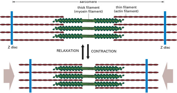
Answer: During muscle contraction, the laterally projecting heads (cross bridges) of the thick myosin myofilaments come in contact with the thin actin myofilaments and rotate on them. This pulls the thin myofilaments towards the middle of the sarcomere past the thick myofilaments. The Z lines come closer together and the sarcomere becomes shorter. Length of the A band remains constant. Myofilaments stay the same length. Free end of actin myofilaments move closer to the centre of the sarcomere, bringing Z lines closer together. I bands shorten and H zone narrows. A similar action in all the sarcomeres results in shortening of the entire myofibril, and thereby of the whole fibre and the whole muscle. A contracted muscle becomes shorter and thicker and its volume remains the same. This theory which is called the sliding filament theory proposed by A.F. Huxley and J. Hansen is the most satisfactory and accepted one.
Section XI Biochemistry of nutrition
1. Note substance, which activates pepsinogen to pepsin:
A. Hydrochloric acid
B. Trypsin
C. Enterokinase
D. Bile acids
E. Adenosine triphosphate