Атлас по рентгенологии травмированных собак и кошек / an-atlas-of-radiology-of-the-traumatized-dog-and-cat
.pdf
132 Radiology of Thoracic Trauma
2
Case 2.64
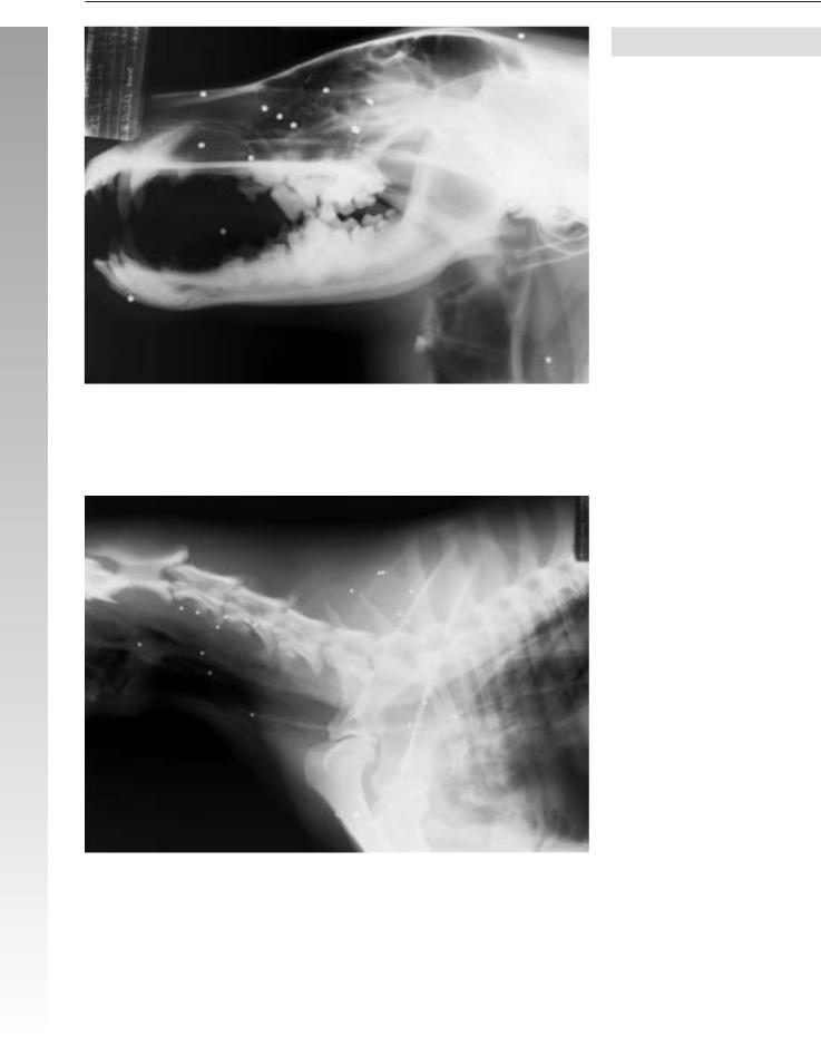
Signalment/History: “Amee” was a 2-year-old, female Borzoi presented in shock following being shot in the neck and shoulder on the left.
Physical examination: The dog was not able to stand and a complete neurologic examination was not carried out. Multiple soft tissue injuries were noted around the head and neck; however, it was not possible to ascertain if there was any thoracic injury.
Radiographic procedure: Lateral radiographs were made of the head and cervical region, with a complete study of the thorax.
Radiographic diagnosis (head and cervical region): Multiple metallic pellets were located in the head and cervical region indicating an injury from a shotgun fired from a short distance. No fluid density was noted in the nasal passages or in the frontal sinuses. Air that had dissected between the soft tissues in the neck permitted identification of both surfaces of the tracheal walls and was the origin of a pneumomediastinum.

Pneumomediastinum 133
2
Radiographic diagnosis (thorax): Subcutaneous emphysema in the cervical region was noted, plus a typical pattern of air within the mediastinum that was indicative of a pneumomediastinum. No evidence of lung injury was noted. Multiple shotgun pellets were present.
Comments: Determining the source of the free air permits a better understanding of the prognosis in such a case. The holes in the skin can be large enough to permit the air to enter the subcutaneous spaces and pass into the mediastinum, although such holes are in themselves usually of little clinical importance.
It was possible in this case that one of the pellets had injured the larynx or trachea permitting air to pass into the mediastinum. This would have been of greater clinical importance. An injury to the esophagus may leak air and may lead to a mediastinitis and be of great importance clinically; however, it is uncommon that an injury of this type would produce such a prominent pneumomediastinum as seen in “Amee”.
Endoscopy is strongly indicated in this type of patient. Several of the pellets were malformed indicating that they had struck bone.
Often only lateral views are made in a deep-chested patient such as “Amee” until more is known of the injury.

134 Radiology of Thoracic Trauma
Case 2.65
2
Signalment/History: “Wendy”, a large 1-year-old, female Scottish Deerhound, had run into a tree the day before.
Physical examination: The dog would not walk on her right forelimb. Crepitus was elicited following palpation of the right shoulder. Movement of the shoulder joint was painful. She was depressed with shallow breathing at the time of examination.
Radiographic procedure: Radiographic studies included multiple views of the thorax plus the region of the right shoulder.
Radiographic diagnosis: Hyperlucent lung fields were noted, but these were possibly due to the body conformation of a deep-chested dog with a thin chest wall. All of the major cranial mediastinal vessels and the tracheal wall could be clearly identified indicating a pneumomediastinum. The pulmonary vessels were also easily seen, but this was thought to be the result of the breed of dog and did not indicate an abnormal lung pattern. The cause of the pneumomediastinum could not be detected radiographically. A comminuted fracture of the right scapula with fragment displacement was seen.
The study included a lateral view of the cervical region and thoracic inlet, neither of which indicated injury to the upper airway or esophagus
Treatment/Management: The scapular fracture was permitted to heal without surgical stabilization of the fracture fragments. The dog was discharged to the referring clinician several days later.
Comments: Hyperlucent lung fields can be the result of the conformation of the thorax or can represent an actual pulmonary hyperinflation. The character of the pulmonary vessels is more easily evaluated in patients in whom the lungs are filled with air. Because of “Wendy’s” deep chest, caution should be used in the evaluation of the cardiac silhouette on the DV/VD views, since minimal obliquity of the thorax markedly influences the appearance of the heart shadow.

Pneumomediastinum 135
2

136 Radiology of Thoracic Trauma
Case 2.66
2
Day 1
Signalment/History: “Shep” was a 1-year-old, male German Shepherd mixed breed, who had been hit by a car 12 hours previously.
Physical examination: The examination was difficult to perform and only demonstrated marked dyspnea.
Radiographic procedure: Because of the dog’s difficulty in breathing, only a lateral thoracic radiograph was made.
Radiographic diagnosis (day 1): The single lateral radiograph was underexposed, but still clearly showed a large pneumothorax characterized by the elevation of the cardiac silhouette away from the sternum and retraction of the lung lobes dorsally from the spine and diaphragm. The ability to visualize both sides of the tracheal wall, the aortic arch, and serosal surface of the air-filled esophagus was indicative of a pneumomediastinum. Collapse of the caudal lung lobes suggested both pulmonary contusion and atelectasis. Liquid-dense, well-cir- cumscribed pulmonary nodules plus air-filled, cyst-like lesions were found in the dorsal lobes caudally. The diaphragm was intact.

Pneumomediastinum 137
2
Day 4
Radiographic diagnosis (day 4): The study on this day showed a complete resorption of the pneumothorax, though persistence of the pneumomediastinum. The lung lesions persisted on the right side caudally. The fluid density nodule remained in its dorsal position.
Treatment/Management: The nodular lesions suggested a more serious lung injury that was slower to repair than just a simple lung contusion following blunt trauma. The etiology of the pneumomediastinum remained undetermined as frequently occurs.

138 Radiology of Thoracic Trauma
2.2.11 Hemomediastinum
Case 2.67
2
Day 1
Signalment/History: “Romo” was a 5-year-old, male Spaniel mixed breed, who had been hit by a car and was referred several days after the accident along with post-trauma radiographs.
Radiographic diagnosis (immediate post-trauma): The radiographs were made on expiration and were underexposed/underdeveloped. However, a large cranial mediastinal fluid density could still be seen suggesting a mediastinal mass probably the result of hemorrhage. The tracheal shadow was shifted toward the right thoracic wall.

Hemomediastinum 139
2
Day 5
Radiographic diagnosis (day 5, lateral view only): Regression of the depth of the cranial mediastinal mass suggested resorption of the blood. The lungs were normal for a dog this age.
Comments: Hemorrhage within the mediastinum is thought to not be as important clinically in the dog as in man, where it pools caudally and does not drain freely resulting in a per-
sistent inflammatory process. In the dog, disappearance of the blood appears to occur rather easily, but does occur at a slower rate than the clearing of pleural fluid. It is helpful to monitor the clearance radiographically since change would confirm the suspicion of mediastinal fluid. A clinically more important abscess, tumor, or hematoma in the mediastinum would not change in size or shape as quickly on the follow-up radiographs.

140 Radiology of Thoracic Trauma
Case 2.68
2

Hemomediastinum 141
Signalment/History: “Raggs” was a moderately obese, 7- year-old, male Poodle with a history of having been hit by a car seven days earlier.
Physical examination: He presented with a right forelimb paralysis due to a probable avulsion of the brachial plexus.
Radiographic procedure: Thoracic radiographs were made to assess additional damage other than the neurological injury.
Radiographic diagnosis: An increase in cranial mediastinal thickness with indistinct borders extended ventrally toward the sternum and suggested mediastinal fluid possibly hemorrhage. Note that the thickness of the cranial mediastinal shadow was greater than the width of the extrathoracic soft tissue, indicating that the width was probably not the result of fat deposition, but was a pathological condition. The generalized increase in pulmonary density was probably due to underinflation of the lungs (note the cranial position of the diaphragm and moderate obesity of the dog). The obesity also caused minimal pleural thickening. A single airgun pellet lay dorsocaudaly within the mediastinum adjacent to the aorta and esophagus. No bony abnormality was present.
Treatment/Management: The mediastinal thickness was probably the result of hemorrhage. The airgun pellet may have been unrelated to the current medical problem and seemed to be in a position that would not cause any of the clinical signs. The absence of bony changes is typical in patients with sus-
pected brachial plexus injuries.
2
“Raggs” was a patient with an old gunshot wound and a more recent history of being hit by a car. Both traumatic events needed to be given consideration in the exploration of the clinical signs. Additional radiographic studies needed to be made of the right forelimb and cervicothoracic spine because of the paralysis.
“Raggs” did not show any marked improvement in his neurological signs and was taken home by the owner without any further radiographic studies being done.
Comments: The rule of measurement of the width of the mediastinum on the DV view in comparison to the width of the extrathoracic soft tissue is a helpful one in determining whether the mediastinal width is the result of fat accumulation or actually represents a pathological condition.
