

Атлас по рентгенологии травмированных собак и кошек / an-atlas-of-radiology-of-the-traumatized-dog-and-cat
.pdf102 Radiology of Thoracic Trauma
Case 2.49
2
Noncontrast
Signalment/History: This cat had no history of trauma, but had been listless for the previous few days with more recently occurring episodes of dyspnea.
Physical examination: Heart and lung sounds could not be heard on the right side.
Radiographic procedure: Radiographs were made of the thorax with the possibility of a contrast study if required.
Radiographic diagnosis: A mass-like lesion lay in the caudal right hemithorax and extended into the caudal left hemithorax with displacement of the heart shadow cranially, dorsally, and to the left.
Because of the failure to identify any air-filled bowel loops in the thoracic cavity, a barium meal was used which clearly showed the displacement of the stomach into the thoracic cavity.
Treatment/Management: The diagnosis was confirmed at surgery.

Diaphragmatic hernia 103
2
Contrast

104 Radiology of Thoracic Trauma
Case 2.50
2

Diaphragmatic hernia 105
2
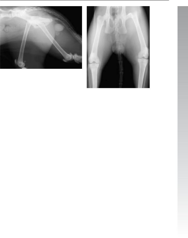
Signalment/History: “Cody” was an 11-month-old, male Labrador Retriever who had been hit by a car four hours earlier.
Physical examination: The dog was dyspneic and appeared to have great pain on palpation of the lumbar spine.
Radiographic procedure: Only lateral radiographs were made because of the suspicion of extensive injuries.
Radiographic diagnosis: The pleural fluid was thought to be hemorrhage. In addition, an increase in fluid density in the lungs was noted. An elevation of the cardiac silhouette suggested either displaced abdominal organs or some other pleural mass. The diaphragm could not be completely identified and provided another feature suggestive of a diaphragmatic hernia. The liver shadow was displaced cranially into the thoracic cavity and the stomach axis was shifted cranially. Neither the spleen nor the urinary bladder could be identified within the abdominal cavity.
A fracture-luxation at L4–5 did not cause marked segmental displacement, but did indicate additional trauma. Following
identification of the spinal fracture, lateral views of the thoracolumbar spine were made permitting further identification of the fracture with small fragments identified within the spinal canal. Retroperitoneal fluid was suggestive of hemorrhage associated with the fractures.
Treatment/Management: The diaphragmatic hernia was repaired and the vertebral fracture/luxation was stabilized surgically.
The status of the urinary system remained in doubt. The urinary bladder appeared intact on a retrograde cystogram using 60 ml of contrast agent. On an excretory urogram using 70 ml of contrast agent injected intravenously, positive contrast agent was extravasated retroperitoneally and peritoneally around the left kidney, suggesting a ureteral avulsion on that side. The renal pelvis on the right was distended indicating obstruction to flow and a possible right ureteral tear as well. The urinary lesions were both treated conservatively because of the owner’s choice not to spend additional money. “Cody” survived and was subsequently released to his owner.

106 Radiology of Thoracic Trauma
Case 2.51
2
Signalment/History: “Morris” was a 1-year-old, male DSH cat that had possibly been traumatized 48 hours earlier. According to the owner, he had been either struck by a car or kicked by a cow.
Physical examination: The cat was dyspneic and could not bear weight on the left pelvic limb.
Radiographic procedure: Studies were made of the thorax and pelvis.
Radiographic diagnosis (thorax): The pericardial sac was dilated and contained gas-filled small bowel loops that extended from the abdomen through the diaphragm into the pericardial sac. The trachea was displaced dorsally by the mass effect. The surrounding lung appeared normal in appearance. Note the appearance of the body of the 7th thoracic vertebral segment. Hemivertebrae are unusual in cats. Could this be a compression fracture?
Diaphragmatic hernia 107
2
Radiographic diagnosis (pelvis): The fracture of the left femoral neck was intertrochanteric and extracapsular. The age of the fracture was difficult to determine because of the fragment position.
Treatment/Management: Pericardio-diaphragmatic hernias often display a change in dynamics following trauma. At surgery, the liver, gall bladder, most of the jejunum, the ileum, and a part of the colon were within the pericardial sac. The radiographic appearance of the lung tissue was normal regardless of its being compressed by the enlarged pericardial sac.

108 Radiology of Thoracic Trauma
2.2.8Pleural air
Case 2.52
2
Day 1
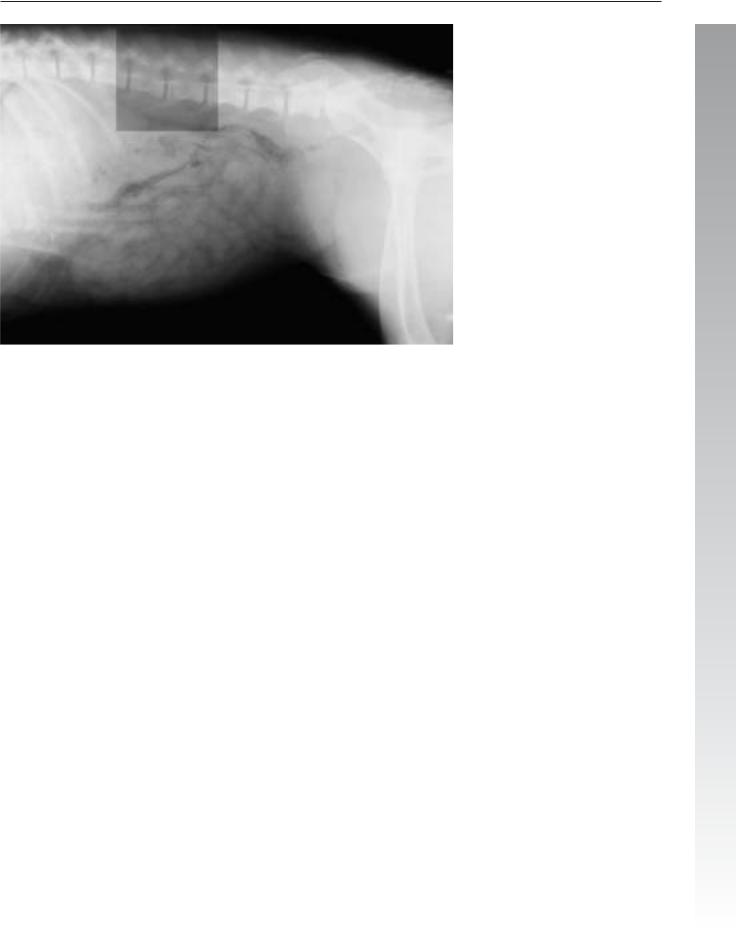
Signalment/History: “Ruff” was a 6-month-old, male Golden Retriever who had been struck by a car 3 days previously.
Physical examination: Palpation of the thorax revealed a probable injury to the caudal ribs on the right. The dog breathed in a careful manner and was unwilling to take a deep breath.
Radiographic procedure: Radiographs were made of the thorax using a technique that would permit evaluation of the ribs.
Radiographic diagnosis (day 1, thorax): Radiographs showed a pneumothorax that was characterized by elevation of the cardiac silhouette away from the sternum and separation of the borders of the caudal lung lobes from the diaphragm. Pulmonary contusion was noted adjacent to the fractures of the 9th, 10th, 11th, 12th, and 13th ribs on the right. The fluid density in the cranial mediastinum adjacent to the sternum was probably a hemomediastinum. A prominent skin fold extended across the caudal right lung field on the DV study.
Pleural air 109
2
Radiographic diagnosis (day 1, abdomen; lateral view only): Abdominal radiographs showed a physeal fracture of L4 with separation and displacement of the cranial end plate. Small bowel loops were distended with fluid. Skin folds were
prominent in the cranial abdomen.

110 Radiology of Thoracic Trauma
2
Day 12
Radiographic diagnosis (day 12, thorax): Radiographs made 11 days later showed clearing of the fluid from the lungs and disappearance of the pleural air.
Treatment/Management: The bowel was possibly distended because of the trauma itself or perhaps due to an injury to the spinal cord. No clinical signs were associated with the small bowel and no treatment was required.
The patient was re-evaluated 11 days after the initial presentation for neurological deficits and was noted to only have pain over the lumbar spine and some hesitancy in walking. He was treated with cage rest for several weeks and was able to walk normally when released.
Comments: Rib fractures are most easily recognized radiographically when the fractures are within the bony portion of the rib and there is a marked displacement of the fragments. Fractures near the costovertebral joints are surrounded by heavy muscle and do not usually show fragment displacement. Fractures near the costochondral junction are difficult to identify because of the cartilage content of the ribs. Fortunately, these types of fracture are not of great clinical importance and when over-looked, probably do not affect the selection of treatment or prognosis of the case.

|
Pleural air 111 |
|
|
|
|
|
Case 2.53 |
|
|
|
|
|
Signalment/History: “Pumpkin”, a 5-month-old, female |
|
|
DSH cat, had been struck by an automobile. |
|
|
Physical examination: The cat was presented in severe re- |
2 |
spiratory distress.
Radiographic procedure: The whole body was radiographed.
Radiographic diagnosis: Extensive pulmonary hemorrhage was noted throughout the lungs. It was unusual that the pneumothorax could be seen on the lateral view, but was difficult to identify on the DV view as it only caused a thin radiolucent line along the left thoracic wall. The diaphragm was intact on both views.
Stress aerophagia had resulted in an air-filled stomach and bowel loops. Distended bowel loops of this degree could be the result of an ileus secondary to loss of blood supply to a portion of the gut or torsion of the mesenteric blood supply.
Identification of the ventral border of the liver ruled out the accumulation of peritoneal fluid. Note the absence of the usually large fat-filled falciform ligament.
Treatment/Management: “Pumpkin” died shortly after the radiographs were made. The necropsy findings were limited to the contused lung with some pleural hemorrhage in addition to fractures of the costal arches. The air-filled bowel was apparently the result of aerophagia.
Comments: Although the degree of pulmonary contusion was not severe, what was important in this patient and led to her death was the fact that all of the lobes were similarly affected.