

Атлас по гематологии (английский язык)
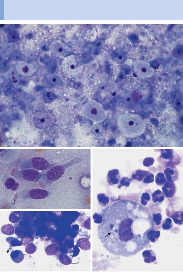
.pdfEpithelioid cells dominate the lymph node biopsy: Boeck disease or tuberculosis
 a
a
 b
b
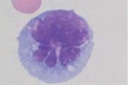
Fig. 64 Boeck disease and tuberculosis. a Lymph node cytology in Boeck disease: a special form of reactive cell pattern with (often predominating) islands and trains of epithelioid cells (arrow), which have ovoid nuclei with delicate chromatin structure and a wide, smoke-gray layer of cytoplasm. b Lymph node cytology in tuberculous lymphadenitis: in addition to lymphocytes and a few epithelial cells (1), enormous syncytes of epithelioid cell nuclei within one cytoplasm (arrow) may be encountered: the Langhans giant cell.
181
Theml, Color Atlas of Hematology © 2004 Thieme
All rights reserved. Usage subject to terms and conditions of license.

182 Cytology of Organ Biopsies and Exudates
Non-Hodgkin Lymphoma
Since the CBC is the first step in any lymph node diagnosis, lymph node biopsy is unnecessary in many cases of non-Hodgkin lymphoma (p. 70), because the most common form of this group of diseases, chronic lymphadenosis, can always be diagnosed on the basis of the leukemic findings of the CBC.
However, when enlarged lymph nodes are found in one or more regions without symptoms of reactive disease, and the blood analysis fails to show signs of leukemia, lymph node biopsy is indicated.
The relatively monotonous lymph node cytology in non-Hodgkin lymphomas and tumor metastases mean that histological differentiation is required.
In contrast to Hodgkin disease, with its conspicuous giant cell forms (p. 177), non-Hodgkin lymphomas display a monotonous picture without any signs of a reactive process (p. 70). Clinically, it is enough to distinguish between small cell forms (which have a relatively good prognosis) and large cell forms (which have a poorer prognosis) to begin with. For a more detailed classification, see page 70f.
Histological analysis may be omitted only when its final results would not be expected to add to the intermediate cytological findings in terms of consequences for treatment.
Metastases of Solid Tumors in Lymph Nodes or Subcutaneous Tissue
When hard nodules are found that are circumscribed in location, biopsy shows aggregates of polymorphous cells with mostly undifferentiated nuclei and a coarse reticular structure of the chromatin (perhaps with welldefined nucleoli or nuclear vacuoles), and the lymphatic cells cannot be classified, there is urgent suspicion of metastasis from a malignant solid tumor, i.e. from a carcinoma in a variety of possible locations or a soft tissue sarcoma.
As a rule, the next step is the search for a possible primary tumor. If this is found, lymph node resection becomes unnecessary.
If no primary tumor is found, lymph node histology is indicated. The histological findings can provide certain clues about the etiology and also helps in the difficult differential diagnosis versus blastic non-Hodgkin lymphoma.
Theml, Color Atlas of Hematology © 2004 Thieme
All rights reserved. Usage subject to terms and conditions of license.

In cases of non-Hodgkin lymphoma and tumor metastases, a tentative diagnosis is possible on the basis of the lymph node cytology
a 
 b
b
c
d |
e |
Fig. 65 Non-Hodgkin lymphoma and tumor metastases. a Lymph node cytology showing small cells with relatively wide cytoplasm (arrow 1) in addition to lymphocytes. There are scattered blasts with wide cytoplasm (arrow 2): lymphoplasmacytic immunocytoma. b Lymph node cytology showing exclusively large blastoid cells with a large central nucleolus (arrow). This usually indicates large-cell nonHodgkin lymphoma (in this case immunoblastic). c–e Metastatic disease from: c uterine carcinoma, d small-cell bronchial carcinoma, and e leiomyosarcoma.
183
Theml, Color Atlas of Hematology © 2004 Thieme
All rights reserved. Usage subject to terms and conditions of license.

184 Cytology of Organ Biopsies and Exudates
Branchial Cysts and Bronchoalveolar
Lavage
Branchial Cysts
A (usually unilateral) swollen neck nodule below the mandibular angle that feels firm to pressure, but is without external signs of inflammation, should suggest the presence of a branchial cyst. Surprisingly, aspiration usually produces a brownish-yellow liquid. In addition to partially cytolysed granulocytes and lymphocytes (cell detritus), a smear of this liquid, or the centrifuged precipitate, shows cells with small central nuclei and wide light cell centers which are identical to epithelial cells from the floor of the mouth. Biopsies from a soft swelling around the larynx show the same picture; in this case it is a retention cyst from another developmental remnant, the ductus thyroglossus.
Cytology of the Respiratory System,
Especially Bronchoalveolar Lavage
Through the development of patient-friendly endoscopic techniques, diagnostic lavage (with 10–30 ml physiological saline solution) and its cytological workup are now in widespread use. This method is briefly mentioned here because of its broad interest for all medical professionals with an interest in morphology; the interested reader is referred to the specialist literature (e.g. Costabel, 1994) for further information. Table 31 lists the most important indications for bronchoalveolar lavage.
Table 31 Clinical indications for bronchoalveolar lavage (according to Costabel 1994)
Interstitial infiltrates |
Alveolar infiltrates |
Pulmonary infiltrates in |
|
|
patients with immune |
|
|
deficiency |
Sarcoidosis (Boeck disease) |
Pneumonia |
HIV Infection |
Exogenous allergic alveolitis |
Alveolar hemorrhage |
Treatment with cytostatic |
Drug-induced alveolitis |
Alveolar proteinosis |
agents |
Idiopathic pulmonary fibrosis |
Eosinophilic pneumonia |
Radiation sickness |
Collagen disease |
Obliterating bronchiolitis |
Immunosuppressive therapy |
Histiocytosis X |
|
Organ transplant |
Pneumoconioses |
|
|
Lymphangiosis carcino- |
|
|
matosa |
|
|
|
|
|
Theml, Color Atlas of Hematology © 2004 Thieme
All rights reserved. Usage subject to terms and conditions of license.
Accessible cysts (e.g., branchial cysts) should be aspirated. Bronchial lavage is a cytological new discipline
a |
|
b |
|
c |
d |
Fig. 66 Cyst biopsy and bronchoalveolar lavage. a Cytology of a lateral neck cyst: no lymphatic tissue, but epithelial cells from the floor of the mouth. b Normal ciliated epithelial cells with typical cytoplasmic processes. c Tumor cell conglomeration in small-cell bronchial carcinoma: conglomeration is typical of tumor cells. d Bronchoalveolar lavage in purulent bronchitis: a macrophage with pigment inclusion (arrow) is surrounded by segmented neutrophilic granulocytes.
185
Theml, Color Atlas of Hematology © 2004 Thieme
All rights reserved. Usage subject to terms and conditions of license.

186 Cytology of Organ Biopsies and Exudates
Cytology of Pleural Effusions and Ascites
Pleural effusions always require cytological diagnostic procedures unless they are secondary to a known disease, such as cardiac insufficiency or pneumonia, and recede on treatment of the primary disease.
Pleura aspirates can be classified as exudates or transudates (the latter usually caused by hydrodynamic stasis). The specific density (measured with a simple areometer) of transudates, which are protein-poor, is between 1008 and 1015 g/l, while for exudates it is greater than 1018 g/l.
Cytological preparation may be done by gentle centrifugation of the aspirate (10 minutes at 300–500 rpm), which should be as fresh as possible; the supernatant is decanted and the sediment suspended in the residual fluid, which will collect on the bottom of the centrifuge tube. Nowadays, however, this procedure has been replaced by cytocentrifugation.
Effusions that are noticeably rich in eosinophilic granulocytes should raise the suspicion of Hodgkin disease, generalized reaction to the presence of a tumor, or an allergic or autoimmune disorder. Purely lymphatic effusions are particularly suggestive of tuberculosis. In addition, all transudates and exudates contain various numbers of endothelial cells (particularly high in cases of bacterial pleuritis) that have been sloughed off from the pleural lining.
Any cell elements that do not fulfill the above criteria should be regarded as suspect for neoplastic transformation, especially if they occur in aggregates. Characteristics that in general terms support such a suspicion include extended size polymorphy, coarse chromatin structure, welldefined nucleoli, occasional polynucleated cells, nuclear and plasma vacuoles, and deep cytoplasmic basophilia. For practical reasons, special diagnostic procedures should always be initiated in these situations.
What was said above in relation to the cell composition of pleural effusions also holds for ascites. Here too, the specific density may be determined and the Rivalta test to distinguish exudate from transudate carried out. Inflammatory exudates usually have a higher cell content; a strong predominance of lymphocytes may indicate tuberculosis. Like the pleura, the peritoneum is lined by phagocytotic endothelial cells which slough off into the ascitic fluid and, depending on the extent of the fluid, may produce a polymorphous overall picture analogous to that of the pleural endothelial cells. It is not always easy to distinguish between such endothelial cells and malignant tumor metastases. However, the latter usually occur not alone but in coherent cell aggregates (“floating metastases”), the various individual elements of which typically show a coarse chromatin structure, wide variation in size, well-defined nucleoli, and deeply basophilic cytoplasm.
Theml, Color Atlas of Hematology © 2004 Thieme
All rights reserved. Usage subject to terms and conditions of license.

Tumor cells can be identified in pleural and ascites smears
a 
 b
b
c 
 d
d
Fig. 67 Pleural effusion and ascites. a Pleural cytology, nonspecific exudate: dormant mesothelial cell (or serosal cover cell) (1), phagocytic macrophage with vacuoles (2), and monocytes (3), in addition to segmented neutrophilic granulocytes
(4). b Cell composition in a pleural aspirate (prepared using a cytocentrifuge): variable cells, whose similarity to cells in acute leukemia should be established by cytochemistry and marker analysis: lymphoblastic lymphoma. c Ascites with tumor cell conglomerate, surrounded with granulocytes and monocytes, in this case of ovarian carcinoma. d Ascites cytology with an island of tumor cells. This kind of conglomeration is typical of tumor cells.
187
Theml, Color Atlas of Hematology © 2004 Thieme
All rights reserved. Usage subject to terms and conditions of license.

188 Cytology of Organ Biopsies and Exudate
Cytology of Cerebrospinal Fluid
The first step in all hemato-oncological and neurological diagnostic assessments of cerebrospinal fluid is the quantitative and qualitative analysis of the cell composition (Table 32).
Table 32 Emergency diagnostics of the liquor (according to Felgenhauer in Thomas 1998)
Pandy’s reaction
Cell count (Fuchs-Rosenthal chamber)
Smear (or cytocentrifuge preparation)
–to analyze the cell differentiation and
–to search for bacteria and roughly determine their types and prevalence
Gram stain
An additional determination of bacterial antigens may be done
Using advanced cell diagnostic methods, lymphocyte subpopulations can be identified by immunocytology and marker analysis and cytogenetic tests carried out on tumor cells.
Prevalence of neutrophilic granulocytes with strong pleocytosis suggests bacterial meningitis; often the bacteria can be directly characterized.
Prevalence of lymphatic cells with moderate pleocytosis suggests viral meningitis. (If clinical and serological findings leave doubts, the differential diagnosis must rule out lymphoma using immunocytological methods.)
Strong eosinophilia suggests parasite infection (e.g., cysticercosis).
A complete mixture of cells with granulocytes, lymphocytes, and monocytes in equal proportion is found in tuberculous meningitis.
Variable blasts, usually with significant pleocytosis, predominate in leukemic or lymphomatous meningitis.
Undefinable cells with large nuclei suggest tumor cells in general, e.g., meningeal involvement in breast cancer or bronchial carcinoma, etc. The cell types are determined on the basis of knowledge of the primary tumor and/or by marker analysis. Among primary brain tumors, the most likely cells to be found in cerebrospinal fluid are those from ependymoma, pinealoma, and medulloblastoma.
Erythrophages and siderophages (siderophores) are monocytes/macrophages, which take up erythrocytes and iron-containing pigment during subarachnoid hemorrhage.
The cytological analysis of the cerebrospinal fluid offers important clues to the character of meningeal inflammation, the presence of a malignancy, or hemorrhage.
Theml, Color Atlas of Hematology © 2004 Thieme
All rights reserved. Usage subject to terms and conditions of license.

Viral, bacterial, and malignant meningitis can be distinguished by means of cerebrospinal fluid cytology
a |
b |
c 
 d
d
e 
 f
f
g 
 h
h
Fig. 68 Cerebrospinal fluid cytology. a Cerebrospinal fluid cytology in bacterial meningitis: granulocytes with phagocytosed diplococci (in this case, pneumococci, arrow). b Cerebrospinal fluid cytology in viral meningitis: variable lymphoid cells. c Cerebrospinal fluid cytology in non-Hodgkin lymphoma: here, mantle cell lymphoma. d Cerebrospinal fluid cytology after subarachnoid hemorrhage: macrophages with phagocytosed erythrocytes. e–h Cerebrospinal fluid cytology in meningeal involvement in malignancy: the origin of the cells cannot be deduced with certainty from the spinal fluid cytology alone: (e) breast cancer, (f) bronchial
carcinoma, (g) medulloblastoma, and (h) acute leukemia. |
189 |
Theml, Color Atlas of Hematology © 2004 Thieme
All rights reserved. Usage subject to terms and conditions of license.

190
References
Begemann, H., M. Begemann: Praktische Hämatologie, 10. Aufl. Thieme, Stuttgart 1998
Begemann, H., J. Rastetter: Klinische Hämatologie, 4. Aufl. Thieme, Stuttgart 1993 Bessis, M.: Blood Smears Reinterpreted. Springer, Berlin 1977
Binet, J.L., A. Auquier, G. Dighiero et al.: A new prognostic classification of chronic lymphocytic leukemia derived from a multivariate survival analysis. Cancer 1981; 48(1): 198-206
Brücher, H.: Knochenmarkzytologie. Thieme, Stuttgart 1986
Classen, M., A. Dierkesmann, H. Heimpel et al.: Rationelle Diagnostik und Therapie in der inneren Medizin. Urben & Schwarzenberg, München 1996
Costabel, M.: Atlas der bronchoalveolären Lavage. Thieme, Stuttgart 1994 Costabel V., J. Guzman: Bronchoalveolar lavage in interstital lung disease. Curr
Opin Pulm Med 2001; 7(5): 255-61
Durie, B.G.M., S.E. Salmon: A clinical staging system for multiple myeloma. Correlation of measured myeloma cell mass with presenting clinical features, response to treatment, and survival. Cancer 1975; 36: 842-854
Heckner, F., M. Freund: Praktikum der mikroskopischen Hämatologie. Urban & Schwarzenberg, München 1994
Heimpel, H., D. Hoelzer, E. Kleihauer, H. P. Lohrmann: Hämatologie in der Praxis. Fischer, Stuttgart 1996
Huber, H., H. Löffler, V. Faber: Methoden der diagnostischen Hämatologie. Springer, Berlin 1994
Jaffe, E. S., N. L. Harris, H. Stein, J. W. Vardiman: World Health Organization Classification of Tumours. Pathology and Genetics of Tumours of Haematopoietic and Lymphoid Tissues. IARC Press, Lyon 2001
Lennert, K., A. C. Feller: Non-Hodgkin-Lymphome. Springer, Berlin 1990 Löffler, H., J. Rastetter: Atlas der klinischen Hämatologie. Springer, Berlin 1999
Murphy, S.: Diagnostic criteria and prognosis in polycythemia vera and essential thombocythemia. Semin Hematol 1999; 36(1 Suppl 2): 9-13
Ovell, S. R., G. Sterrett, M. N. Walters, D. Whitaker: Fine Needle Aspiration Cytology. Churchill Livingstone, Edinburgh London 1992
Pearson, T.C., M Messinezy: The diagnostic criteria of polycythaemia rubra vera. Leuk Lymphoma 1996;22(Suppl 1): 87-93
Pralle, H. B.: Checkliste Hämatologie, 2. Aufl. Thieme, Stuttgart 1991
Schmoll, H. J., K. Höffken, K. Possinger: Kompendium internistische Onkologie, 3. Bde., 3. Aufl. Springer, Berlin 1999
Theml, H., W. Kaboth, H. Begemann: Blutkrankheiten. In Kühn, H. A., J. Schirmeister: Innere Medizin, 5. Aufl. Springer, Berlin 1989
Theml, H., H. D. Schick: Praktische Differentialdiagnostik hämatologischer und onkologischer Krankheiten. Thieme, Stuttgart 1998
Zucker-Franklin, D., M. F. Greaves, C. E. Grossi, A. M. Marmont: Atlas der Blutzellen, 2. Aufl. Fischer, Stuttgart 1990
Theml, Color Atlas of Hematology © 2004 Thieme
All rights reserved. Usage subject to terms and conditions of license.