

Атлас по гематологии (английский язык)
.pdf Conspicuous erythrocyte inclusions suggest malaria
a 
b |
c |
d 
 e
e
Fig. 57 Blood analysis in malaria. a Trophozoites in Plasmodium falciparum (falciparum or malignant tertian malaria) infection. Simple signet-ring form (1), dou- ble-invaded erythrocyte with basophilic stippling (2). Erythrocyte with basophilic stippling without plasmodium (3) and segmented neutrophilic granulocyte with toxic granulation (4). b and c Plasmodium falciparum: single invasion with delicate trophozoites (1), multiple invasion (2). d and e In the gametocyte stage of falciform malaria, the pathogens appear to reside outside the erythrocyte, but remnants of the erythrocyte membrane may be seen (arrow, e). For a systematic overview of malarial inclusions, see p. 159.
161
Theml, Color Atlas of Hematology © 2004 Thieme
All rights reserved. Usage subject to terms and conditions of license.

162 Erythrocyte and Thrombocyte Abnormalities
Polycythemia Vera (Erythremic
Polycythemia) and Erythrocytosis
Increases in erythrocytes, hemoglobin, and hematocrit above the normal range due to causes unrelated to hematopoiesis (i.e., the majority of cases) are referred to as secondary erythrocytosis or secondary polycythemia. However, it should be remembered that the “normal” range of values is quite wide, especially for men, in whom the normal range can be as much as 55% of the hematocrit!
Table 28 Causes of secondary erythrocytosis
Reduced O2 transport capacity |
– |
Carboxyhemoglobin formation in chronic |
|
|
smokers |
|
– High altitude hypoxia, COPD |
|
|
– Heart defect (right–left shunt) |
|
|
– Hypoventilation (e. g., obesity hypoventilation) |
|
Reduced O2 release from hemoglobin |
– |
Congenital 2,3-diphosphoglycerate deficiency |
Renal hypoxia |
– |
Hydronephrosis, renal cyst |
|
– |
Renal artery stenosis |
Autonomous erythropoietin biosynthesis |
– |
Renal carcinoma |
|
– |
Adenoma |
|
|
|
COPD = Chronic obstructive pulmonary disease
An autonomous increase in erythropoiesis, i.e., polycythemia vera, represents a very different situation. Polycythemia vera—which one might call a “primary erythrocytosis“—is a malignant stem cell abnormality of unknown origin and is a myeloproliferative syndrome (p. 112). Leukocyte counts (with increased basophils) and thrombocyte counts often rise simultaneously, and a transition to osteomyelosclerosis often occurs in the later stages.
Revised Criteria for the Diagnosis of Polycythemia Vera (according to Pearson and Messinezy, 1996)
—A1 elevated erythrocyte numbers
—A2 absence of any trigger of secondary polycythemia
—A3 palpable splenomegaly
—A4 clonality test, e.g., PRV-1 marker (polycythemia rubra vera 1)
—B1 thrombocytosis (!400 "109/l)
—B2 leukocytosis (!10 "109/l)
—B3 splenomegaly (sonogram)
—B4 endogenous erythrocytic colonies
Diagnosis in the presence of: A1 + A2 + A3/A4 or A1 + A2 + 2 "B
Bone marrow cytology shows increased erythropoiesis in both polycythemia vera and secondary polycythemia. Polycythemia vera is the more likely diagnosis when megakaryocytes, granulopoiesis, basophils, and eosinophils are also increased.
Theml, Color Atlas of Hematology © 2004 Thieme
All rights reserved. Usage subject to terms and conditions of license.

|
|
Bone marrow analysis contributes to the differential diagnosis |
|
|
|
between secondary erythrocytosis and polycythemia vera |
|
a |
|
|
b |
|
|
||
c |
|
|
|
d |
|
|
e |
Fig. 58 Polycythemia vera and secondary erythrocytosis. a In reactive secondary erythrocytosis there is usually only an increase in erythropoiesis. b In polycythemia vera megakaryopoiesis (and often granulopoiesis) are also increased. c Bone marrow smear at low magnification in polycythemia vera, with a hyperlobulated megakaryocyte (arrow). d Bone marrow smear at low magnification in polycythemia vera, showing increased cell density and proliferation of megakaryocytes. e In polycythemia vera, iron staining shows no iron storage particles.
163
Theml, Color Atlas of Hematology © 2004 Thieme
All rights reserved. Usage subject to terms and conditions of license.

164 Erythrocyte and Thrombocyte Abnormalities
Thrombocyte Abnormalities
EDTA in blood collection tubes (e.g., lavender top tubes) can lead to aggregation (pseudothrombocytopenia). A control using citrate as anticoagulant is required.
The clinical sign of spontaneous bleeding in small areas of the skin and mucous membranes (petechial bleeding), which on injury diffuses out to form medium-sized subcutaneous ecchymoses, is grounds for suspicion of thrombocyte (or vascular) anomalies.
Thrombocytopenia
Thrombocytopenias Due to Increased Demand (High Turnover)
A characteristic sign of this pathology can be that among the few thrombocytes seen in the blood smear, there is an increased presence of larger, i.e., less mature cell forms.
The bone marrow in these thrombocytopenias shows raised (or at least normal) megakaryocyte counts. Here too, there may be an increased presence of less mature forms with one or two nuclei (Figs. 60, 61).
Drug-induced immunothrombocytopenia As in drug-induced agranulocytosis, the process starts with an antibody response to a drug, or its metabolites, and binding of the antibody complex to thrombocytes. Rapid thrombocyte degradation by macrophages follows (Table 29). The most common triggers are analgesic/anti-inflammatory drugs and antibiotics.
Theml, Color Atlas of Hematology © 2004 Thieme
All rights reserved. Usage subject to terms and conditions of license.

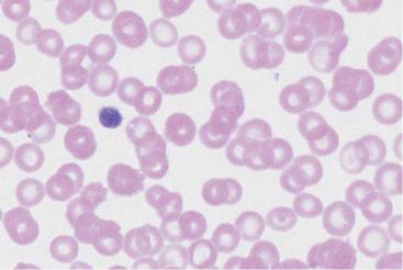
Thrombocytes: increases, reductions, and anomalies may be seen
 a
a
 b
b
Fig. 59 Forms of thrombocytopenia. a This blood smear shows normal size and density of thrombocytes. b In this blood smear thrombocyte density is lower and size has increased, a feature typical of immunothrombocytopenia. continued !
165
Theml, Color Atlas of Hematology © 2004 Thieme
All rights reserved. Usage subject to terms and conditions of license.

166 Erythrocyte and Thrombocyte Abnormalities
Table 29 Most common triggers of drug-induced immunothrombocytopenia
Analgesics |
Quinine |
Isoniazid |
Antibiotics |
Digitalis preparations |
Methyldopa |
Anticonvulsive drugs |
Furosemide |
Spironolactone |
Arsenic, e. g., in water |
Gold salts |
Tolbutamide |
Quinidine |
Heparin! |
|
|
|
|
“Idiopathic” immunothrombocytopenia. The name is largely historical, because often a trigger is found.
Postinfectious = acute form, usually occurs in children after rubella, mumps, or measles.
Essential thrombocytopenia, thrombocytopenic purpura (Werlhof disease)
Thrombocyte antibodies develop without an identifiable trigger. This is thus a primary autoimmune disease specifically targeting thrombocytes.
Secondary immunothrombocytopenias, e.g., in lupus erythematosus and other forms of immune vasculitis, lymphomas, and tuberculosis.
Post-transfusion purpura occurs mostly in women about one week after a blood transfusion, often after earlier transfusions or pregnancy.
Thrombocytopenia in microangiopathy. This group includes thrombotic– thrombocytopenic purpura (see p. 144) and disseminated intravascular coagulation.
Thrombocytopenia in hypersplenism of whatever etiology.
Fig. 59 Continued. c Pseudothrombocytopenia. The thrombocytes are not lying ! free and scattered around, but agglutinated together, leading to a reading of thrombocytopenia from the automated blood analyzer. d Giant thrombocyte (as large as an erythrocyte) in thrombocytopenia. Döhle-type bluish inclusion (arrow)
in the normally granulated segmented neutrophilic granulocyte: May-Hegglin anomaly.
Theml, Color Atlas of Hematology © 2004 Thieme
All rights reserved. Usage subject to terms and conditions of license.

Thrombocytes: increases, reductions, and anomalies may be seen
c 
 d
d
 e
e
Fig. 59 e Large thrombocyte (1) in thrombocytopenia. Thrombocyte-like fragments from destroyed granulocytes (cytoplasmic fragments) (2), which have the same structure and staining characteristics as the cytoplasm of band granulocytes
(3). Clinical status of sepsis with disseminated intravascular coagulation. In automated counters cytoplasmic fragments are included in the thrombocyte fraction.
167
Theml, Color Atlas of Hematology © 2004 Thieme
All rights reserved. Usage subject to terms and conditions of license.

168 Erythrocyte and Thrombocyte Abnormalities
Thrombocytopenias Due to Reduced Cell Production
In this condition, blood contains few, usually small, pyknotic (“old”) thrombocytes. Only a very few megakaryocytes are found in the bone marrow, and these have a normal appearance.
Causes
Chronic alcoholism. There may be some overlap with increased turnover and folic acid deficiency.
Chemical and radiological noxae. Cytostatics naturally lead directly to a dose-dependent reduction in megakaryocyte counts. A large therapeutic radiation burden in the area of blood-producing bone marrow has the same result, which may persist for many months.
Virus infections. Measles, mononucleosis (Epstein–Barr virus, cytomegalovirus), rubella, and influenza may (usually in children) trigger thrombocytopenias of various types. In these cases, the virus affects the megakaryocytes directly. However, antibodies to thrombocytes may also arise in the course of these infections (p. 166), so that the pathomechanism of the parainfectious thrombocytopenias described by Werlhof in children may lie in impaired production and/or increased degradation of thrombocytes.
Neoplastic and aplastic bone marrow diseases. All neoplasms of the bone marrow cell series (e.g., leukemia, lymphoma, and plasmacytoma), together with their precursor forms (e.g., myelodysplasia), lead to progressive thrombocytopenia, as do panmyelophthisis and bone marrow infiltration by metastases from solid tumors.
Vitamin deficiency. Folic acid and vitamin B12 deficiencies from various causes (p. 152) also affect the rapidly proliferating megakaryocytes. In these cases, thrombocytopenia is often present before anemia and leukocytopenia in circulating blood, while the bone marrow shows copious megakaryocytes that have been blocked from maturation.
Constitutional diseases. An amegakaryocytic thrombocytopenia without any of the above causes is rare. It is seen in children with congenital radial aplasia; in adults it tends usually to be an early sign of leukemia, myelodysplastic syndrome, or aplastic anemia.
Wiscott-Aldrich syndrome (thrombocytopenia, immune deficiency, and eczema) is an X-chromosomal recessive disease in boys and presents with thrombocytopenia with ineffective megakaryopoiesis.
The May-Hegglin anomaly (dominant hereditary transmission) is characterized by thrombocytopenia with giant thrombocytes and granulocyte inclusions, which resemble Döhle bodies (endoplasmatic reticulum aggregates).
Theml, Color Atlas of Hematology © 2004 Thieme
All rights reserved. Usage subject to terms and conditions of license.
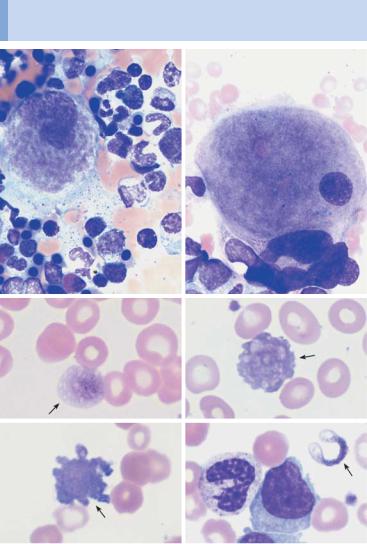
Variant forms of thrombocyte and megakaryocyte morphology in the bone marrow are diagnostic aids in thrombocytopenia
a |
b |
c |
d |
e |
f |
Fig. 60 Morphology of thrombocytes and megakaryocytes. a Bone marrow in thrombocytopenia due to increased turnover (e.g., immunothrombocytopenia). Mononuclear “young” megakaryocytes clearly budding a thrombocyte (irregular, cloudy cytoplasm structure). b In thrombocytopenia against a background of myelodysplasia, the bone marrow shows various megakaryocyte anomalies: here, too small a nucleus surrounded by too wide cytoplasm. c–f In myelodysplasia (c and d) and acute myeloid leukemia (e and f), bizarre anomalous thrombocyte shapes (arrows) may occasionally be found.
169
Theml, Color Atlas of Hematology © 2004 Thieme
All rights reserved. Usage subject to terms and conditions of license.

170 Erythrocyte and Thrombocyte Abnormalities
Thrombocytosis (Including
Essential Thrombocythemia)
Reactive. Thrombocytosis in the form of a constant elevation of thrombocyte counts above an upper normal range of 300 000–450 000/µl, may be reactive, i.e., may appear in response to various tumors (particularly bronchial carcinoma), chronic inflammation (particularly ulcerative colitis, primary chronic polyarthritis), bleeding, or iron deficiency. The pathology of this form is not known.
Essential Thrombocythemia
Essential thrombocythemia, by contrast, is a myeloproliferative disease (see p. 114) in which the main feature of increased thrombocytes is accompanied by other signs of this group of diseases that may vary in severity, such as leukocytosis and an enlarged spleen. Severe thrombocythemia may also be seen in osteomyelosclerosis, polycythemia vera, and chronic myeloid leukemia, and for this reason the following specific diagnostic criteria have been suggested:
Diagnostic criteria for essential thrombocytopenia (according to Murphy et al.)
Thrombocytes !600 "109/l (with control)
Normal erythrocyte mass or Hb #18.5 g/dl !, 16.5 g/dl "
No significant bone marrow fibrosis
No splenomegaly
No leukoerythroblastic CBC
Absence of morphological or cytogenetic criteria of myelodysplasia
Secondary thrombocytosis (iron deficiency, inflammation, neoplasia, trauma, etc.) excluded
Large thrombocytes are found in the peripheral blood smear. However, these also occur in polycythemia vera and osteomyelosclerosis.
Bone marrow cytology will show markedly elevated megakaryocyte counts, with the cells often forming clusters and often with hypersegmented nuclei.
Fig. 61 Essential thrombocythemia. a Increased thrombocyte density and mar- ! ked anisocytosis in essential thrombocythemia. b Large thrombocytes (1) and a micro(mega)karyocyte nucleus (2) in essential thrombocythemia. Micro(mega)karyocytes are characterized by a small, very dense and often lobed nucleus with narrow, uneven cytoplasm, the processes of which correspond to thrombocytes (arrow).
Theml, Color Atlas of Hematology © 2004 Thieme
All rights reserved. Usage subject to terms and conditions of license.