

Атлас по гематологии (английский язык)
.pdf Cytoplasmic processes the main feature of hairy cell leukemia
a 
 b
b
 d
d
c |
e |
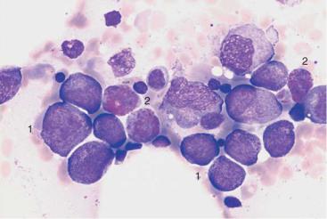
Fig. 26 Hairy cell leukemia and splenic lymphoma. a and b Ovaloid nuclei and finely “fraying” cytoplasm are characteristics of cells in hairy cell leukemia (HCL). c Occasionally, the hairy cell processes appear merely fuzzy. d and e When the cytoplasmic processes look thicker and much less like hair, diagnosis of the rare splenic lymphoma with villous lymphocytes (SLVL) must be considered. Here, too, the next diagnostic step is analysis of cell surface markers.
81
Theml, Color Atlas of Hematology © 2004 Thieme
All rights reserved. Usage subject to terms and conditions of license.

82 Abnormalities of the White Cell Series
Monoclonal Gammopathy (Hypergammaglobulinemia),
Multiple Myeloma*, Plasma Cell Myeloma, Plasmacytoma
Plasmacytoma is the result of malignant transformation of the most mature B-lymphocytes (Fig. 1, p. 2). For this reason the diagnostics of this disease will be discussed here, even though migration of its specific cells into the blood stream (plasma cell leukemia) is extremely rare (1–2%).
Immunoelectrophoresis of serum and urine is performed when electrophoresis shows very discrete gammaglobulin, or globulin, fractions, or when hypogammaglobulinemia is found (in light-chain plasmacytoma). A wide range of possibilities arises for the differential diagnosis of monoclonal transformed cells (Table 10).
The presence of more than 10% of plasma cells, or atypical plasma cells in the bone marrow, is an important diagnostic factor in the diagnosis of plasmacytoma. For more criteria, see p. 84.
Table 10 Differential diagnosis of monoclonal hypergammaglobulinemia
Type |
|
Characteristics |
Benign disorders |
|
|
Essential hypergammaglobuline- |
Usually in advanced age |
|
mia = MGUS (monoclonal gam- |
$ 10%, plasma cells found in the |
|
mopathy of unknown significance) |
bone marrow, no progression, |
|
|
|
normal polyclonal Ig |
Symptomatic hypergammaglobu- |
All ages (otherwise as above) |
|
linemia, secondary to |
|
|
– |
Infections |
|
– |
Tumors |
|
– |
Autoimmune disease |
|
Malignant diseases
Plasmocytoma
(usually IgG, A or light-chain [Bence Jones protein], rarely IgM, D, E) 90% disseminated (multiple myeloma), 5% solitary, 5% extramedullary (like a lymphoma or ENT tumor)
–Osteolysis or X-ray with severe osteoporosis
–Plasmocytosis of the bone marrow " 10%
–Monoclonal gammaglobulin in serum/urine with progression
Lymphoma |
– Enlarged lymph nodes |
e. g., immunocytoma, CLL |
– Usually blood lymphocytosis |
(potentially all lymphomas of the |
– Monoclonal immunoglobulin, |
B-cell series) |
usually IgM |
|
|
*The current WHO classification suggests “multiple myeloma” (MM) for generalized plasmacytoma and “plasmacytoma” only for the rare solitary or nonosseous form of plasmacytoma.
Theml, Color Atlas of Hematology © 2004 Thieme
All rights reserved. Usage subject to terms and conditions of license.

Plasmacytoma cannot be diagnosed without bone marrow analysis
 a
a
 b
b
Fig. 27 Reactive plasmacytosis and plasmacytoma. a Bone marrow cytology with clear reactive features in the granulocyte series: strong granulation of promyelocytes (1) and myelocytes (2), eosinophilia (3), and plasma cell proliferation (4): reactive plasmacytosis (magnification #630). b Extensive (about 50%) infiltration of the bone marrow of mostly well-differentiated plasma cells: multiple myeloma (magnification #400).
83
Theml, Color Atlas of Hematology © 2004 Thieme
All rights reserved. Usage subject to terms and conditions of license.

84 Abnormalities of the White Cell Series
Variability of Plasmacytoma Morphology
It is not easy to visually distinguish malignant cells from normal plasma cells. Like lymphocytes, normal plasma cells have a densely structured nucleus. Plasma cell nuclei with radial chromatin organization, known as “wheel-spoke nuclei,” are mostly seen during histological analysis.
The following attributes suggest a malignant character of plasma cells: the cells are unusually large (Fig. 28c), they contain crystalline inclusions or protein inclusions (“Russell bodies”) (Fig. 28b), or they have more than one nucleus (Fig. 28c).
In the differential diagnosis, they must be distinguished from hematopoietic precursor cells (Fig. 28d), osteoblasts (Fig. 20c), and blasts in acute leukemias (see Fig. 31, p. 97).
Bone marrow involvement may be focal or in rare cases even solitary. Aside from cytological tests, bone marrow histology assays are therefore indicated. Sometimes, the biopsy must be obtained from a clearly identified osteolytic region.
Although plasmacytomas progress slowly, staging criteria are available (staging according to Salmon and Durie) (Table 11).
Therapy may be put on hold in stage I. Smoldering indolent myeloma can be left for a considerable time without the introduction of therapy stress. However, chemotherapy is indicated once the myeloma has exceeded the stage 1 criteria.
Table 11 Staging of plasmacytomas according to Salmon and Durie
Stage I
All the following are present:
–Hb " 10 g/dl
–Serum calcium is normal
–X-ray shows normal bone structure or solitary skeletal plasmacytoma
–IgG $ 5 g/dl* IgA $ 3 g/dl*
Light chains in the urine* $ 4 g/24 h
Stage II
Findings fulfill neither stage I nor stage III criteria
Stage III
One or more of the following are present:
–Hb $ 8.5 g/dl
–Serum calcium is elevated
–X-ray shows advanced bone lesions
–IgG " 7 g/dl* IgA " 5 g/dl*
Light chains in the urine: " 12 g/24 h
* Monoclonal in each case.
Theml, Color Atlas of Hematology © 2004 Thieme
All rights reserved. Usage subject to terms and conditions of license.

Atypias and differential diagnoses of multiple myeloma
a |
b |
c 
 d
d
Fig. 28 Atypical cells in multiple myeloma. a Extensive infiltration of the bone marrow by loosely structured, slightly dedifferentiated plasma cells with wide cytoplasm in multiple myeloma. b In multiple myeloma, vacuolated cytoplasmic protein precipitates (Russell bodies) may be seen in plasma cells but are without diagnostic significance. c Binuclear plasma cells are frequently observed in multiple myeloma (1). Mitotic red cell precursor (2). d Differential diagnosis: red cell precursor cells can sometimes look like plasma cells. Proerythroblast (1) and basophilic erythroblast (2).
85
Theml, Color Atlas of Hematology © 2004 Thieme
All rights reserved. Usage subject to terms and conditions of license.

86 Abnormalities of the White Cell Series
Relative Lymphocytosis Associated with Granulocytopenia (Neutropenia) and Agranulocytosis
Neutropenia is defined by a decrease in the number of neutrophilic granulocytes with segmented nuclei to less than 1500/µl (1.5 #109/l). A neutrophil count of less than 500/µl (0.5 #109/l) constitutes agranulocytosis. Absolute granulocytopenias with benign cause develop into relative lymphocytoses.
In the most common clinical picture, drug-induced acute agranulocytosis, the bone marrow is either poor in cells or lacks granulopoietic precursor cells (aplastic state), or shows a “maturation block” at the myelo- blast–promyelocyte stage. The differential diagnosis of pure agranulocytosis versus aplasias of several cell lines is outlined on page 146.
Classification of Neutropenias and Agranulocytoses
1.Drug-induced:
a)Dose-independent—acute agranulocytosis. Caused by hypersensitivity reactions: for example to pyrazolone, antirheumatic drugs (anti-inflammatory agents), antibiotics, or thyrostatic drugs.
b)Relatively dose-dependent—subacute agranulocytosis (observed for carbamazepine [Tegretol], e.g. antidepressants, and cytostatic drugs).
c)Dose-dependent—cytostatic drugs, immunosuppressants.
2.Infection-induced:
a)E.g., EBV, hepatitis, typhus, brucellosis.
3.Autoimmune neutropenia:
a)With antibody determination of T-cell or NK-cell autoimmune response
b)In cases of systemic lupus erythematosus (SLE), Pneumocystis carinii pneumonia (PCP), Felty syndrome
c)In cases of selective hypoplasia of the granulocytopoiesis (“pure white cell aplasia”)
4.Congenital and familial neutropenias:
Various pediatric forms; sometimes not expressed until adulthood, e.g. cyclical neutropenia
5. Secondary neutropenia in bone marrow disease:
Myelodysplastic syndromes (MDS), e.g., acute leukemia, plasmacytoma, pernicious anemia
Theml, Color Atlas of Hematology © 2004 Thieme
All rights reserved. Usage subject to terms and conditions of license.

Bone marrow diagnosis is indicated in cases of unexplained agranulocytosis
 a
a
 b
b
Fig. 29 The bone marrow in agranulocytosis. a In the early phase of agranulocytosis the bone marrow shows only red cell precursor cells (e.g., 1), plasma cells (2), and lymphocytes (3); in this sample a myeloblast—a sign of regeneration—is already present (4). b Bone marrow in agranulocytosis during the promyelocytic phase, showing almost exclusively promyelocytes (e.g., 1); increased eosinophilic granulocytes (2) are also present.
87
Theml, Color Atlas of Hematology © 2004 Thieme
All rights reserved. Usage subject to terms and conditions of license.

88 Abnormalities of the White Cell Series
Monocytosis
If mononuclear cells stand out in showing an unusually elaborate nuclear structure with ridges and lobes and a wider cytoplasmic layer with very delicate granules (for characteristics see p. 46, for cell function, see p. 6), and this is in the context of relative ("10%) or absolute monocytosis (cell count "900/µl), a series of possible triggers must be considered (Table 12). If the morphology does not clearly identify the cells as monocytes, then esterase assays should be done in a hematological laboratory using unstained smears.
Table 12 Possible causes of monocytosis
Infection
Nonspecific monocytosis occurs in many bacterial infections during recuperation from or in the chronic phase of:
–Mononucleosis (aside from stimulated lymphocytes there are also monocytes)
–Listeriosis
–Acute viral hepatitis
–Parotitis epidemica (mumps)
–Chickenpox
–Recurrent fever
–Syphilis
–Tuberculosis
–Endocarditis lenta
–Brucellosis (Bang disease)
–Variola vera (smallpox)
–Rocky mountain spotted fever
–Malaria
–Paratyphoid fever
–Kala-azar
–Thypus fever
–Trypanosomiasis (African sleeping sickness)
Chronic reactive condition of the immune system, e. g., in:
–Autoimmune diseases
–Chronic dermatoses
–Regional ileitis
–Sarcoidosis
Paraneoplasm
(as attempted immune defense),
e.g., in:
–Large solid tumors
–Lymphogranulomatosis
Neoplasm
–Chronic myelomonocytic leukemia (CMML, p. 107)
–Acute monocytic leukemia (p. 100)
–Acute myelomonocytic leukemia (p. 98)
Theml, Color Atlas of Hematology © 2004 Thieme
All rights reserved. Usage subject to terms and conditions of license.

Conspicuously large numbers of monocytes usually indicate a reactive disorder. Only a monotonous predominance of monocytes suggests leukemia
a |
b |
 c
c
Fig. 30 Reactive monocytosis and monocytic leukemia. a Reactive and neoplastic monocytes are morphologically indistinguishable; here two relatively condensed monocytes in reactive monocytosis are shown. b Whenever monocytes are found exclusively, a malignant etiology is likely: in this case AML M5b according to the FAB classification (see p. 100). Auer bodies (arrow). c Monocytes of different degrees of maturity, segmented neutrophilic granulocytes (1), and a small myeloblast (2) in chronic myelomonocytic leukemia (CMML, see p. 107).
89
Theml, Color Atlas of Hematology © 2004 Thieme
All rights reserved. Usage subject to terms and conditions of license.

90 Abnormalities of the White Cell Series
Acute Leukemias
Acute leukemias are described in this place for morphological reasons, because they involve a predominance of mononuclear cells (p. 63). Al- though—or perhaps because—the term “leukemia” is relatively imprecise, an overview seems required (Table 13).
The cellular phenomenon common to the different forms of leukemia is the rapidly progressive reduction in numbers of mature granulocytes, thrombocytes, and erythrocytes. Simultaneously, the leukocyte count usually increases due to the occurrence of atypical round cells.
Table 13 Overview of all forms of leukemia
Type of leukemia |
Classification/clinical findings |
Acute leukemias (AL#AML, ALL) |
|
– Myeloid M0-7 (incl. monocytic, ery- |
Always acute disease, often with |
throid, megakaryoblastic leukemias) |
fever and tendency to hemorrhage |
– Lymphocytic |
There may be lymphoma and thy- |
|
mus infiltrates |
|
|
Chronic myeloid leukemia (CML) |
|
– Persistent leukemic diseases of the |
Chronic disease, usually with |
myeloproliferative system (p. 114) |
splenomegaly |
|
|
Chronic lymphocytic leukemia (CLL) |
|
and other leukemic lymphomas |
|
–B-CLL (90%), T-CLL (p.74f.)
–Prolymphocytic leukemia (p.77)
–Hairy cell leukemia (p.80)
–Chronic lymphocytic leukemia, (rarely) prolymphocytic leukemia and hairy cell leukemia are primary leukemic lymphomas with a chronic course. All other non-Hodgkin lymphomas can develop secondary leukemic disease
Chronic myelomonocytic leukemia (CMML)
–Leukemic form of myelodysplasia (p.106) is classified between myelodysplasia and myeloproliferative diseases
Subacute disease with transitions into acute forms of myeloid leukemia (secondary AML following MDS)
Theml, Color Atlas of Hematology © 2004 Thieme
All rights reserved. Usage subject to terms and conditions of license.