


Zinc Fingers
They play a key part in regulating the activity of genes in many species, from yeast to humans. Fewer than 10 years ago no one knew they existed
by Daniela Rhodes and Aaron Klug
One of the most fascinating questions in biology today asks how genes are turned on in multi-
cellular organisms. If a gene is to be activated, several proteins known as transcription factors must attach themselves to a segment of the gene called the promoter. This assembly forms a kind of Òon switchÓ: it enables an enzyme to transcribe a second genetic segment from DNA into RNA. In most cases, the resulting RNA molecule serves as a template for synthesis of a speciÞc protein, or string of amino acids; sometimes RNA itself is the Þnal product. Yet scientists have continued to wonder exactly how a transcription factor picks out its particular docking site on a promoter, distinguishing that site from the masses of other DNA found in a cell.
Answers are now beginning to emerge. It turns out that many transcription factors include small projections called zinc Þngers that are perfectly suited to DNA recognition. Our laboratory at the Medical Research Council in Cambridge, England, Þrst identiÞed a zinc Þnger in 1985Ñin a transcription factor obtained from a frog. Since then, more than 200 proteins, many of them transcription factors, have been shown to incorpo-
DANIELA RHODES and AARON KLUG both work at the Medical Research Council Laboratory of Molecular Biology in Cambridge, England. Rhodes, who holds a Ph.D. in biochemistry from the University of Cambridge, joined the council in 1969. She has been senior scientist since 1990. Klug, the 1982 winner of the Nobel Prize in Chemistry, began working at the Laboratory of Molecular Biology in 1962 and is now its director. The Nobel
Prize recognized his development of electron microscopy techniques for determining the structure of complexes of biological molecules. It also honored his elucidation of the structure and assembly of proteinÐnucleic acid complexes in viruses and in chromosomes.
rate such zinc Þngers. And several other transcription factors contain related structures, or motifs. Recently a number of laboratories, among them ours, have also begun to decipher just how zinc Þngers and their relatives manage to select and grip their speciÞc binding sites on DNA.
Of course, zinc Þngers are not the only structures transcription factors exploit for interacting with DNA. Other important examples bear such names as helix-turn-helix motifs (discovered before zinc Þngers, in 1981), homeodomains and leucine zippers [see ÒMolecular Zippers in Gene Regulation,Ó by Steven Lanier McKnight; SCIENTIFIC AMERICAN, April 1991, and ÒSmart Genes,Ó by Tim Beardsley, August 1991]. The zinc Þnger, however, is by far the most prevalent DNA-binding motif.
Ultimately, research into the problem of DNA recognition should advance inquiry into the larger question of how development unfolds in multicellular organisms. Although every cell in an embryo carries the same genes, some cells diÝerentiate to become, say, neurons, whereas others become skin cells. Their fates vary because diÝerent combinations of genes are turned on in the cells as the embryo grows, leading to synthesis of the specialized proteins that give diÝerentiated cells their distinctive properties. Knowledge of how transcription factors recognize their speciÞc binding sites on DNA is central to an understanding of such selective gene activation.
We uncovered the existence of zinc Þngers after becoming intrigued by results from the
laboratories of Robert G. Roeder, then at Washington University, and Donald D. Brown of the Carnegie Institution of Washington in Baltimore. By 1980 Roeder and Brown and their associates had for the Þrst time dissected the steps leading to transcription of a gene in an organism more advanced than bacteria.
56 SCIENTIFIC AMERICAN February 1993
As part of that work, they demonstrated that in the frog Xenopus laevis a protein called transcription factor IIIA (TFIIIA) is one of at least three factors required to activate the gene that gives rise to 5S RNA. 5S RNA is a constituent of the ribosomes on which molecules of messenger RNA (the typical products of gene transcription) are translated into protein.
The investigators further found that TFIIIA binds to a relatively long patch of DNA, encompassing a particular sequence of about 45 base pairs, or ÒrungsÓ on the familiar DNA Òladder.Ó (DNA is made up of two strands of nucleotides, which themselves consist of the sugar deoxyribose, a phosphate group and one of four distinguishing bases: adenine, cytosine, guanine or thymine. The two strands are attached to each other through their bases, so that adenine always pairs with thymine, and cytosine pairs with guanine.)
The length of the TFIIIA-docking site surprised us because TFIIIA is itself rather small. Transcription factors of the same size that had earlier been identiÞed in bacteria attach themselves to much shorter tracts of DNA, on the order of 15 base pairs long. How, we asked, could this small TFIIIA molecule span such an extended stretch of DNA? Fortunately, the problem seemed tractable. Although transcription factors tend to be produced in scarce amounts, TFIIIA
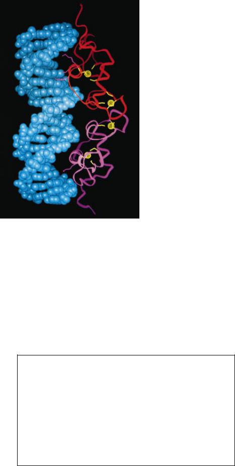
THREE ZINC FINGERS (protrusions) extending from a transcription factor, or gene-regulating protein (red ), have fastened themselves to the wide, major groove of a DNA molecule (double helix). Zinc Þngers connect transcription factors to their target genes mainly by binding to speciÞc sequences of DNA base pairsÑthe ÒrungsÓ in the twisted DNA Òladder.Ó Zinc Þngers are so named both because they can grasp DNA and because a zinc ion at the core ( yellow spheres) plays a critical role in determining their structure.
Copyright 1993 Scientific American, Inc.

is abundant in the ovaries of immature frogs. There it is stored as a complex with the 5S RNA it helps to generate. The abundance gave us conÞdence to think we could gather enough of the TFIIIAÐ5S RNA complex to isolate the protein. Having done that, we might learn something about the three-dimen- sional organization of the protein and about how it binds to its target site on the 5S RNA gene.
The plan was sound, but we soon en-
DNA
countered a diÛcultyÑone that would prove fortunate because it set us directly on a path toward discovery of the zinc Þnger. In 1982 Jonathan Miller, then a research student in our laboratory, applied a known recovery technique to extract the TFIIIAÐ5S RNA complex from frog ovaries. He obtained disappointingly little of it. It turned out that the method he used was eliminating a metal needed to hold the complex together. After Miller modiÞed the extraction
procedure and procured a good supply of the complex, he showed that the lost metal was zinc. Each TFIIIAÐ5S RNA unit incorporated between seven and 11 zinc ions, an unusually large number. Other experiments led us further toward the zinc Þnger. When an enzyme called a protease chopped TFIIIA into successively smaller fragments, the fragments shrank by increments of about three kilodaltons (a measure of molecular weight). They ended up as three-kilodal-
 ZINC
ZINC
ZINC
FINGER
BASE
PAIR
TRANSCRIPTION
FACTOR
CELL
NUCLEUS
Copyright 1993 Scientific American, Inc.

ton units that resisted further attack, presumably because they were tightly folded. Collectively, these results suggested that TFIIIA was built almost entirely from a string of tandem three-ki- lodalton segments (representing about 30 amino acids per segment), each of which was folded around a zinc ion into a small, compact DNA-binding domain.
If we were right, the discovery would mean we had come across a novel kind of transcription factor. All others that had been studied in similar detail had
been found to interact with DNA as dimers, or pairs, in which each protein in the dimer made contact with DNA through just one DNA-binding motif. Our Þndings implied that TFIIIA would stretch out along the double helix, touching it at several points instead of just one or two. Such multiple contacts would also explain how TFIIIA could interact with a very long segment of DNA.
As we were considering how to substantiate our model, RoederÕs laboratory published the amino acid sequence of
How Zinc Fingers Were Discovered
One of the authors (Klug) deduced in 1985 that certain stretches of amino acids can fold independently around a zinc ion, forming modules that would come to be called zinc fingers (bracketed regions at top). The gray line represents a string of amino acids; the small, colored circles represent amino acids that Klug correctly thought might participate in the folding. A major clue (bottom) to the folding pattern came from inspection of the sequence of amino acids (capital letters) in the protein TFIIIA. The bulk of the protein can be arranged into nine successive sections, or sequence units (numbered), that exhibit important similarities: they include, at virtually identical positions, a pair of cysteine amino acids (gold C’s), a pair of histidine amino acids (red H’s) and (with the possible exception of section 7) three hydrophobic amino acids (green letters). (Asterisks mark unimportant breaks in the pattern.) These observations, added to biochemical findings, led to the proposal that the cysteine and histidine pairs in every module would combine with a single zinc ion (large yellow spheres in top image), causing the amino acids between those pairs to loop out as shown. At the same time, the three hydrophobic amino acids would somehow help stabilize the arrangement.
ZINC-FINGER MODULE |
LINKER ZINC-FINGER MODULE |
||
|
|
HYDROPHOBIC |
|
|
|
AMINO ACID |
|
F |
L |
F |
L |
|
|
||
CYSTEINE |
|
HISTIDINE |
|
|
|
|
|
C |
H |
C |
H |
|
Zn |
|
Zn |
C |
H |
C |
H |
Y |
|
Y |
|
|
ZINC |
|
|
1 |
Y I C S F A D C G A A Y N K N W K L Q * A H L C * K H |
||
2 T G E K * P F P C K E E G C E K G F T S L H H L T * R H S L * T H 3 T G E K * N F T C D S D G C D L R F T T K A N M K * K H F N R F H 4 N I K I C V Y V C H F E N C G K A F K K H N Q L K * V H Q F * S H 5 T Q Q L * P Y E C P H E G C D K R F S L P S R L K * R H E K * V H 6 A G * * * * Y P C K K D D S C S F V G K T W T L Y L K H V A E C H 7 Q D * * * L A V C * * D V C N R K F R H K D Y L R * D H Q K * T H 8 E K E R T V Y L C P R D G C D R S Y T T A F N L R * S H I Q S F H 9 E E Q R * P F V C E H A G C G K C F A M K K S L E * R H S V * V H
58 SCIENTIFIC AMERICAN February 1993
TFIIIA. In that sequence, we found support for our proposal: the Þrst three quarters of the protein formed a continuous run of nine similar units of about 30 amino acids. Moreover, a pair of cysteine amino acids and a pair of histidine amino acids resided at virtually identical positions within each unit [see bottom illustration in box at left]. This last Þnding was consistent with the notion that each unit contained its own zinc ion, because zinc in proteins is generally found bound to four amino acids, often four cysteines or some combination of cysteines and histidines.
By 1985 these results led one of us (Klug) to propose formally that the invariant cysteines and histidines were used to fold each unit independently into a DNA-binding minidomainÑlater called a zinc Þnger because it was used to grip the DNA double helix. He suggested that the pair of cysteines near one end of the unit and the pair of histidines near the other end bound the same zinc atom, causing the intervening stretch of amino acids to loop out. Thus, in each 30-amino-acid unit, about 25 amino acids would fold into a structured domain (a Þnger); the remaining amino acids would serve as a linker between consecutive Þngers [see top illustration in box at left].
Shortly thereafter, measurements we made with Gregory P. Diakun of the Science and Engineering Research Council Daresbury Laboratory near Manchester, England, proved that each of the nine units did indeed contain a zinc ion bound to two cysteines and two histidines. TFIIIA was therefore fashioned almost entirely out of nine consecutive zinc Þngers. All had the same basic architecture but were chemically distinct because of variations in the amino acids that did not participate in building the framework of the Þnger module.
But did the zinc Þngers in fact contact DNA independently, as was predicted? To Þnd out, Louise Fairall in our group, like investigators elsewhere, conducted what are called footprinting studies. A protein is allowed to attach to DNA. Then enzymes or other agents that attack DNA are applied. Any site that resists cleavage can be assumed to have been protected by the bound protein, indicating that the spared spot is a site of protein-DNA interaction. By 1986 the footprinting data conÞrmed that TFIIIA makes repeated contacts with DNA.
Hence, TFIIIA was the novelty we suspected it to be: it connected to a speciÞc region on DNA by exploiting a string of independent DNA-binding modules. The economy of the modular arrangement was beautiful. Cells were already
Copyright 1993 Scientific American, Inc.

FINGER STRUCTURE (left) has been known in detail since the late 1980s. The ribbon represents the carbon-nitrogen backbone of the amino acid chain. The left half of the backbone folds back on itself to form a two-strand substructure known as a beta sheet (V-shaped region). The right half twists into an alpha helix (spiral). Binding of zinc ( yellow sphere) by cysteines in the beta sheet ( yellow lines) and histidines in the helix (red lines) draws the halves together near the base of the Þnger. It also brings hydrophobic amino acids (green) close to one another at the Þngertip (top of image), where their mutual
attraction helps to keep the structure intact. At the right, three tandem zinc Þngers (red ribbon trisected by white lines) from the gene-regulating protein Zif268 have each made contact (magenta) with bases in the major groove of DNA (blue), collectively attaching to almost a full turn of the double helix. Five of the six base contacts are visible in this view. Yellow lines and rings represent the connections between zinc and the cysteines and histidines. The Zif268 image is based on an x-ray crystallographic analysis conducted at Johns Hopkins University by Nikola P. Pavletich and Carl O. Pabo.
known to build a large repertoire of on switches for genes by combining in various permutations a limited set of transcription factors. That is, one gene might be activated by a combination of proteins a, b and c, whereas another gene might make use of just a and b or of a, b and d. By such a strategy, organisms avoid having to produce a unique transcription factor for each of the enormous number of genes that are active in cells. The zinc-Þnger studies revealed that the combinatorial principle can also operate within a transcription factor. A cell can produce a vast collection of distinct transcription factors by varying the choice, order and number of independent DNA-binding modules in the proteins. The particular combination of zinc fingers in a transcription factor enables the factor to recognize a specific DNA sequence and no other.
The eÛciency of the combinatorial approach led us to suggest that the zinc-Þnger motif might turn
up in many proteins. But the extent of its occurrence in eukaryotesÑorganisms more advanced than bacteriaÑis astonishing. Peter F. R. Little of Imperial College, London, estimates that as much as 1 percent of the DNA in hu-
man cells speciÞes zinc Þngers. In chromosome 19 the Þgure is as high as 8 percent. The zinc ÞngerÐcontaining proteins that have been identiÞed so far carry from as few as two to as many as 37 tandem Þngers.
To understand how a zinc Þnger recognizes a speciÞc sequence of base pairsÑwhich adopts a precise conformationÑone needs to know the detailed three-dimensional structure of the Þnger module. Most proteins include local regions of ÒsecondaryÓ structure that fold together to yield the overall, three-dimensional shape of the protein. The most common secondary structures are the alpha helix (in which the backbone of the protein twists into a characteristic spiral) and the beta strand (in which the backbone is fully extended) [see ÒThe Protein Folding Problem,Ó by Frederic M. Richards; SCIENTIFIC AMERICAN, January 1991].
Jeremy M. Berg of Johns Hopkins University deciphered the important features of the three-dimensional architecture on theoretical grounds in 1988, but his model was not conÞrmed until 1989. Then Peter E. Wright and his colleagues at the Scripps Clinic and Research Foundation in La Jolla, Calif., determined the structure of a zinc Þnger from the Xeno-
pus protein XÞn. They did it by applying nuclear magnetic resonance spectroscopy (NMR), a technique that can be used to solve the three-dimensional structure of small proteins in solution. Soon after, other laboratories and also our group identiÞed the same design in other zinc-Þnger proteins.
As Berg predicted, the characteristic amino acid sequence of the zinc finger folds into a compact shape by forming two prominent substructures along the way. One part of the sequence (comprising, say, the left half of a vertical protrusion) adopts the shape of a small beta ÒsheetÓ; it is composed of two beta strands that form a sheet when the second strand folds back onto the Þrst one [see left illustration above]. The other part of the sequence (the ÒrightÓ half) twists into an alpha helix. The two cysteines reside at the bottom of the beta sheet, and the two histidines reside at the bottom of the helix. All four amino acids are joined through a zinc atom that essentially pins together the beta sheet and helix.
The NMR analysis also helped to clarify the role of a few additional amino acids. When originally examining the sequence of TFIIIA, we noted that the putative Þngers each included a set of
SCIENTIFIC AMERICAN February 1993 59
Copyright 1993 Scientific American, Inc.
three hydrophobic amino acids in virtually identical positions. (Hydrophobic substances often associate with one another in the interior of a protein in preference to water in the surroundings.) The invariance suggested that those amino acids had an important structural role. Although they are fairly far apart from one another in linear representations of the amino acid sequence, we thought they might somehow interact in three-dimensional space and thus assist in the folding of the minidomain. Consistent with the Berg model, the NMR results showed that when the zinc-Þnger module folds up, the hydrophobic amino acids do indeed come close enough to one another for their mutual attraction to come into play. They form a hydrophobic core that helps the module to maintain its shape.
In parallel with our eÝorts to understand the architecture of zinc Þngers, we and others were also pondering a more general problem. Many experiments led to the conclusion that the zinc Þngers in TFIIIA, which constitute the bulk of the protein, were solely responsible for the ability of the factor to recognize the promoter of the 5S RNA gene. But increasing numbers of proteins were being discovered in which only a few zinc Þngers were embedded in a large protein. Could such short runs of zinc Þngers direct these proteins to promoters, without assistance from oth-
er parts of the protein?
In our own eÝorts to answer these questions, we concentrated on a threeÞngered yeast transcription factor called SWITCH 5 (SWI5). With our colleague Kyoshi Nagai, we isolated the region containing the Þngers and exposed it to the promoter of the target gene for the protein. Sure enough, the isolated protein segment bound to the promoter avidly, implying that the zinc Þngers are alone responsible for DNA binding. Interestingly, we found as well that at least two linked Þngers had to be present for the SWI5 protein to attach with reasonable strength to its correct target site on DNA. By then applying NMR to the Þrst two zinc Þnger motifs of SWI5, we and our colleagues David Neuhaus and Yukinobu Nakeseko conÞrmed that adjacent zinc Þngers do not meld with each other; zinc Þngers are truly independent Òreading headsÓ joined by ßexible linkers.
The precise points of contact between zinc Þngers and DNA had yet to be identiÞed, however. Nikola P. Pavletich and Carl O. Pabo, both then at Johns Hopkins, made the initial breakthrough in 1991. First, they obtained crystals of
the complex formed by the DNA and the DNA-binding domain of a transcription factor called Zif268. By then carrying out an x-ray crystallographic analysis, they were able to determine the detailed structure of the complex. Zif268, which in common with SWI5 includes a run of three zinc Þngers, participates in the early development of mice.
The x-ray analyses revealed that the Zif268 zinc-Þnger region curls around almost one turn of the DNA helix (more or less tracing the letter ÒCÓ), Þtting itself into the major groove. (The major groove is the wider of two parallel gullies that spiral around the long axis of the DNA double helix, much as red-and- white ribbons of color encircle old-fash- ioned barber poles.) The Þngers make contacts with successive, three-base-pair sites on the DNA, and they approach the DNA in much the same orientation. That is, the alpha helix of each Þnger points into the major groove, abutting one of its walls.
More speciÞcally, the Þrst and third Þngers of Zif268 bind to DNA identically: an amino acid in the Þrst turn of the alpha helix contacts the Þrst base pair of the corresponding binding site on DNA, and an amino acid in the third turn of the helix contacts the third base pair of that same DNA site. The second Þnger also makes two contacts through the alpha helix, but this time amino acids on the Þrst and second turn contact the Þrst and second base pairs of the corresponding binding site on DNA. (In each instance, one amino acid contacts one DNA base in a pair.) In addition, both the alpha helix and the beta sheet in the Þngers bind to phosphate groups in the chains of sugar and phosphate that make up the ÒsidesÓ of the DNA ladder. These added links help to stabilize the attachment of zinc Þngers to DNA.
So far no other complexes of zinc Þngers and DNA have been solved by x-ray crystallography. Nevertheless, Grant H. Jacobs in our laboratory has good evidence that many zinc Þngers bind to DNA in much the same way as Zif268 does. Jacobs has compared the amino acid sequences of more than 1,000 zincÞnger motifs. He Þnds that the amino acids in three positions are particularly variable. These highly variable positions are precisely those that are used to make contacts in the Zif268 complex, namely, those falling on the Þrst, second and third turns of the alpha helix. Such similarity raises the exciting possibility that zinc-Þnger modules might one day be designed at will to recognize selected DNA sequencesÑa feat that could be important both for the study
62 SCIENTIFIC AMERICAN February 1993
of gene regulation and for medicine. Of course, there are limits to how much one can extrapolate from the Zif268 model and from statistical analyses. Proteins with many zinc Þngers would be expected to interact with DNA somewhat diÝerently. For instance, if the Zif268 pattern of binding applied to TFIIIA, this protein, with its nine fingers, would wind around the DNA for three turns, like thread on a spool. This extensive wrapping could well hamper the factor from coming oÝ the DNA when detachment became necessary. Indeed, footprinting data obtained by us and others suggest that TFIIIA does not twist continuously around the DNA. The Þrst three Þngers of TFIIIA almost certainly clasp onto a single turn of the DNA, and it is very likely that the last three Þngers do the same. But the bulk of the protein lies on just one face of the double helix; hence, it crosses the narrow, minor groove at least twice. The varied DNA-binding patterns of separate regions of TFIIIA probably reßect the fact that the amino acid sequences of the TFIIIA Þngers diÝer more from one another than do those of the Þngers in
Zif268-type proteins.
From an evolutionary standpoint, there is good reason to think that multiÞngered DNA-binding domains arose by duplication of some ancestral gene that speciÞed a small protein of 30 or so amino acids. We think, too, that the 30-amino-acid chain may have been among the earliest of proteins to evolve. Such a protein would, after all, have been simple to produce. Once synthesized, it would easily and safely pick up zinc (which is a relatively inert metal) from its surroundings and would then fold without assistance into a stable conformation. So folded, it would acquire the ability to bind to DNA or RNA. Such attributes almost certainly help to explain why zinc Þngers are now prevalent throughout the animal and plant kingdoms. Any species acquiring the genetic blueprint for a particular, autonomously folding zinc Þnger would instantly acquire the ability to bind to a new stretch of DNA. That property, in turn, could give rise to new cellular functions, such as the ability to transcribe some previously silent gene and thus to produce a novel enzyme or other valuable protein.
As we and others were gaining insight into the structure and function of classic zinc Þngers, tan-
talizing Þndings began to suggest that the motif we had initially discovered in TFIIIA was not the only zinc-centered structure devoted to DNA recognition.
Copyright 1993 Scientific American, Inc.

|
FINGERLIKE UNIT |
|
FINGERLIKE UNIT |
|||
Y |
|
|
DIMERIZATION |
|
|
|
|
|
REGION |
|
|
|
|
G |
H |
|
|
N |
|
|
S |
Y |
|
|
K |
R |
|
A |
G |
|
D |
|
R |
|
|
|
|
|
|
||
Y |
V |
|
I |
|
K |
|
|
|
|
|
|
||
D |
W |
|
T |
|
S |
CYSTEINE |
|
|
|
|
|||
|
|
|
|
|
|
|
N |
S |
|
C |
|
C |
|
C |
C |
DNA-BINDING |
|
|
||
Q |
|
|
|
|||
AMINO ACIDS |
|
|
|
|||
V |
E |
|
N |
Zn |
Q |
|
|
T |
|
|
|||
Zn |
|
|
A |
|
||
|
LINKER |
A |
|
|
||
A |
G |
|
|
|||
|
P |
|
|
|
||
|
|
|
|
|
|
|
C |
C |
|
C |
ZINC |
C |
|
M K E T R Y |
|
K A F F K R S I Q G H N D Y M |
|
|
R L R K C Y E V G M M K G |
|
DNA-BINDING DOMAIN of the estrogen receptor (a transcription factor that must be bound by estrogen to act on a gene) is built from an amino acid sequence (capital letters) that has here been divided into two zinc-Þnger-like units (blue and green regions). Such diagrams initially led investigators to assume that the two units, like classical zinc Þngers, each recognize separate base sequences in the DNA. In fact, only the
three amino acids colored dark blue are thought to interact with DNA bases, which means the Þrst Þngerlike unit makes the main contact with DNA. The second unit serves a diÝerent function than the Þrst: it carries Þve amino acids (dark green) that enable one receptor molecule to combine, or dimerize, with a second receptor molecule. Such pairing is required if estrogen receptors are to attach securely to DNA.
Three-Dimensional View of the DNA-Binding Domain of the Estrogen Receptor
The detailed structure of the
DNA-binding domain of the es-
trogen receptor was determined
in 1990 by one of the authors
(Rhodes) and her collaborators John W. R. Schwabe and David Neu-
haus. This work and biochemical data enabled them to pinpoint
the parts of the three-dimensional structure that perform the critical tasks of recognizing DNA and
pairing with other receptor molecules. The two units in the domain (light blue and light green) adopt similar conformations. An irregularly structured loop (hatched)
that includes two cysteines (C’s) is followed by an alpha helix (stippled spiral) carrying the third and fourth cysteines. Binding of zinc
(yellow) by the cysteines ties the  terminal segments of the irregu-
lar region to the base of the helix.  So folded, the two units mesh
  through their helices. The amino
acids responsible for recognizing specific bases (dark blue) fall on the helix in the first unit; those responsible for forming a dimer
(dark green) reside in the irregular loop of the second unit.
SCIENTIFIC AMERICAN February 1993 |
63 |
Copyright 1993 Scientific American, Inc.

By 1987 investigators had elucidated the amino acid sequences of several members of a large family of transcription factors known as nuclear hormone receptors. Such factors must be bound by a particular steroid or thyroid hormone or vitamin before they can activate a gene. In examining the newly determined sequences, workers saw that every one of them bore a conserved, or highly similar, domain of about 80 amino acids. This domain consistently included two, and always two, units whose amino acid sequence was reminiscent of the zinc Þnger. As was true of zinc Þngers, each unit, or motif, contained two pairs of potential zinc-binding amino acids; here, however, the zinc binders were exclusively cysteines instead of cysteines and histidines. These resemblances of the sequences to TFIIIA zincÞnger motifs implied that the cysteinerich, 80-amino-acid segment of the factors was the DNA-binding domain.
Pierre Chambon and Stephen Green of INSERM in Strasbourg conÞrmed that assumption in the late 1980s. Soon after, Paul B. Sigler, then at the University of Chicago, and Keith R. Yamamoto of the University of California at San Francisco and their associates established that each of the two segments of the DNA-binding domain incorporates a zinc atom. Naturally, we and others expected that, as is true of TFIIIA-type zinc Þngers, the conÞgurations of the two motifs would resemble each other,
and the motifs would form independent DNA-binding modules.
The assumption turned out to be partly wrong. Structural analyses would eventually show that the two units do fold similarly. But, before that, some striking biochemical work would demonstrate that the units do not function as independent DNA-reading heads. By substituting one amino acid for another and examining the eÝect on DNA binding, Chambon, Ronald M. Evans of the Salk Institute for Biological Studies in San Diego and Gordon M. Ringold, formerly of Stanford University, and their associates found that the Þrst motif serves as the primary DNA-rec- ognition unit. At about the same time, Evans and his co-worker Kazuhiko Umesono, again applying the substitution method, uncovered at least one function of the second motif. To understand that function, one must Þrst know something general about how steroid receptors interact with DNA.
Such receptors bind to DNA as pairs, or dimers, of identical molecules. Each protein in a pair recognizes half of a two-part binding site that is known as a palindrome, because the halves are identical if read in opposite directionsÑ that is, along opposite strands of the DNA [see illustration below]. The base sequence of the half site recognized by one type of transcription factor (such as the estrogen receptor) can exactly match that recognized by another fac-
|
|
|
|
|
|
|
|
|
INTERVENING |
|
|
|
|
|
|||
|
|
|
HALF SITE |
|
BASE PAIRS |
|
HALF SITE |
||||||||||
a |
5' |
|
|
|
|
|
|
|
|
|
|
|
|
|
|
|
3' |
|
|
|
|
|
|
|
|
|
|
|
|
|
|
|
|||
|
|
|
|
|
|
|
|
|
|
|
|
|
|
|
|||
|
|
|
|
|
|
|
|
|
|
|
|
|
|
|
|||
|
|
|
|
|
|
|
|
|
|
|
|
|
|
|
|||
|
|
A G A A C A |
|
|
|
|
|
|
T G T T C T |
|
|||||||
|
|
T C T T G T |
' ' ' |
|
A C A A G A |
|
|||||||||||
b |
3' |
|
|
|
|
|
|
|
|
|
|
|
|
|
|
|
5' |
5' |
|
|
|
|
|
|
|
|
|
|
|
|
|
|
|
3' |
|
|
|
|
|
|
|
|
|
|
|
|
|
|
|
|
|||
|
|
A G G T C A |
|
|
|
|
|
|
T G A C C T |
|
|||||||
|
|
T C C A G T |
' ' ' |
|
A C T G G A |
|
|||||||||||
c |
3' |
|
|
|
|
|
|
|
|
|
|
|
|
|
|
|
5' |
5' |
|
|
|
|
|
|
|
|
|
|
|
|
|
3' |
|
||
|
|
|
|
|
|
|
|
|
|
|
|
|
|
||||
|
|
|
|
|
|
|
|
|
|
|
|
|
|
||||
|
|
|
A |
G |
G T C A T G A C C T |
|
|
||||||||||
|
|
|
T |
C |
C A G T A C T G G A |
|
|
||||||||||
|
3' |
|
|
|
|
|
|
|
|
|
|
|
|
|
5' |
|
|
|
|
|
BASE PAIR |
|
|
|
|
|
|
|
|
|
|
||||
|
|
|
|
|
|
DIRECTION OF READING |
|||||||||||
|
|
|
|
|
|
|
|
|
|
|
|||||||
DOCKING SITES ON DNA, or response elements, that are recognized by the glucocorticoid (a), estrogen (b) and thyroid (c) receptors include two half sites (shaded regions). The half sites in any element are alike, if their base pairs (linked letters) are read along opposite strands of the DNA (arrows in c), in the 5′ to 3′ direction. Receptors bind to the response elements as dimers, or pairsÑone molecule to each half site. To bind successfully, they must be able to distinguish both the base sequence of, and the spacing between, half sites. The diÝerences between response elements can be subtle: element b diÝers from a by only two base pairs (red letters), and it diÝers from c only in the number of bases separating the half sites.
64 SCIENTIFIC AMERICAN February 1993
tor (such as the thyroid receptor). In that case, the only diÝerence between the two binding sites is the number of base pairs separating the half sites in each palindrome.
Consequently, for a transcription factor to Þnd its corresponding docking site on DNA, the protein must contain regions dedicated to picking out a speciÞc half-site base sequence and also to measuring the distance between half sites. Evans and Umesono found that a part of the second motif is responsible for measuring such spacing.
Despite this progress, one could not fit these pieces of information together to explain how se-
quence-speciÞc recognition took place. That explanation could come only from knowing the three-dimensional conÞguration of the DNA-binding domains of receptor proteins and thus seeing where on the structure the functionally important amino acids would lie. In 1990, by applying NMR, Robert Kaptein and his colleagues at the University of Utrecht solved the structure of the DNA-binding domain of the rat glucocorticoid (cortisone) receptor. Shortly thereafter, John W. R. Schwabe and Neuhaus in our laboratory and one of us (Rhodes) solved that of the human estrogen receptor.
As could be deduced from their similar amino acid compositions, the DNAbinding domains of the glucocorticoid and estrogen receptors were found to adopt much the same structure. Each of the two zinc ÞngerÐcontaining motifs within the domain consists of two parts: an irregularly looped string of amino acids (instead of the beta sheet in classic zinc Þngers), followed by an alpha helix. The loop carries two of the zincbinding sites, and the other two reside at the beginning of the helix that follows. Yet instead of remaining separate, as standard zinc Þngers would, the two motifs merge into a single structural unit. In this arrangement, the helices cross perpendicularly at their midpoints, a conÞguration that is created by the mutual attraction of invariant and relatively invariant hydrophobic amino acids.
With the three-dimensional structure of the DNA-binding domain known, we proceeded to map onto it the locations of amino acids that had earlier been shown to be critical for DNA recognition. Groups led by Chambon, Evans and Ringold had identiÞed three amino acids in the Þrst Þngerlike motif that were responsible for recognizing the base sequence of a half site. Those amino acids turn out to reside on one face of the helix in the motif, leading
Copyright 1993 Scientific American, Inc.
us to call that substructure the DNA-recognition helix. This information also told us something more about the function of the second motif: by crossing the DNA-recognition helix of the Þrst motif, the helix of the second motif serves as a backing strut to hold the recognition helix in place. The separation of function between the two motifs suggests that the second Þnger arose from duplication of the Þrst but that once the second unit appeared, it was pressed into service for new tasks.
Mapping in three dimensions told us as well how the second motif performs the vital role of discriminating the spacing between half sites on DNA. Evans and Umesono had established that the amino acids responsible for such discrimination lie between the Þrst two cysteines of the second motif. In the three-di- mensional conÞguration, these amino acids map to the loop preceding the helix, where they would be available to link one molecule to its partner. Computer modeling of the interaction between DNA and the DNA-binding regions of the glucocorticoid and estrogen receptors then enabled KapteinÕs and our group, respectively, to see that the pairing of proteins through the predicted connection would orient the dimer properly. The two recognition helices on the dimer would be arranged so that the spacing be-
tween them would match the spacing between the appropriate half sites in the DNA.
Sigler and his colleague Ben F. Luisi, working together at Yale University, in collaboration with Yamamoto and Leonard P. Freedman of the University of California at San Francisco have since conÞrmed this picture by x-ray crystallography. They have also learned that each protein in the dimer makes several contacts with the phosphates on either side of the major groove. These contacts position the DNA-recognition helix so that it can reach deep into the major groove to form bonds with base pairs in the half site. Overall, then, studies of the nuclear hormone receptor class of zinc-Þnger motifs indicate that, despite some structural similarity to TFIIIA-type zinc Þngers, these motifs function more like the DNA-recogni- tion motifs of other transcription factors, such as the helix-turn-helix and leucine zipper. That is, by folding together instead of remaining distinct, the
motifs help nuclear hormone receptors to form the dimers that enable such factors to recognize their speciÞc binding sites on DNA.
When knowledge of the structure of a molecule reveals something about the way in which it works, that information
may also oÝer insight into disease. In the case of zinc Þngers, researchers have learned that a renal cancer called WilmÕs tumor arises from a genetic mutation that interferes with the proper binding to DNA of the zincÞnger region in a particular protein. Moreover, some of the symptoms that can follow from insufÞcient intake of zinc in the dietÑsuch as delayed sexual developmentÑcan now be attributed to the inability of estrogen and androgen receptors to fold properly in the absence of zinc.
Clearly, the two classes of zinc Þngers we have discussed vary profoundly in both their structure and the way
in which they interact with DNA. We have no doubt that still more variety will be discovered in the extended family of zinc-Þnger proteins. Nature continues to surprise and amuse us with the ingenuity of the designs it has evolved to enable proteins to recognize speciÞc base sequences in DNA. For instance, an increasing number of amino acid sequences include what seem to be zincbinding motifs, although the spacing between the pairs of cysteines or histidines, or the numbers of pairs, diÝers from that in the standard zinc Þnger. One unusual example is the yeast protein GAL4; it bears six cysteines that fold around two zinc atoms. We also expect to Þnd that some
zinc Þngers or their cousins are involved in activities other than transcription, such as transporting, processing or otherwise acting on DNA or even RNA; recall, for instance, that TFIIIA binds to RNA as well as to DNA. We still have much to learn.
FURTHER READING
REPETITIVE ZINC-BINDING DOMAINS IN THE |
|
RECEPTORS HAVE A NOVEL STRUCTURAL |
PROTEIN TRANSCRIPTION FACTOR IIIA |
|
MOTIF FOR DNA RECOGNITION. John W. R. |
FROM XENOPUS OOCYTES. J. Miller, A. D. |
|
Schwabe and Daniela Rhodes in Trends in |
McLachlan and A. Klug in EMBO Journal, |
|
Biochemical Sciences, Vol. 16, No. 8, pages |
Vol. 4, No. 6, pages 1609Ð1614; 1985. |
|
291Ð296; August 1991. |
ZINC FINGERS: A NOVEL PROTEIN MOTIF FOR |
|
CRYSTALLOGRAPHIC ANALYSIS OF THE |
NUCLEIC ACID RECOGNITION. Aaron Klug |
|
INTERACTION OF THE GLUCOCORTICOID |
and Daniela Rhodes in Trends in Biochem- |
|
RECEPTOR WITH DNA. B. F. Luisi, W. X. |
ical Sciences, Vol. 12, No. 12, pages 464Ð |
|
Xu, Z. Otwinowski, L. P. Freedman, K. |
469; December 1987. |
|
R. Yamamoto and P. B. Sigler in Nature, |
ZINC FINGERÑDNA RECOGNITION: CRYSTAL |
|
Vol. 352, No. 6335, pages 497Ð505; Au- |
STRUCTURE OF A ZIF268-DNA COMPLEX AT |
|
gust 8, 1991. |
° |
|
A STRUCTURAL TAXONOMY OF DNA-BIND- |
2.1A. Nikola P. Pavletich and Carl O. Pabo |
|
|
in Science, Vol. 252, pages 809Ð817; May |
|
ING DOMAINS. Stephen C. Harrison in Na- |
10, 1991. |
|
ture, Vol. 353, No. 6346, pages 715Ð719; |
BEYOND ZINC FINGERS: STEROID HORMONE |
|
October 24, 1991. |
|
|
|
Copyright 1993 Scientific American, Inc. |
SCIENTIFIC AMERICAN February 1993 65 |