

1radiation_people_and_the_environment
.pdfChapter 6 / System of radiological protection
radiation workers is well below (i.e. by a factor of ten or more) the 20 mSv per year that ICRP has recommended. Some groups of workers receive doses a few times the average, and some workers receive more than 20 mSv/a, but the number doing so is a very small percentage of the total. Analysis by UNSCEAR shows that the average annual dose to workers from man-made sources is 0.6 mSv, whereas the average annual dose to workers from enhanced natural sources (e.g. in mining) is higher at 1.8 mSv.
In most countries the annual doses to individual members of the public from practices that cause exposure have been brought below 0.3 mSv in a year — the primary dose constraint recommended by ICRP for the public. Even the groups of people who are most exposed to radioactive discharges from nuclear facilities, because they live nearby or have particular eating habits, typically receive annual doses that are a fraction of this constraint.
Dose constraints or guidance levels are also appropriate for medical exposures of |
|
|||
patients, the objective being to minimize doses in a sensible way. Some routine medi- |
|
|||
cal procedures can give significant doses (i.e. several mSv) and, importantly, can vary |
|
|||
greatly from hospital to hospital. The use of guidance levels can provide a practical |
|
|||
means of reducing doses to patients without a reduction in the diagnostic information |
International dose |
|||
available to physicians. |
|
|
limits and |
|
|
|
|
constraints |
|
Limitation of doses |
|
|
(mSv/a) |
|
|
|
|
||
Parameters |
Workers |
Public |
||
|
||||
The third requirement for practices is an obligation |
Effective dose |
|
|
|
not to expose individuals and their descendants |
|
|
||
Prime limit |
20a |
1 |
||
to an unacceptable degree of risk. This is fulfilled |
||||
Constraints |
–b |
0.3c |
||
by imposing strict dose limits and applying the |
||||
|
|
|
||
principle of optimization of protection. The BSS |
Equivalent dose |
|
|
|
specify dose limits for workers of 20 mSv per year |
Lens of eye |
150a |
15 |
|
(averaged over a five-year period, with no more |
Area of skind |
500a |
50 |
|
than 50 mSv in any year) and for members of the |
Extremitiese |
500a |
50 |
|
public of 1 mSv in a year. |
||||
Notes
aFor students and apprentices, three-tenths of these values.
bThere are no agreed international values; constraints should be set according to the particular circumstance (e.g. type of industry or operation).
cProspective value for a single new source of exposure.
dAveraged over any 1 cm2 of skin regardless of area exposed.
eForearms and ankles as well as hands and feet.
RADIATION, PEOPLE AND THE ENVIRONMENT 27

Chapter 6 / System of radiological protection
|
|
These prime limits, expressed in terms of effective dose, are intended to control |
|
|
the incidence of serious effects such as cancer and hereditary harm that involve an |
Organizations |
element of probability. Another set of limits, expressed in terms of equivalent dose, is |
|
sponsoring the |
to protect the eyes, skin and extremities against other forms of damage. |
|
International |
|
|
Basic Safety |
There are two common misconceptions about dose limits. The first is that they mark an |
|
Standards |
abrupt change in biological risk, a line of demarcation between safe and unsafe. It should |
|
|
|
be clear from the discussion on dose and risk that this is not so. It should also be apparent |
Food and |
from the fact that there are different dose limits for workers and members of the public. |
|
Agriculture |
These limits differ because higher risks are deemed more acceptable for workers, who |
|
Organization* |
receive a benefit from their employment, than for members of the public, whose risk is |
|
|
|
involuntary. The second misconception is that keeping doses below the limits is the only |
International Atomic |
important requirement in radiological protection. On the contrary, the overriding require- |
|
Energy Agency* |
ment is to keep doses as low as reasonably achievable. This is reflected in the increasing |
|
|
|
emphasis on investigation levels, which are, of course, set below dose limits. |
International Labour |
|
|
Organization* |
The International Basic Safety Standards |
|
Nuclear Energy |
|
|
Agency of the |
The BSS, published in 1996, are based primarily on the ICRP system of radiological |
|
OECD |
protection described above. These standards lay down detailed requirements for |
|
|
|
occupational, medical and public exposures, and specify dose limits and exemptions. |
Pan American |
They also specify requirements for ensuring the safety of radioactive sources and for |
|
Health |
dealing with nuclear emergencies. IAEA Safety Guides give more detailed guidance |
|
Organization* |
on how the requirements should be met in particular situations. Most countries apply |
|
|
|
these standards in their own legislation and regulatory requirements. |
World Health |
|
|
Organization* |
Regulatory infrastructure |
|
* denotes |
|
|
United Nations |
The BSS specify technical, scientific and administrative requirements for the safe use of |
|
Agency |
radiation. However, these requirements presuppose that certain basic arrangements are |
|
|
|
in place to control uses of radiation. These basic arrangements are sometimes referred |
|
|
|
|
|
to as ‘infrastructure for safety’, and include such things as laws and regulations on the |
|
|
use of radiation and radioactive materials, and a regulatory body responsible for making |
|
|
sure these are followed. In countries with nuclear power programmes, this infrastructure |
|
|
has normally been developed. But this infrastructure is necessary (albeit on a smaller |
|
|
scale) for any uses of radiation, not just nuclear power. Almost all countries make some |
|
|
use of radiation in medicine or industry. Around the time the BSS were published, the |
|
|
IAEA realized that many countries without nuclear power programmes did not have |
|
|
a proper safety infrastructure, and so a major project was initiated to assist them in |
|
|
improving their capabilities to manage these uses of radiation safely. |
28 RADIATION, PEOPLE AND THE ENVIRONMENT

Chapter 7 / Natural radiation
Chapter 7 Natural radiation
Natural ionizing radiation pervades the whole environment. Cosmic rays reach the Earth from outer space. The Earth itself is radioactive. Natural activity is present in food and drink and in the air. We are all exposed to natural radiation to a greater or lesser extent, and for most people it is the major source of radiation exposure. Nevertheless, humans, animals and plants have evolved in this background of natural radiation, and the general view is that it is not a significant risk to health — but there are exceptions.
Cosmic radiation
Cosmic rays are mainly protons of uncertain origin in space and very high energies that reach our atmosphere in fairly constant numbers. It is known, however, that some protons with lower energies come from the sun and are given off in bursts during solar flares. Protons are charged particles, so the number entering the atmosphere is affected by the Earth’s magnetic field — more come in near the poles than the equator
— so the dose rate increases with latitude. As they penetrate the atmosphere, the cosmic rays initiate complex reactions and are gradually absorbed so that the dose rate decreases as altitude decreases. Cosmic radiation is a mixture of many different types of radiation, including protons, alpha particles, electrons and other various exotic (high energy) particles. At ground level, cosmic radiation is primarily muons, neutrons, electrons, positrons and photons, and most of the dose comes from muons and electrons. UNSCEAR has calculated that the annual effective dose from cosmic rays at ground level is about 0.4 mSv, on average, allowing for variations in altitude and latitude.
Most people live at low altitudes, and so experience similar annual doses from cosmic radiation (apart from some variation with latitude). However, there are some significant population centres at considerable altitude (for example, Quito and La Paz in the Andes, Denver in the Rocky Mountains, Lhasa in the Himalayas), where residents may receive annual doses several times higher than those people living at sea level.
The annual value for La Paz, for example, is five times the global average. The type of building in which a person lives may also affect the dose from cosmic rays to a slight degree. The intensity of cosmic rays at altitudes where aircraft fly is much greater than on the ground. At cruising altitude on an intercontinental flight, the dose rate can reach 100 times that on the ground. General air travel gives rise to a further annual dose of 0.01 mSv on average to some populations (the doses to some individual ‘frequent
RADIATION, PEOPLE AND THE ENVIRONMENT 29

Chapter 7 / Natural radiation
Annual effective doses from natural radiation
Based on Table 1 of UNSCEAR 2000 Report to UN General Assembly
30
fliers’ will be much higher than this average), but this does not affect the world average of 0.4 mSv.
Gamma radiation
All materials in the Earth’s crust contain radionuclides. Indeed, energy from natural activity deep in the Earth contributes to the shaping of the crust and the maintenance of internal temperatures. This energy comes mainly from the decay of the radioactive isotopes of uranium, thorium and potassium.
Uranium is dispersed throughout rocks and soils in low concentrations of a few parts per million (ppm). Where it exceeds 1000 ppm or so in an ore, it may be economical to mine it for use in nuclear reactors. Uranium-238 is the parent of a long series of radionuclides of several elements, which decay in succession until the stable nuclide lead-206 is reached. Among the decay products in the
series is an isotope of the radioactive gas radon, namely radon-222, which can reach the atmosphere, where it continues to decay. Thorium is similarly dispersed in the ground. Thorium-232 is the parent of another radioactive series, which gives rise to radon-220, another isotope of radon, sometimes called thoron. Potassium is far more common than either uranium or thorium and makes up 2.4 per cent by weight of the Earth’s crust. The radionuclide potassium-40, however, constitutes only 120 ppm of stable potassium.
Source |
Worldwide average |
Typical range |
|
Dose (mSv) |
Dose (mSv) |
||
|
|||
|
|
|
|
Cosmic radiation |
0.4 |
0.3–1.0 |
|
Gamma radiation |
0.5 |
0.3–0.6 |
|
Radon inhalation |
1.2 |
0.2-10 |
|
Internal irradiation |
0.3 |
0.2–0.8 |
|
|
|
|
|
Total (rounded) |
2.4 |
1.0–10 |
|
|
|
|
RADIATION, PEOPLE AND THE ENVIRONMENT
Chapter 7 / Natural radiation
The radionuclides in the ground emit penetrating gamma rays that irradiate us more or less uniformly. Since most building materials are extracted from the Earth, they too are mildly radioactive, and people are irradiated indoors as well as out of doors. The doses they receive are affected both by the geology of the area where they live and the structure of the buildings in which they live, but the average effective dose from natural gamma rays is about 0.5 mSv in a year. Actual values vary appreciably. Some people may receive doses a few times higher or lower than the average. In a few places where the ground naturally contains relatively high concentrations of radionuclides, such as Kerala in India and parts of France and Brazil, the dose can be up to 20 times the global average. Although in general there is little that can be done to affect this dose, it would be sensible where possible to avoid building in locations or with materials with unusually high activity.
Radon inhalation
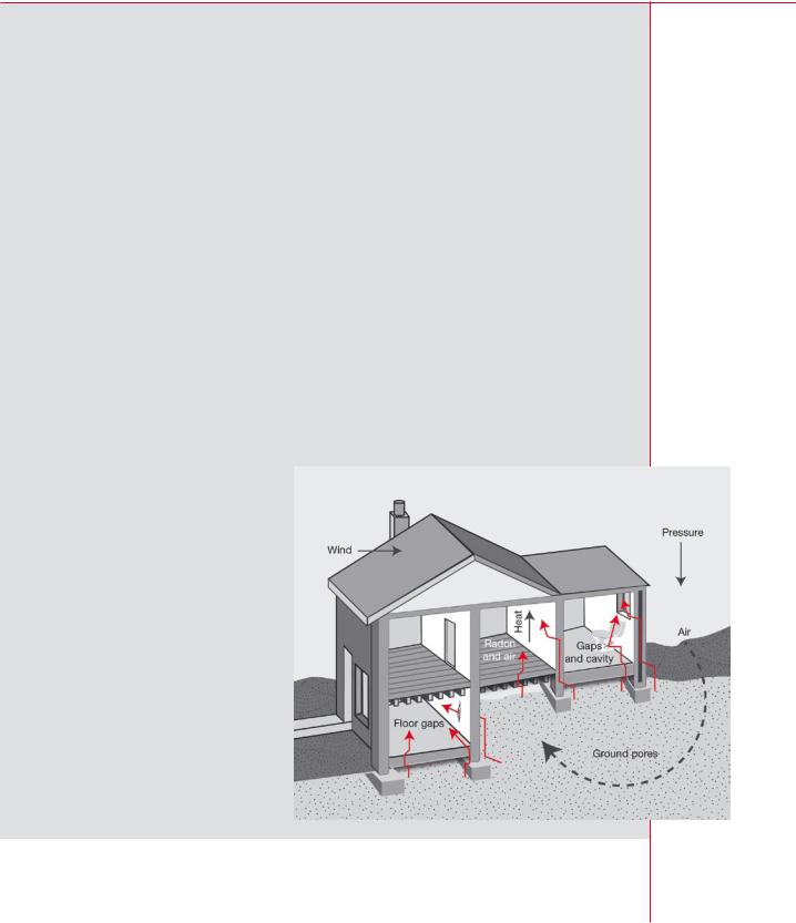
Radon gas is a particularly significant source of exposure to natural radiation. This is because the immediate decay products of radon-222 are radionuclides with short half-lives, which attach themselves to fine particles in the air, are inhaled, irradiate the tissues of the lung with alpha particles, and increase the risk of lung cancer. The same is true of radon-220 (thoron), but the degree of exposure of the lung is much less. When radon gas enters the atmosphere from the ground, it disperses in the air, so concentrations out of doors are low. When the gas enters a building, predominantly through the floor from the ground, the concentration of activity builds up within the enclosed space.
If buildings are well ventilated this accumulation of radon will not be marked. However, in many — generally colder — countries, buildings are constructed with more emphasis on retaining heat and preventing draughts. They are, therefore, often poorly ventilated, and radon concentrations indoors can be many times higher than those outdoors. Radon concentrations in buildings are also very dependent on the local geology and can vary a great deal between different parts of a country and even from building to building in the same area.
RADIATION, PEOPLE AND THE ENVIRONMENT
How
radon enters a home
31

Chapter 7 / Natural radiation
The worldwide average annual effective dose from the decay products of radon is estimated to be about 1.2 mSv. There are, however, pronounced variations about this value. In some countries (e.g. Finland) the national average is several times higher, and in particular homes in many countries occupants have received effective doses of the order of hundreds of mSv in a year. Given this, ICRP and IAEA have recommended the use of Action Levels (expressed in Bq m-3) above which householders are advised to reduce radon levels in their homes. Typically these Action Levels should be in the range 200–600 Bq m-3, which is about ten times the average value for the radon concentration in homes.
Anyonefindinghighradonlevelsintheirhomescanreduceitbypreventingairfromtheground entering the building. The most effective way to do so is to reduce the air pressure under the house with a small fan. As mentioned in Chapter 5, this circumstance is an example of intervention, in the ICRP sense, to reduce human exposure to ionizing radiation.
Radionuclides are Internal irradiation
found naturally in
the diet Other radionuclides from the uranium and thorium series, in particular lead-210 and polonium-210, are present in air, food, and water, and so irradiate the body internally. Potassium-40 also comes into the body with the normal diet. It is the main source of internal irradiation apart from the radon decay products. In addition, the interactions of cosmic rays with the atmosphere create a number of radionuclides, such as carbon-14, which also contribute to internal irradiation.
The average effective dose from these sources of internal irradiation is estimated to be 0.3 mSv in a year, with potassium-40 contributing about half. Information on how the total varies from one person to another is limited, although it is known that the potassium content of the human body is controlled by biological processes. The amount of potassium, and hence potassium-40, varies with the amount of muscle in the body, and is about twice as high in young men as in older women. There is little anyone could do to affect internal irradiation from the other radionuclides except by avoiding any food and water with a high radioactive content.
Total doses
The total average effective dose from natural radiation is about 2.4 mSv in a year, but doses can vary a great deal. Some national averages exceed 10 mSv in a year, and in some regions individual doses may exceed 100 mSv in a year, usually because of homes with particularly high levels of radon and its decay products.
Average doses are useful measures for comparing the health significance of radiation from natural and artificial sources, but they may need to be supplemented by additional data when there are, as with indoor radon, large variations about the average. The most helpful step might be to describe the frequency with which doses of a certain magnitude occur in the circumstances of interest.
32 RADIATION, PEOPLE AND THE ENVIRONMENT

Chapter 8 / Medical uses of radiation
Chapter 8 Medical uses of radiation
Ionizing radiation has two very different uses in medicine — for diagnosis and therapy. Both are intended to benefit patients and, as with any use of radiation, the benefit must outweigh the risk. We have touched on this matter of justification in Chapter 6.
Most people at some time in their lives have an X ray examination to help the physician diagnose disease or damage in the body. A much less common diagnostic procedure involves the administration of radionuclides to patients so that detectors outside the body can be used to observe how organs are functioning. Physicians use either of these procedures if they cannot make a diagnosis without them. Radiation doses are generally low, although they can be appreciable in certain procedures.
Much higher doses are required to treat malignant diseases or malfunctioning organs sometimes in combination with other forms of treatment. A beam of radiation may be used to irradiate the affected part of the body or a fairly high activity of a radionuclide may be administered to the patient.
The use of X rays for examining patients is called diagnostic radiology and the use of pharmaceuticals labelled with radionuclides for diagnosis or therapy is called nuclear medicine. When radiation beams are used to treat patients, the procedure is called radiotherapy.
Population |
Number of examinations |
Average annual |
per physician |
per 1000 people per year |
effective dose, mSv |
|
|
|
< 1000 |
920 |
1.2 |
1000–3000 |
150 |
0.14 |
3000–10 000 |
20 |
0.02 |
> 10 000 |
< 20 |
0.02 |
|
|
|
World average |
330 |
0.4 |
|
|
|
RADIATION, PEOPLE AND THE ENVIRONMENT
Radiation exposures from diagnostic medical procedures (UNSCEAR)
Taken from Table 2 of UNSCEAR 2000 Report
to the General Assembly
33
Chapter 8 / Medical uses of radiation
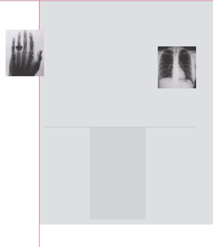
First X ray of hand (Frau Röntgen)
Typical doses to patients from conventional X ray and computed tomography examinations
Derived from data in UNSCEAR 2000 Report, Annex D, Vol. 1 Tables 15 and 19
Diagnostic radiology
In a conventional X ray examination, radiation from a machine passes through the patient. X rays penetrate flesh and bone to different degrees and produce images of the internal structures of the body on photographic film. In some cases, the images are captured and processed electronically. The value of these images explains why doctors conduct as many as one diagnostic X ray per person per year in developed
countries.
The parts of the body most frequently examined are the chest, limbs, and teeth, each accounting for about 25 per cent of the total number of examinations. Doses are fairly low — about 0.1 mSv from a chest examination, for example. Effective doses from other types of examination, such as the lower spine, are higher because organs and tissues that are more sensitive to radiation are exposed to a greater degree. Examinationsofthelowerbowelusingabariumenemaresult in a substantial effective dose around 6 mSv; only 1 per cent or so of all examinations are of this type.
Examination |
Conventional X ray |
Computed tomography |
|
dose (mSv) |
dose (mSv) |
||
|
|||
|
|
|
|
Head |
0.07 |
2 |
|
Teeth |
< 0.1 |
– |
|
Chest |
0.1 |
10 |
|
Abdomen |
0.5 |
10 |
|
Pelvis |
0.8 |
10 |
|
Lower spine |
2 |
5 |
|
Lower bowel |
6 |
– |
|
Limbs and joints |
0.06 |
– |
|
|
|
|
34 RADIATION, PEOPLE AND THE ENVIRONMENT
Chapter 8 / Medical uses of radiation
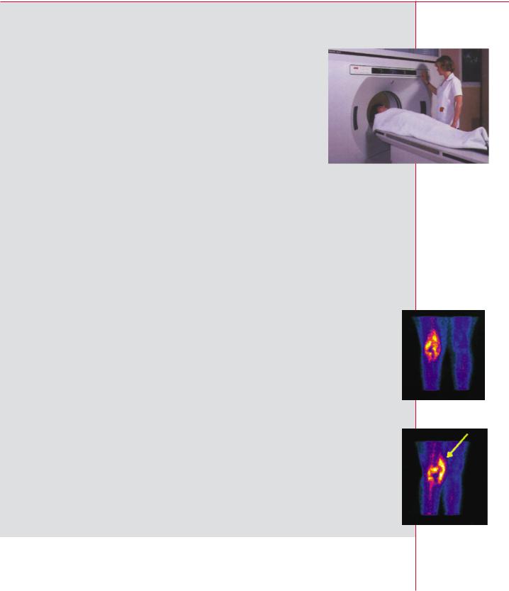
The use of computed tomography (CT) has increased considerably in recent years to |
|
the point where approximately 5 per cent of all procedures in diagnostic radiology in |
|
developed countries are CT scans. With this technique, a fan-shaped beam of X rays |
CT scanning |
is rotated around the patient and registered on the opposite side |
|
by a row of detectors. An image of a slice or section through the |
|
patient is then reconstructed by a computer and conveys supe- |
|
rior diagnostic information. However, doses in CT can be an order |
|
of magnitude or more higher than those from conventional X ray |
|
examinations. |
|
CT examinations are significant contributors to collective dose |
|
from medical diagnosis, and in some countries their contribu- |
|
tion is above 40 per cent of the total. Examinations of the lower |
|
bowel contribute about 10 per cent of the total collective dose |
|
and chest examinations about one per cent. It is clear from |
|
these figures that some relatively infrequent procedures can give a far greater dose |
|
to the population than the more common examinations. This is why a CT scan is not |
|
used where an ordinary X ray examination would suffice for a sound diagnosis. |
|
The diagnostic procedure that gives the highest doses, however, is interventional |
|
radiology. This is where a physician performing a procedure inside the patient’s body |
Technetium-99m |
uses a series of X rays to ‘see’ into the patient in real time. This allows a procedure on |
scintigram of a |
an internal organ to be carried out without the major surgery that might otherwise have |
patient with a right |
been needed to gain access to the organ. However, these procedures can give patient |
knee prosthesis |
doses in the range 10–100 mSv and, if not carefully controlled can lead to similarly |
and signs of |
high doses to surgeons. In some cases, the doses from such procedures have been |
infection (arrow) |
high enough to cause deterministic effects in patients and surgeons. |
|
Nuclear medicine
For a diagnostic procedure in nuclear medicine, the patient is given a radionuclide in a carrying substance, such as a pharmaceutical, which is preferentially taken up by the tissue or organ under study. Administration may be by injection, ingestion, or inhalation. The radionuclide emits gamma rays.
Most of the diagnostic procedures make use of the radionuclide technetium-99m. It has a half-life of 6 hours, gives off gamma rays with an energy of 0.14 MeV, can be conveniently prepared in the hospital, and readily labels a variety of carrying substances. A special detector called a gamma camera is used to observe how the organs or tissue behave or how quickly the radionuclide moves.
Individual doses from technetium scans are comparable to those in diagnostic radiology. The collective dose from nuclear medicine is, however, lower by more than an order of magnitude, because the number of procedures is much lower.
RADIATION, PEOPLE AND THE ENVIRONMENT 35

Chapter 8 / Medical uses of radiation
Typical doses to patients from common investigations of organs in nuclear medicine
Rounded values, derived from data in UNSCEAR 2000 Report, Vol.1, Annex D, Table 42
Organ scan |
Effective dose |
|
(mSv) |
||
|
||
|
|
|
Brain |
7 |
|
Bone |
4 |
|
Thyroid, lung |
1 |
|
Liver, kidney |
1 |
When radionuclides are used for treatment rather than diagnosis, much greater activities are given to the patient and much higher doses are given to the target tissues or organs. The treatment of an overactive thyroid gland — hyperthyroidism — is probably the most common therapeutic procedure, the radionuclide used being iodine-131.
Although the radionuclides used for these procedures have short halflives, medical staff need to take account of the fact that activity will remain in the body
of a patient to whom a radionuclide has been given for some time after the procedure. This might need to be taken into account, especially after therapeutic procedures, in deciding when he or she can be discharged from hospital. Family and friends of the patient may also sometimes be advised by the hospital to take appropriate precautions against inadvertent exposure from this residual activity.
Radiotherapy
This technique is used to cure cancers or at least to alleviate the most distressing symptoms, by killing the cancerous cells. A beam of high energy X rays, gamma rays or electrons is directed towards the diseased tissue so as to give it a high dose while sparing the surrounding healthy tissue. If a tumour is deep in the body, the beam is pointed at it from several directions so as to reduce the incidental damage. Another form of treatment, in which a radiation source is placed in or on the body for a short period, is used for some cancers: it is called brachytherapy. As radiotherapy doses are strong, such treatment is only used when the outlook for a cure or relief is good and when other methods of treatment would be less effective.
Although radiotherapy can cure the original cancer, it may possibly cause cancer in other tissues or adverse hereditary effects in subsequent generations. Most people who receive radiotherapy are, however, past the age to have children and too old for delayed cancers to occur. So the aim of radiotherapy is to maximize the effectiveness of treatment while minimizing the adverse side-effects.
Tumours require absorbed doses of tens of gray to kill the cancer cells effectively. Prescribed doses to tissues are typically in the range 20–60 Gy. Considerable care is required to deliver accurate doses: too low or too high doses may lead to incomplete treatment or unacceptable side-effects. Rigorous quality assurance procedures are needed to make sure that equipment is properly set up and maintained. If this
36 RADIATION, PEOPLE AND THE ENVIRONMENT