

1radiation_people_and_the_environment
.pdfChapter 2 / Atoms and radiation
Types of radiation
Most of the common types of radiation come from radioactive materials, but some types of radiation are produced in other ways. The most important example is that of X rays that are normally produced by firing a beam of electrons at a metal target (usually tungsten). The electrons in the metal atoms absorb energy from the electron beam — in scientific terms, the metal atoms become ‘excited’ — and then release the energy in the form of X rays as they ‘relax’. The radiation, therefore, comes from the metal atoms but, unlike radioactivity, it is not from the nucleus. Because of how they are produced, there is no half-life for an X ray. Once the beam is switched off, the X rays disappear.
Alpha radiation (α) is a positively charged helium nucleus emitted by a larger unstable nucleus. It is a relatively massive particle, but it only has a short range in air (1–2 cm) and can be absorbed completely by paper or skin. Alpha radiation can, however, be hazardous if it enters the body by inhalation or ingestion, because large exposures can result in nearby tissues, such as the lining of the lung or stomach.
Beta radiation (β) is an electron emitted by an unstable nucleus. Beta particles are much smaller than alpha particles and can penetrate further into materials or tissue. Beta radiation can be absorbed completely by sheets of plastic, glass,ormetal.Itdoesnotnormallypenetrate beyond the top layer of skin. However large exposures to high-energy beta emitters can cause skin burns. Such emitters can also be hazardous if inhaled or ingested.
RADIATION, PEOPLE AND THE ENVIRONMENT 7

Chapter 2 / Atoms and radiation
Gamma radiation (γ) is a very high energy photon (a form of electromagnetic radiation like light) emitted from an unstable nucleus that is often emitting a beta particle at the same time. Gamma radiation causes ionization in atoms when it passes through matter, primarily due to interactions with electrons. It can be very penetrating and only a substantial thickness of dense materials such steel or lead can provide good shielding. Gamma radiation can therefore deliver significant doses to internal organs without inhalation or ingestion.
X rays are high-energy photons, like gamma radiation, and are produced artificially by the rapid slowing down of an electron beam. X rays are similarly penetrating and, in the absence of shielding by dense materials, can deliver significant doses to internal organs.
Neutron radiation (n) is a neutron emitted by an unstable nucleus, in particular during atomic fission and nuclear fusion. Apart from a component in cosmic rays, neutrons are usually produced artificially. Because they are electrically neutral particles, neutrons can be very penetrating and when they interact with matter or tissue, they cause the emission of beta and gamma radiation. Neutron radiation therefore requires heavy shielding to reduce exposures.
8RADIATION, PEOPLE AND THE ENVIRONMENT
Cosmic radiation comes from deep space. It is a mixture of many different types of radiation, including protons, alpha particles, electrons and other various exotic (high energy) particles. All these energetic particles interact strongly with the atmosphere and, as a result, cosmic radiation at ground level becomes primarily muons, neutrons, electrons, positrons and photons. Most of the dose at ground level comes from muons and electrons.
Chapter 3 / Radiation and matter
Chapter 3 Radiation and matter
When radiation passes through matter, it deposits energy in the material concerned. Alpha and beta particles, being electrically charged, deposit energy through electrical interactions with electrons in the material. Gamma rays and X rays lose energy in a variety of ways, but each involves liberating atomic (orbiting) electrons, which then deposit energy in interactions with other electrons. Neutrons also lose energy in various ways, the most important being through collisions with nuclei that contain protons. The protons are then set in motion and, being charged, they again deposit energy through electrical interactions. So in all cases, the radiation ultimately produces electrical interactions in the material.
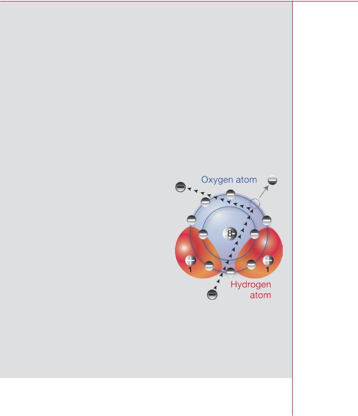
In some cases, an electron in the material may receive enough energy to escape from an atom leaving the atom or molecule thus formed positively charged. The figure illustrates this process for a molecule of water. The molecule has ten protons and ten electrons altogether, but only nine atomic electrons remain after a charged particle passes by; the molecule as a whole is left with one excess positive charge.
The process by which a neutral atom or molecule becomes charged is called ionization and the resulting entity an ion. Once removed
from an atom, an electron may in turn ionize other atoms or molecules. Any radiation that causes ionization — either directly, as with alpha and beta particles or indirectly as with gamma rays, X rays, and neutrons — is known as ionizing radiation. Charged particles passing through atoms may also give energy to the atomic electrons without actually removing them; this process is called excitation.
RADIATION, PEOPLE AND THE ENVIRONMENT
Ionization of a water molecule by a charged particle
9

Chapter 3 / Radiation and matter
|
|
Ionization in tissue |
|
|
Each time a charged particle ionizes or excites an atom, it loses energy until it no longer |
|
|
has enough energy to interact; the final result of these energy losses is a minute rise in |
|
|
the temperature of the material of which the atom is a part. In this way, all the energy |
|
|
deposited in biological tissues by ionizing radiation is eventually dissipated as heat |
|
|
through increased vibrations of the atomic and molecular structures. It is the initial |
Diagram of |
ionization and the resulting chemical changes that cause harmful biological effects. |
|
a cell |
|
|
|
|
The basic unit of biological tissue is the cell, which has a |
|
|
control centre called the nucleus. The nucleus of a cell is an |
|
|
intricate structure and not to be confused with the nucleus |
|
|
of an atom. About 80 per cent of the cell consists of water, |
|
|
the other 20 per cent being complex biological compounds. |
|
|
When ionizing radiation passes though cellular tissue, it |
|
|
produces charged water molecules. These break up into |
|
|
entities called free radicals, such as the free hydroxyl radical |
|
|
(OH), which is composed of an oxygen atom and a hydrogen |
|
|
atom. Free radicals are highly reactive chemically and can |
|
|
alter important molecules in the cell. |
|
|
One particularly important molecule is deoxyribonucleic |
|
|
acid, DNA, found mainly in the nucleus of the cell. |
|
|
DNA controls the structure and function of the cell and |
|
|
passes on copies of itself: its molecules are large and the structures that carry them, |
|
|
chromosomes, are visible through the microscope. We still do not fully understand all |
Diagram of |
the ways in which radiation damages |
|
DNA |
cells, but many involve changes to |
|
|
|
the DNA. There are two ways in |
|
|
which this can happen. Radiation |
|
|
may ionize a DNA molecule lead- |
|
|
ing directly to a chemical change, |
Ionizing radiation |
or the DNA may be changed indi- |
|
and tissue |
rectly when it interacts with a free |
|
|
|
hydroxyl radical produced in the |
Charged particles |
water of the cell by the radiation. In either case, the chemical change can cause a |
|
Ú |
|
harmful biological effect leading to the development of cancers or inherited genetic |
Electrical |
defects. Chapter 5 has more detail on radiation effects. |
|
interactions |
|
|
ÚA most important property of the various types of ionizing radiation is their ability to
Ionization occurs penetrate matter. The depth of penetration for a particular type of radiation increases
Úwith its energy, but varies from one type of radiation to another for the same amount
Chemical changes |
of energy. With charged particles such as alpha and beta particles, the depth of |
|
Ú |
|
penetration also depends on the mass of the particle and its charge. For equal energies, |
Biological effects |
a beta particle will penetrate to a much greater depth than an alpha particle. Alpha |
|
|
|
particles can scarcely penetrate the dead, outer layer of human skin; consequently, |
10 |
|
|
|
RADIATION, PEOPLE AND THE ENVIRONMENT |
|

Chapter 3 / Radiation and matter |
|
|
|
|
|
|
|
radionuclides that emit them are not hazardous unless they are taken into the body |
|
|
|
through breathing or eating or through a skin wound. Beta particles penetrate about |
|
|
|
a centimetre of tissue, so radionuclides that emit them are hazardous to superficial |
|
Hierarchy of |
|
tissues, but not to internal organs unless they too are taken into the body. For indi- |
|
dose quantities |
|
rectly ionizing radiation, such as gamma rays and neutrons, the degree of penetration |
|
|
|
depends on the nature of their interactions with tissue. Gamma rays can pass through |
|
Absorbed dose |
|
the body, so radionuclides that emit them may be hazardous whether on the outside |
|
Energy imparted |
|
or the inside. X rays and neutrons can also pass through the body. |
|
by radiation to unit |
|
|
|
mass of tissue |
|
Dose quantities |
|
Ú |
|
|
Equivalent dose |
|
|
|
|
Absorbed dose |
|
We cannot detect ionizing radiation directly through our senses, but we can detect and |
|
weighted for the |
|
measure it by other means: these include established methods based on photographic |
|
harm of different |
|
films, geiger–müller tubes, and scintillation counters, as well as newer techniques using |
|
types of radiation |
|
thermoluminescent materials and silicon diodes. We can interpret the measurements |
|
Ú |
|
we make in terms of the energy that the radiation concerned would have deposited |
|
Effective dose |
|
throughout the human body or in a particular part of the body. When direct measure- |
|
Equivalent dose |
|
ments are not possible — when, for instance, a radionuclide is deposited in an internal |
|
weighted for the |
|
organ — we can calculate the dose absorbed by that organ provided that we know the |
|
harm to different |
|
amount of activity retained in the organ. |
|
tissues |
|
The amount of energy that ionizing radiation deposits in a unit mass of matter, such |
|
Ú |
|
|
Collective |
|
|
as human tissue, is called the absorbed dose. It is expressed in a unit called the gray, |
|
effective dose |
|
symbol Gy, where 1 gray is equal to 1 joule per kilogram. Submultiples of the gray are |
|
Effective dose to |
|
often used, such as the milligray, mGy, which is one-thousandth of a gray. The gray is |
|
a group from a |
|
named after the English physicist Harold Gray (pictured on page 13). |
|
source of radiation |
|
Types of ionizing radiation differ in the way in which they interact with biological materi- |
|
|
|
|
|
|
|
als, so that equal absorbed doses (meaning equal amounts of energy deposited) do |
|
|
|
not necessarily have equal biological effects. For instance, 1 Gy to tissue from alpha |
|
|
|
radiation is more harmful than 1 Gy from beta radiation because an alpha particle, being |
|
|
|
slower and more heavily charged, loses its energy much more densely along its path. So |
|
|
|
in order to put all the different types of ionizing radiation on an equal basis with respect |
|
|
|
to their potential for causing harm, we need another quantity. This is the equivalent dose. |
|
|
|
It is expressed in a unit called the sievert, symbol Sv. Submultiples of the sievert are |
|
|
|
commonly used, such as the millisievert, mSv, which is one-thousandth of a sievert. The |
|
|
|
sievert is named after the Swedish physicist Rolf Sievert (pictured on page 13). |
|
|
|
Equivalent dose is equal to the absorbed dose multiplied by a factor that takes into |
|
|
|
account the way in which a particular type of radiation distributes energy in tissue so |
|
|
|
that we can allow for its relative effectiveness to cause biological harm. For gamma rays, |
|
|
|
X rays, and beta particles, this radiation-weighting factor is set at 1, so the absorbed |
|
|
|
dose and equivalent dose are numerically equal. For alpha particles, the factor is set at |
|
|
|
20, so that the equivalent dose is deemed to be 20 times the absorbed dose. Values of |
|
|
|
the radiation weighting factor for neutrons of various energies range from 5 to 20. |
|
|
|
RADIATION, PEOPLE AND THE ENVIRONMENT |
|
11 |
|
|
|
|
|

|
|
|
Chapter 3 / Radiation and matter |
|
||
|
|
|
|
|
||
|
|
|
|
Defined in this way, the equivalent dose provides an index of the likelihood of harm to |
||
|
Calculation of |
|
a particular tissue or organ from exposure to various types of radiation regardless of |
|||
|
effective dose |
|
their type or energy. So 1 Sv of alpha radiation to the lung, for example, would create |
|||
|
|
|
|
the same risk of inducing fatal lung cancer as 1 Sv of beta radiation. The risk to the |
||
|
Consider a |
|
various parts of the human body varies from organ to organ. For example, the risk of |
|||
|
circumstance |
|
fatal malignancy per unit equivalent dose is lower for the thyroid than for the lung. |
|||
|
in which a |
|
Moreover, there are other important types of harm such as non-fatal cancers or the |
|||
|
radionuclide |
|
risk of serious hereditary damage caused by irradiation of the testes or ovaries. These |
|||
|
causes |
|
|
|
effects are different both in kind and |
|
|
exposure of |
|
|
|
in magnitude and we must take |
|
|
|
|
|
|||
|
the lung, the |
|
Tissue or organ |
Tissue weighting |
them into account when assessing |
|
|
liver, and the |
|
the overall detriment to the health of |
|||
|
|
factor |
||||
|
surfaces of the |
|
|
human beings arising from exposure |
||
|
|
|
|
|||
|
|
|
|
|||
|
bones. |
|
Gonads |
0.20 |
to radiation. |
|
|
Suppose that |
|
Bone marrow (red) |
0.12 |
We can deal with all these complexi- |
|
|
|
Colon |
0.12 |
|||
|
the equivalent |
|
ties by taking the equivalent dose in |
|||
|
doses to the |
|
Lung |
0.12 |
each of the major tissues and organs |
|
|
tissues are, |
|
of the body and multiplying it by a |
|||
|
|
Stomach |
0.12 |
|||
|
respectively, |
|
weighting factor related to the risk |
|||
|
|
|
|
|||
|
100, 70, and |
|
Bladder |
0.05 |
associated with that tissue or organ. |
|
|
300 mSv. |
|
Breast |
0.05 |
The sum of these weighted equivalent |
|
|
|
|
|
doses is a quantity called the effec- |
||
|
|
|
|
Liver |
0.05 |
|
|
The |
|
tive dose: it allows us to represent |
|||
|
effective |
|
Oesophagus |
0.05 |
the various dose equivalents in the |
|
|
dose is |
|
body as a single number. The effec- |
|||
|
|
Thyroid |
0.05 |
|||
|
calculated as |
|
tive dose also takes account of the |
|||
|
(100 × 0.12) + |
|
Skin |
0.01 |
energy and type of radiation, and |
|
|
(70 × 0.05) + |
|
Bone surface |
0.01 |
therefore gives a broad indication |
|
|
(300 × 0.01) = |
|
of the detriment to health. Moreover, |
|||
|
|
Remainder |
0.05 |
|||
|
18.5 mSv |
|
it applies equally to external and |
|||
|
|
|
|
|||
|
The calculation |
|
|
|
internal exposure and to uniform or |
|
|
|
|
|
|||
|
|
Whole body total |
1.00 |
non-uniform irradiation. |
||
|
shows that the |
|
|
|
|
|
|
risk of harmful |
|
|
|
It is sometimes useful to have a mea- |
|
|
effects from |
|
sure of the total radiation dose to groups of people or a whole population. The quantity |
|||
|
this particular |
|
used to express this total is the collective effective dose. It is obtained by adding, for |
|||
|
pattern of radia- |
|
all exposed people, the effective dose that each person in that group or population has |
|||
|
tion exposure |
|
received from the radiation source of interest. For example, the effective dose from all |
|||
|
will be the |
|
sources of radiation is, on average, 2.8 mSv in a year. Since the world population is |
|||
|
same as the risk |
|
about 6000 million, the annual collective effective dose to the whole population is the |
|||
|
from 18.5 mSv |
|
product of these two numbers — about 17 000 000 man sievert, symbol man Sv. |
|||
|
received uniformly |
|
|
|
|
|
|
throughout the |
|
It is common for effective dose to be abbreviated to dose and collective effective dose |
|||
|
whole body. |
|
to collective dose. This will be the case in the following chapters except where exactness |
|||
|
|
|
|
is essential. |
|
|
|
12 |
RADIATION, PEOPLE AND THE ENVIRONMENT |
|
|||
|
|
|
|
|
|
|

Chapter 4 / Sources of ionizing radiation
|
Absorbed dose |
|
is expressed in a |
|
unit called the gray, |
|
named after the |
|
English physicist |
|
Harold Gray |
|
(1905–1965) |
Chapter 4 Sources of ionizing radiation |
|
Ionizing radiation enters our lives in a variety of ways. It arises from natural processes, |
|
such as the decay of uranium in the Earth, and from artificial procedures like the use |
|
of X rays in medicine. So we can classify radiation as natural or artificial according |
|
to its origin. Natural sources include cosmic rays, gamma rays from the Earth, radon |
|
decay products in the air, and various radionuclides found naturally in food and drink. |
|
Artificial sources include medical X rays, fallout from the testing of nuclear weapons in |
|
the atmosphere, discharges of radioactive waste from the nuclear industry, industrial |
|
gamma rays, and miscellaneous items such as consumer products. Later chapters |
|
have more information on both classes of source. |
|
Each source of radiation has two important characteristics, the dose that it delivers to |
|
human beings and the ease with which we can do something to affect such doses. |
|
Until recently, radiation from natural sources seemed both unremarkable and unalter- |
|
able — a background phenomenon. We now know, however, that doses from the |
|
decay products of radon gas (itself a product of uranium decay) in the home can be |
|
remarkably high in some areas, although it is fairly easy to reduce them in existing |
|
homes and to avoid high concentrations of the gas when building new homes. In |
|
contrast, we cannot do much to change our exposure to the other natural sources of |
|
radiation. This basic background of cosmic rays, gamma rays, and natural radioactivity |
Rolf Sievert |
within the body gives rise to an annual dose of about 1 mSv or more to an average |
(1896–1966) |
citizen of the world. A comparable dose (at least) from radon decay products is also |
|
unavoidable in practice for most people. |
Equivalent dose is |
|
expressed in a unit |
It is easier, in most cases, to control artificial sources of radiation because we can alter |
called the sievert, |
or terminate the procedure producing the radiation, but there is always a balance to |
named after the |
be made. It is important, for instance, to pay attention to the doses from medical X ray |
Swedish physicist |
examinations, but it would be unwise to reduce them where this would lead to a loss |
|
of essential diagnostic information. |
|
The United Nations Scientific Committee on the Effects of Atomic Radiation (UNSCEAR) was established in 1955 to estimate the potential health risks from radioactive fallout from atmospheric nuclear weapons tests. Today, UNSCEAR regularly publishes data on doses from all sources. The results of the latest review, published in 2000, are reflected in the pie chart on the next page. The annual dose, averaged over the population of the world, is about 2.8 mSv in total. Over 85 per cent of this total is
RADIATION, PEOPLE AND THE ENVIRONMENT 13

Chapter 4 / Sources of ionizing radiation
Compiled from data in Tables 1 and 2 of UNSCEAR 2000
Report to the UN General Assembly
Average radiation exposure from all sources = 2.8 mSv/a
Average annual doses to the world population from all sources of radiation
from natural sources with about half coming from radon decay products in the home. Medical exposure of patients accounts for 14 per cent of the total, whereas all other artificial sources — fallout, consumer products, occupational exposure, and discharges from the nuclear industry — account for less than 1 per cent of the total value.
The greatest variations in dose arise from radon decay products in the home, which can give annual doses of 10 mSv or more. Annual doses for those exposed to radiation at work are, at present, limited by law in most countries to 50 mSv or less, but only a small fraction of the workforce exceeds 20 mSv. It is unlikely that many members of the public receive more than a fraction of 1 mSv in a year from incidental exposure to artificial sources. Doses to patients in some diagnostic procedures may be around 10 mSv. For consumer products that contain radioactive material, such as smoke alarms and luminous watches, annual doses are at most 1 µSv (1 millionth of a sievert), although less common items, such as gas mantles containing thorium, may cause as much as 0.1 mSv in a year in certain circumstances.
Source |
Dose (mSv) |
|
|
Natural |
|
Cosmic |
0.4 |
Gamma rays |
0.5 |
Internal |
0.3 |
Radon |
1.2 |
Artificial |
|
Medical |
0.4 |
Atmospheric nuclear testing |
0.005 |
Chernobyl |
0.002 |
Nuclear Power |
0.0002 |
|
|
Total (rounded) mSv |
2.8 |
14 RADIATION, PEOPLE AND THE ENVIRONMENT
Chapter 5 / Radiation effects
Chapter 5 Radiation effects |
|
Radiation doses of different sizes, delivered at different rates to different parts of the |
|
body, can cause different types of health effect at different times. |
|
A very high dose to the whole body can cause death within weeks. For example, an |
|
absorbed dose of 5 gray or more received instantaneously would probably be lethal, |
|
unless treatment were given, because of damage to the bone marrow and the gastro- |
|
intestinal tract. Appropriate medical treatment may save the life of a person exposed |
|
to 5 gray, but a whole body dose of, say, 50 gray would almost certainly be fatal even |
|
with medical attention. A very high dose to a limited area of the body might not prove |
|
fatal, but other early effects could occur. For example, an instantaneous absorbed |
|
dose of 5 gray to the skin would probably cause erythema — painful reddening of the |
|
skin — within a week or so, whereas a similar dose to the reproductive organs might |
|
cause sterility. These types of effect are called deterministic effects: they occur only if |
|
the dose or dose rate is greater than some threshold value, and the effect occurs earlier |
|
and is more severe as the dose and dose rate increase. Deterministic effects in an |
|
individual can be identified clinically to be the result of radiation exposure (although on |
|
the few occasions when they have occurred as a result of accidents — see Chapter 14 |
|
— they have not always been immediately recognized as such). |
Deterministic |
|
effects on vision |
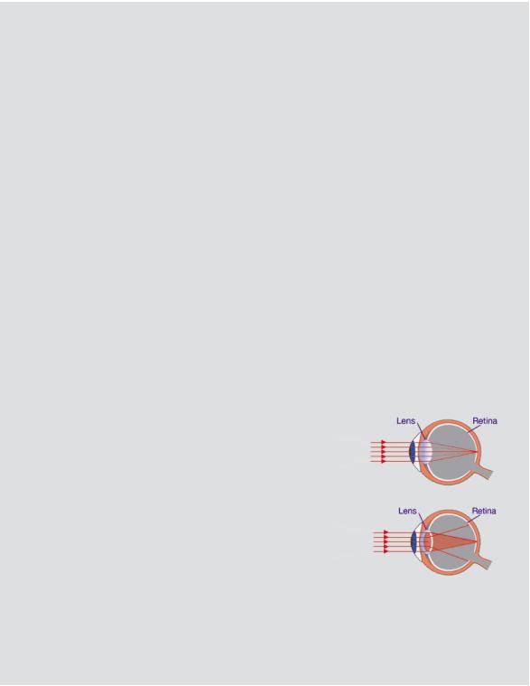
One type of deterministic effects occurs a longer |
Normal lens — |
time after exposure. Such effects are not usually |
light is focussed |
fatal, but can be disabling or distressing because |
normally on the |
the function of some parts of the body may be |
retina |
impaired or other non-malignant changes may |
|
arise. The best-known examples are cataracts |
Lens with cataract |
(opacity in the lens of the eye) and skin damage |
— opacity of the |
(thinning and ulceration). High absorbed doses of |
lens blocks or |
several gray are normally required to induce these |
distorts light from |
conditions. |
being focussed on |
|
the retina, resulting |
|
in reduced vision |
If the dose is lower, or is delivered over a longer period of time, there is a greater |
|
opportunity for the body cells to repair, and there may be no early signs of injury. Even |
|
so, tissues may still have been damaged in such a way that the effects appear only |
|
later in life (perhaps decades later), or even in the descendants of the irradiated person. |
|
These types of effect are called stochastic effects: they are not certain to occur, but the |
|
RADIATION, PEOPLE AND THE ENVIRONMENT |
15 |
|
|

Chapter 5 / Radiation effects
likelihood that they will occur increases as the dose increases, whereas the timing and severity of any effect does not depend on the dose. Because radiation is not the only known cause of most of these effects, it is normally impossible to determine clinically whether an individual case is the result of radiation exposure or not.
Induction of cancers
The most important of these stochastic effects is cancer, which is always serious and often fatal. Although the exact cause of most cancers remains unknown or poorly understood, exposure to agents such as tobacco smoke, asbestos and ultraviolet radiation, as well as ionizing radiation, are known to play a role in inducing certain types of cancer. The development of cancer is a complex, multistage process that usually takes many years. Radiation appears to act principally at the initiation stage, by introducing certain mutations in the DNA of normal cells in tissues. These mutations allow a cell to enter a pathway of abnormal growth that can sometimes lead to the development of a malignancy.
Follicular Given that we cannot distinguish between those cancer cases resulting from radiation Carcinoma of exposure and those with other causes, how can we calculate the risk of cancer from Thyroid radiation? In practice, we have to use epidemiology — the statistical study of the inci- A.K. Padhy/IAEA dence (the number of cases and their distribution) of specific disorders in specific population groups. Suppose that we know the number of people in an irradiated group and the doses they have received. Then by observing the occurrence of cancer in the group and comparing with the doses and the number of cancers expected in an otherwise similar but unirradiated group, we can estimate the raised risk of cancer per unit dose. This is commonly called a risk factor. It is most important to include data for large groups of people in these calculations so as to minimize the statistical uncertainties in the estimates and take account of factors,
such as age and gender, that affect the spontaneous development of cancer.
Not all cancers are fatal. Average mortality from radiation-induced thyroid cancer is about 10 per cent (although it is much lower — less than 1 per cent — for the cases caused in children and teenagers by the Chernobyl accident), from breast cancer about 50 per cent, and from skin cancer about 1 per cent. Overall, the total risk of inducing cancer by uniformly irradiating the whole body is about half as great again as the risk of inducing a fatal cancer. In radiological protection the risk of fatal cancer is of more concern because of its extreme significance. The use of fatal cancer risks also makes it easier to compare them with the other fatal risks encountered in life. In contrast, comparisons of non-fatal risks are fraught with difficulty.
Risk assessments
The main sources of information on the additional risk of cancer following exposure of the whole body to gamma radiation are studies of the survivors of the atomic bombs
16 RADIATION, PEOPLE AND THE ENVIRONMENT