

Human_Histology
.pdf38 Textbook of Human Histology
A
B C D E
F G H
I
Figs. 4.1A to I: Scheme to show various ways in which the secretory elements of a gland may be organized. (A) Unicellular gland; (B to G)
Multicellular glands with a single duct are simple glands; (H and I) Multicellular glands with branching duct system are compound glands
Alveolar glands: Glands with secretory unit flaskshaped. However, it may be noted that the terms acini and alveoli are often used as if they were synonymous. Glands in which the secretory elements are greatly distended are called saccular glands.
Note: Combinations of the above may be present in a single gland. From what has been said above it will be seen that an exocrine gland may be:
Unicellular
Simple tubular
Simple alveolar (or acinar)
Compound tubular
Compound alveolar
Compound tubulo-alveolar (or racemose)
Some further subdivisions of these are shown in Figures
4.1A to I.
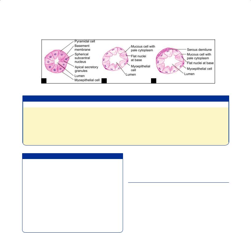
Nature of their secretions: mucous glands and serous glands.
Mucous glands: Cell of mucous acini are tall with flat nuclei at their bases. The lumen of these acini is larger than the serous acini. In mucous glands the secretion contains mucopolysaccharides. The
secretion collects in the apical parts of the cells. As a result nuclei are pushed to the base of the cell, and may be flattened. In classroom slides stained with hematoxylin and eosin, the secretion within mucous cells remains unstained so that they have an “empty” look. However, the stored secretion can be brightly stained using a special procedure called the periodic acid Schiff (PAS) method. Unicellular cells secreting mucus are numerous in the intestines, they are called goblet cells because of their peculiar shape (Figs. 4.2A to C).
Serous glands: Cells of serous acini are triangular in shape with a rounded nucleus. Their nuclei are centrally placed. The secretions of serous glands are protein in nature. The cytoplasm of these cells is granular and often stains bluish with hematoxylin and eosin. The lumen of their acini is small. A comparison of mucous and serous acini has been given in Table 4.1.
Note: Some glands contain both serous and mucous elements (mixed).
Chapter 4 Glands 39
A B C
Figs. 4.2A to C: Types of acini. (A) Serous; (B) Mucous; (C) Mucous acini with serous demilune (Schematic representation)
Table 4.1: Comparison between serous and mucous acini
Serous acini |
Mucous acini |
Triangular cells with rounded nucleus at the base. Cell |
Tall cells with flat nucleus at the base. Cell boundaries are |
boundaries are indistinct |
distinct |
Contain zymogen granules |
Contain mucoid material |
Darkly stained with H & E (because of the presence of zymogen |
Lightly stained and appear empty with H & E |
granules, the color varies from pink to dark purple) |
|
Thin watery secretion |
Thick mucoid secretion |
Example: Parotid gland |
Example: Sublingual gland |
Added Information
Epithelia in secretory portions of glands show specialza tions of structure depending upon the nature of secretion as follows:
Cells that are protein secreting (e.g. hormone producing cells) have a well developed rough endoplasmic reti culum(ER),andasupranuclearGolgicomplex.Secretory granules often fill the apical portions of the cells. The staining characters of the granules differ in cells produc ing different secretions (the cells being described as acidophil, basophil, etc.).
Mucin secreting cells have a well developed rough ER (where the protein component of mucin is synthesized) and a very well developed Golgi complex (where proteins are glycosylated).
Steroid producing cells are characterized by the presence of extensive smooth ER and prominent mitochondria.
The manner in which their secretions are poured out of the cells:
Merocrine: In most exocrine glands secretions are thrown out of the cells by a process of exocytosis, the cell remaining intact, this manner of secretion is described as merocrine, e.g. goblet cell (sometimes also called eccrine or epicrine) (Fig. 4.3A).
Apocrine: In some glands the apical parts of the cells are shed off to discharge the secretion, this manner of secretion is described as apocrine. An example of apocrine secretion is seen in some atypical sweat glands and in mammary glands (Fig. 4.3B).
Holocrine: In some glands, the entire cell disintegrates while discharging its secretion. This manner of discharging secretion is described as holocrine, and is seen typically in sebaceous glands (Fig. 4.3C).
STRUCTURAL ORGANIZATION
Exocrine Glands
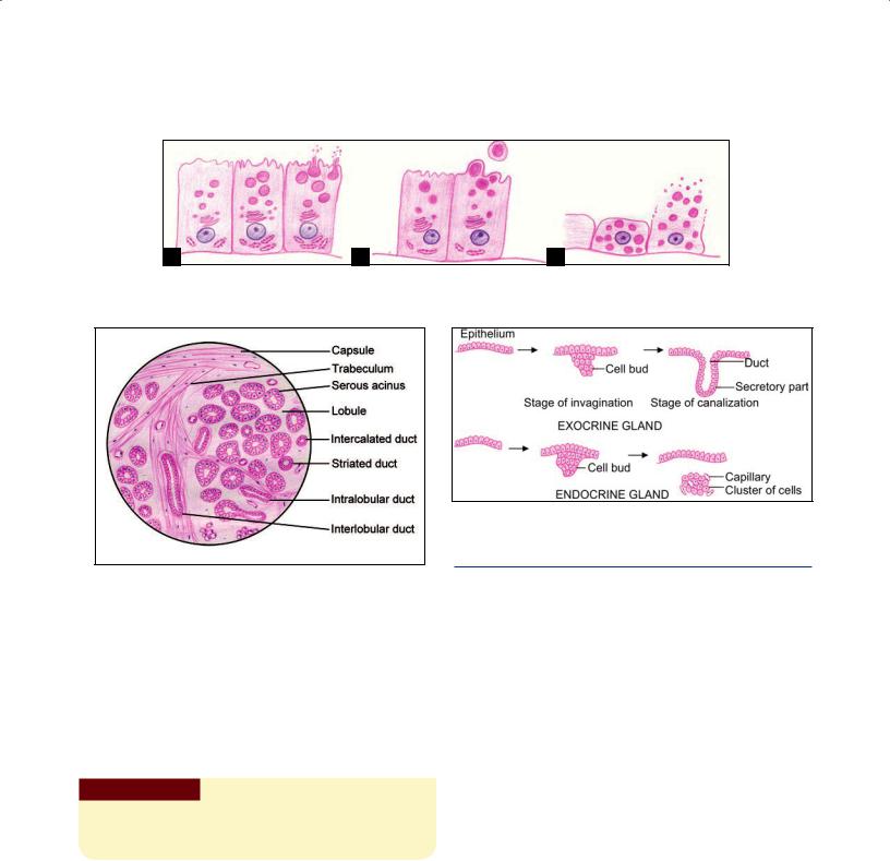
All exocrine glands have basically the same structural organization consisting of three components—parenchyma, stroma, and duct system.
Parenchyma: The secretory cells of a gland constitute its parenchyma.
Stroma: The connective tissue in which the parenchyma lies is called the stroma. The glandular tissue is often divisible into lobules separated by connective tissue septa. Aggregations of lobules may form distinct lobes. The connective tissue covering the entire gland forms a capsule for it. Blood vessels and nerves pass along connective tissue septa to reach the secretory elements. Their activity is under nervous or hormonal control (Fig. 4.4).
Duct system: The ducts convey the secretory product of the gland. When a gland is divided into lobes the ducts draining it may be intralobular (lying within a lobule), interlobular (lying in the intervals between lobules), or interlobar (lying between adjacent lobes), in increasing order of size (Fig. 4.4).
40 Textbook of Human Histology
A B C
Figs. 4.3A to C: Types of glands based on the manner in which their secretions are poured out of the cells.
(A) Merocrine; (B) Apocrine; (C) Holocrine (Schematic representation)
Fig. 4.5: Development of glands (Schematic representation)
DEVELOPMENT OF GLANDS
Fig. 4.4: Structural organization of an exocrine gland (Schematic representation)
Endocrine Glands
Endocrine glands are usually arranged in cords or in clumps that are intimately related to a rich network of blood capillaries or of sinusoids. In some cases (for example the thyroid gland) the cells may form rounded follicles.
Endocrine cells and their blood vessels are supported by delicate connective tissue, and are usually surrounded by a capsule.
Clinical Correlation
Neoplasms can arise from the epithelium lining a gland. A benign growth arising in a gland is an adenoma; and a malignant growth is an adenocarcinoma.
Glands, both exocrine and endocrine develop as diverticula of the epithelium. As shown in the Figure 4.5, the exocrine develops as a solid bud from the epithelium into the underlying connective tissue. Soon it elongates, undergoes canalization and displays a secretory and conducting portion. The conducting part forms the duct and is continuous with the epithelium. Hence, an endocrine gland discharges its secretions through a duct.
Endocrine gland also develops in a similar manner to exocrine gland but with further development it breaks the continuity with overlying epithelium. It appears as a clump of cells. Soon these groups of cells get surrounded by blood vessels into which they pour their secretions.
Note: In this chapter we have considered the general features of glands. Further details of the structure of exocrine and endocrine glands will be considered while studying individual glands.
Chapter
5
General Connective Tissue
INTRODUCTION |
Flowchart 5.1: Basic components of connective tissue |
The term connective tissue is applied to a tissue that fills the interstices between more specialized elements.
Connective tissue serves to hold together, and to sup port, different elements within an organ. For this reason, such connective tissue is to be found in almost every part of the body. It is conspicuous in some regions and scanty in others. This kind of connective tissue is referred to as general connective tissue to distinguish it from more specialized connective tissues that we will consider sepa rately.
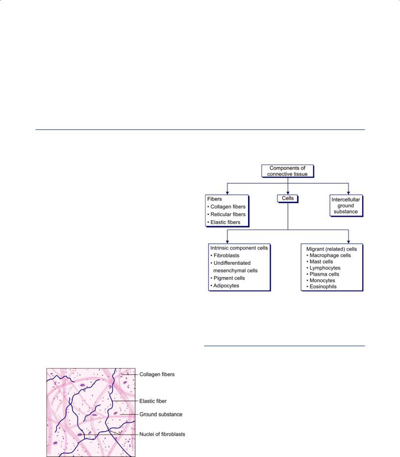
Components of Connective Tissue
(Fig. 5.1 and Flowchart 5.1)
Cells
Fibers
Extracellular matrix
Many tissues and organs of the body are made up mainly
of aggregations of closely packed cells, e.g. epithelia, and solid organs like the liver. In contrast, cells are relatively few in connective tissue, and are widely separated by a prominent intercellular substance. Which is in the form of a ground substance within which there are numerous fibers (Fig. 5.2). Connective tissue can assume various
Fig. 5.1: Stretch preparation of omentum showing loose areolar tissue (Schematic representation)
forms depending upon the nature of the ground substance, and of the type of fibers and cells present. It is also referred to as the extracellular matrix.
FIBERS OF CONNECTIVE TISSUE
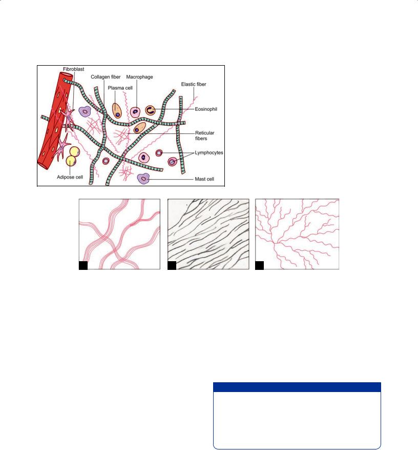
The most conspicuous components of connective tissue are the fibers within it. These are of three main types: collagen, reticular and elastic.
Collagen fibers are most numerous. They can be classified into various types. Reticular fibers were once described as a distinct variety of fibers, but they are now regarded as one variety of collagen fiber.
Collagen Fibers
Collagen fibers are most abundant type of connective tissue fibers. With the light microscope collagen fibers are seen in bundles (Figs. 5.2 and 5.3A). The bundles may be straight or wavy depending upon how much they are stretc hed. The bundles are made up of collections of individual collagen fibers which are 1–12 µm in diameter.
42 Textbook of Human Histology
Fig. 5.2: Components of loose connective tissue (Schematic representation)
A B C
Figs. 5.3A to C: Fibers of connective tissue. (A) Collagen fibers; (B) Reticular fibers; (C) Elastic fibers (Schematic representation)
The bundles often branch, or anastomose with adjacent bundles, but the individual fibers do not branch.
With the EM each collagen fiber is seen to be made of fibrils that are 20–200 nm in diameter. Each fibril consists of a number of microfibrils (3.5 nm in diameter). At high magnifications of the EM each fibril shows characteristic cross striations (or periods) after every 67 nm interval (in unfixed tissue).
Staining Characters
Bundles of collagen fibers appear white with the unaided eye. In sections stained with hematoxylin and eosin, collagen fibers are stained light pink. With special meth ods they assume different colors depending upon the dye used. Two commonly used methods are Masson’s trichrome with which the fibers stain blue and the Van Gieson method with which they stain red. After silver impregnation the fibers are stained brown.
Chemical Nature
Microfibrils of collagen are chains of tropocollagen molecules. Each molecule of tropocollagen is 300 nm in length. Within a fiber, the molecules of tropocollagen are arranged in a regular overlapping pattern which is responsible for producing the cross striated appearance of the fibers.
Each molecule of tropocollagen is made up of three polypeptide chains. The chains are arranged in the form of a triple helix. The polypeptide chains are referred to as procollagen.
Added Information
Each procollagen chain consists of a long chain of amino acids that are arranged in groups of three (triplets). Each triplet contains the amino acid glycine. The other two amino acids in each triplet are variable. Most commonly these are hydroxyproline and hydroxylysine. Variations in the amino acid pattern give rise to several types of collagen as described below.
Physical Properties
Collagen fibers are so called because they are made up mainly of a protein called collagen. Collagen is made up of molecules of tropocollagen.
Collagen fibers can resist considerable tensile forces (i.e. stretching) without significant increase in their length. At the same time they are pliable and can bend easily.

Chapter 5 General Connective Tissue 43
Added Information
The mechanism of the production of collagen fibers by fibroblasts has been extensively studied. Amino acids necessary for synthesis of fibers are taken into the cell. Under the influence of ribosomes located on rough endoplasmic reticulum, the amino acids are bonded together to form polypeptide chains (a-chains) (Flowchart 5.2). A procollagen molecule is formed by joining together of three such chains. Molecules of procollagen are transported to the exterior of the cell where they are acted upon by enzymes (released by the fibroblast) to form tropocollagen. Collagen fibers are formed by aggregation of tropocollagen molecules. Vitamin C and oxygen are necessary for collagen formation, and wound repair may be interfered with if either of these is deficient. In this connection it may be noted that fibroblasts are themselves highly resistant to damaging influences and are not easily destroyed.
There are observations that indicate that the orientation of collagen fibers depends on the stresses imposed on the tissue. If fibroblasts growing in tissue culture are subjected to tension in a particular direction, the cells exposed to the tension multiply faster than others; they orientate themselves along the line of stress; and lay down fibers in that direction. It follows that in the embryo collagen fibers would tend to be laid down wherever they are required to resist a tensile force. In this way tendons, ligaments, etc., will tend to develop wherever they are called for. (This cannot, of course, be a complete explanation.
Genetic factors must play a prominent role in development of these structures).
Flowchart 5.2: Scheme to show how collagen fibers are synthesized
When polarized light is thrown on the fibers the light is split into two beams that are refracted in different directions. This is called birefringence: it is an indication of the fact that each collagen fiber is made up of finer fibrils.
Collagen fibers swell and become soft when treated with a weak acid or alkali. They are destroyed by strong acids. (This fact is sometimes made use of to soften collagen fibers to facilitate preparation of anatomical specimens). On boiling, collagen is converted into gelatin.
Production of Collagen Fibers
Tropocollagen is synthesized by fibroblast and released into extracellular space, where they get polymerized to form collagen fibers. Collagen is not only synthesized by fibro blast but by other cells also, e.g. Chondroblasts in cartilage, osteoblast in bone and smooth muscle in blood vessel.
Varieties of Collagen and Their Distribution
Several types of collagen (more than 25) are recognized depending upon the diameter of fibers, the prominence of cross striations, and other features.
Type I: These are collagen fibers of classical descrip tion having the properties described above. They are found in connective tissue, tendons, ligaments, fasciae, aponeuroses, etc. They are also present in the dermis of the skin, and in meninges. They form the fibrous basis of bone and of fibrocartilage. Type I fibers are of large diameter (about 250 nm) and have prominent cross striations.
Type II: These are of two subtypes. The larger of these are about 100 nm in diameter, while the narrower fibers are about 20 nm in diameter. In type II collagen striations are less prominent than in type I. Type II collagen fibers form the fibrous basis of hyaline cartilage. Fine type II fibers are also present in the vitreous body.
Type III: These form the reticular fibers described below.
Type IV: This type of collagen consists of short filaments that form sheets. It is present in the basal laminae of basement membranes. It is also seen in the lens capsule.
Type V: This type of collagen is found in blood vessels and fetal membranes.
44 Textbook of Human Histology
Fig. 5.4: Reticular fibers (black) forming a network in the liver. The white spaces represent sinusoids (Schematic representation)
Reticular Fibers
These fibers are a variety of collagen fibers and are com posed of collagen type III. They show periodicity (striations) of 67 nm. They differ from typical (Type I) collagen fibers as follows:
They are much finer and have uneven thickness.
They form a network (or reticulum) by branching, and by anastomosing with each other. They do not run in bun dles (Fig. 5.3B).
They can be stained specifically by silver impregnation, which renders them black. They can thus be easily distinguished from type I collagen fibers which are stained brown. Because of their affinity for silver salts reticular fibers are sometimes called argentophil fibers (Fig. 5.4).
Reticular fibers contain more carbohydrates than type I fibers (which is probably the reason why they are argen tophil).
Reticular fibers provide a supporting network in lymp hoid organs like the spleen, lymph nodes and bone mar row; most glands, including the liver (Fig. 5.4); and the kidneys. Reticular fibers form an essential component of all basement membranes. They are also found in relation to smooth muscle and nerve fibers. Reticular fibers are synthesized by fibroblasts and reticular cells (special variety of fibroblasts).
Elastic Fibers
Elastic fibers are much fewer than those of collagen. They run singly (not in bundles), branch and anastomose with other fibers (Fig. 5.3C).
Elastic fibers are thinner than those of collagen (0.1– 0.2 µm) (see Fig. 5.1). In some situations elastic fibers are thick (e.g. in the ligamenta flava). In other situations (as in walls of large arteries) they form fenestrated membranes.
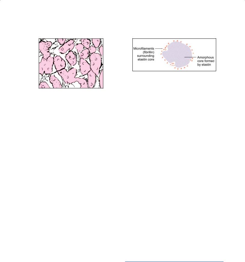
With the EM each elastic fiber is seen to have a central amorphous core and an outer layer of fibrils (Fig. 5.5).
Fig. 5.5: Electron microscopy appearance of an elastic fiber as seen in transverse section (Schematic representations)
The outer fibrils are made up of a glycoprotein called fibri llin. Periodic striations are not present in elastic fibers.
Staining Characters
Elastic fibers do not stain with the usual stains for collagen. They can be demonstrated by staining with orcein, with aldehyde fuchsin, and by Verhoeff’s method.
Chemical Nature
Elastic fibers are composed mainly of a protein called elastin that forms their central amorphous core. Elastin is made up of smaller units called tropoelastin. Elastin con tains a high quantity of the amino acids valine and alanine. Another amino acid called desmosine is found exclusively in elastic tissue. The outer fibrils of elastic fibers are com posed of the glycoprotein fibrillin.
Production of Elastic Fibers
Elastic fibers of connective tissue are produced by fibro blasts. In some situations elastic tissue can be formed by smooth muscle cells.
Physical Properties
As their name implies elastic fibers can be stretched (like a rubber band) and return to their original length when tension is released. They are highly refractile and are, therefore, seen as shining lines in unstained preparations. Relaxed elastic fibers do not show birefringence, but when stretched the fibers become highly birefringent.
Unlike collagen, elastic fibers are not affected by weak acids or alkalies, or by boiling. However, they are digested by the enzyme elastase.
CELLS OF CONNECTIVE TISSUE
Various types of cells are present in connective tissue. These can be classed into two distinct categories (see Flowchart 5.1).
Cells that are intrinsic components of connective tissue:
A B
Figs. 5.6A and B: Structure of a fibroblast. (A) Profile view; (B)
Structure view (Schematic representation)
In typical connective tissue the most important cells are fibroblasts. Others present are undifferentiated mesen chymal cells, pigment cells, and fat cells. Other varieties of cells are present in more specialized forms of connective tissues.
Cells that belong to the immune system and are iden tical or closely related with certain cells present in blood and in lymphoid tissues.
These include macrophage cells (or histiocytes), mast cells, lymphocytes plasma cells, monocytes, and eosinophils.
Fibroblasts
These are the most numerous cells of connective tissue. They are called fibroblasts because they are concerned with the production of collagen fibers. They also produce reti cular and elastic fibers. Where associated with reticular fibers they are usually called reticular cells.
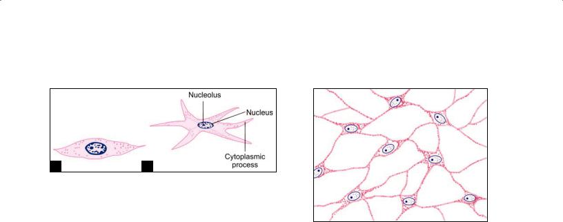
Fibroblasts are present in close relationship to collagen fibers. They are “fixed” cells, i.e. they are not mobile. In tissue sections these cells appear to be spindle shaped, and the nucleus appears to be flattened. When seen from the sur face the cells show branching processes (Figs. 5.6A and B). The nucleus is large, euchromatic, and has prominent nucleoli.
The amount of cytoplasm and of organelles varies depending upon activity. In inactive fibroblasts that are the cytoplasm is scanty, organelles are few, and the nucleus may become heterochromatic. Inactive fibroblasts are often called fibrocytes. In contrast to fibrocytes active fibroblasts have abundant cytoplasm (characteristic of cells actively engaged in protein synthesis). The endoplasmic reticulum, the Golgi complex and mitochondria become much more conspicuous.
Fibroblasts become very active when there is need to lay down collagen fibers. This occurs, for example, in wound repair. When the need arises fibroblasts can give rise, by division, to more fibroblasts. They are, however, regarded to be specialized cells and cannot convert them selves to other cell types.
Chapter 5 General Connective Tissue 45
Fig. 5.7: Mesenchymal cells (Schematic representation)
Myofibroblasts
Under EM some cells resembling fibroblasts have been shown to contain actin and myosin arranged as in smooth muscle, and are contractile. They have been designated as myofibroblasts. In tissue repair such cells probably help in retraction and shrinkage of scar tissue.
These cells are pleuripotent and develop into new cells when stimulated. They are found along the periphery of blood vessels and hence, also called as adventitial cells.
Undifferentiated Mesenchymal Cells
Embryonic connective tissue is called mesenchyme. It is made up of stellate small cells with slender branching processes that join to form a fine network called as undifferentiated mesenchymal cells (Fig. 5.7).
Note: It is from such a tissue that the various elements of mature connective tissue are derived. As more specialized types of cells (e.g. fibroblasts) are formed they lose the ability to transform themselves into other types. At one time it was believed that fibroblasts were relatively undifferentiated cells, and that when the need arose they could transform themselves into other types. However, it is now believed that mature fibroblasts are not able to do so. It is also believed that some undifferentiated mesenchymal cells persist as such and these are the cells from which other types can be formed when required.
Pigment Cells
Pigment cells are easily distinguished as they contain brown pigment (melanin) in their cytoplasm (Fig. 5.8). They are most abundant in connective tissue of the skin, and in the choroid and iris of the eyeball.
Along with pigment containing epithelial cells they give the skin, the iris, and the choroid their dark color. Varia tions in the number of pigment cells, and in the amount of pigment in them accounts for differences in skin color of different races, and in different individuals.
46 Textbook of Human Histology
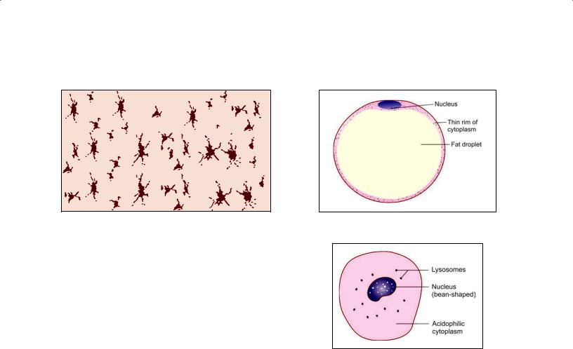
Fig. 5.8: Pigment cells (Schematic representation) |
Fig. 5.9: Fat cell (Schematic representation) |
Of the many cells that contain pigment in their cyto plasm only a few are actually capable of synthesizing melanin. Such cells, called melanocytes, are of neural crest origin.
The remaining cells are those that have engulfed pig ment released by other cells. Typically, such cells are star shaped (stellate) with long branching processes. In contrast to melanocytes such cells are called chromatophores or melanophores. They are probably modified fibroblasts.
Pigment cells prevent light from reaching other cells. The importance of this function in relation to the eyeball is obvious. Pigment cells in the skin protect deeper tissues from the effects of light (specially ultraviolet light). The darker skin of races living in tropical climates is an obvious adaptation for this purpose.
Fat Cells (Adipocytes)
Although some amount of fat (lipids) may be present in the cytoplasm of many cells, including fibroblasts, some cells store fat in large amounts and become distended with it (Fig. 5.9). These are called fat cells, adipocytes, or lipo cytes. Aggregations of fat cells constitute adipose tissue.
Each fat cell contains a large droplet of fat that almost fills it (Fig. 5.9). As a result the cell becomes rounded (when several fat cells are closely packed they become polygonal because of mutual pressure).
The cytoplasm of the cell forms a thin layer just deep to the plasma membrane.
The nucleus is pushed against the plasma membrane and is flattened resembling a signet ring. Adipocytes are incapable of division.
Macrophage Cells
Macrophage cells of connective tissue are part of a large series of cells present in the body that have similar func tions. These collectively form the mononuclear phagocyte system.
Fig. 5.10: Macrophage cell (histiocyte) (Schematic representation)
Macrophage cells of connective tissue are also called histiocytes or clasmatocytes (Fig. 5.10). They have the ability to phagocytose (eat up) unwanted material. Such material is usually organic: it includes bacteria invading the tissue, and damaged tissues. Macrophages also phago cytose inorganic particles injected into the body (e.g. India ink).
In ordinary preparations of tissue it is difficult to dis tinguish macrophages from other cells. However, if an animal is injected with India ink (or trypan blue, or lithium carmine) particles of it are taken up into the cytoplasm of macrophages, thus making them easy to recognize.
Macrophages are usually described as “fixed” when they are attached to fibers; or as motile (or “free”). Fixed macro phages resemble fibroblasts in appearance, but free macro phages are rounded. However, all macrophages are capable of becoming mobile when suitably stimulated. The nuclei of macrophages are smaller, and stain more intensely than those of fibroblasts. They are often kidney shaped (Fig. 5.10). With the EM the cytoplasm is seen to contain numerous lysosomes that help in “digesting” material phagocytosed by the macrophage. Sometimes macrophages may fuse together to form multinucleated giant cells.
Apart from direct phagocytic activity, macrophages play an important role in immunological mechanisms.
Chapter 5 General Connective Tissue 47
|
|
|
|
|
|
|
|
|
A |
B |
|||
|
|
|
Figs. 5.12A and B: Schematic representation |
|||
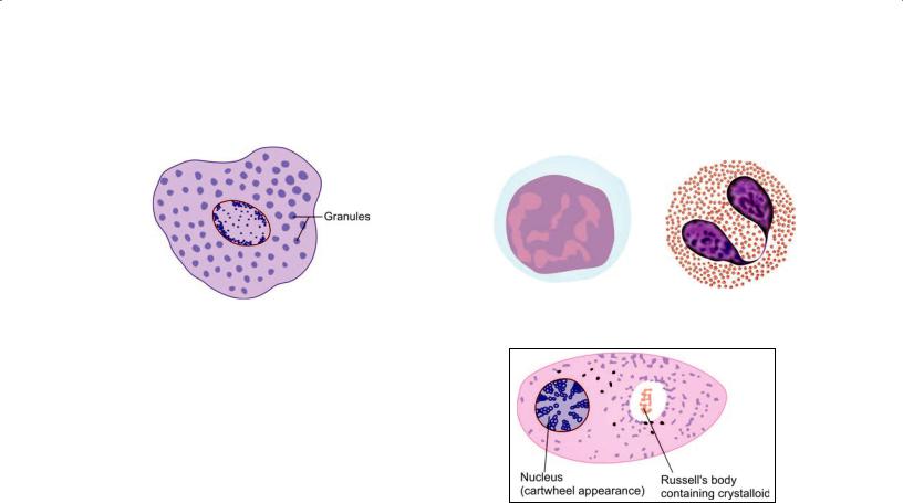
Fig. 5.11: Mast cell (Schematic representation) |
||||||
(A) Lymphocyte; (B) Eosinophil |
||||||
|
|
|
||||
Mast Cells
These are small round or oval cells (mastocytes or histami nocytes) (Fig. 5.11). The nucleus is small and centrally placed. Irregular microvilli (filopodia) are present on the cell surface.
The distinguishing feature of these cells is the presence of numerous granules in the cytoplasm. The granules can be demonstrated with the PAS stain. They also stain with dyes like toluidine blue or alcian blue: with them the nuclei stain blue, but the granules stain purple to red (When components of a cell or tissue stain in a color different from that of the dye used, the staining is said to be metachro matic). On the basis of the staining reactions the granules are known to contain acid mucopolysaccharides.
With the EM the “granules” are seen to be vesicles, each of which is surrounded by a membrane (in other words they are membrane bound vesicles).
The granules contain various substances. The most important of these is histamine. Release of histamine is associated with the production of allergic reactions when a tissue is exposed to an antigen to which it is sensitive (because of previous exposure).
Apart from histamine mast cells may contain various enzymes, and factors that attract eosinophils or neut rophils.
Mast cells differ considerably in size and in number from species to species, and at different sites in the same animal.
They are most frequently seen around blood vessels and nerves. Mast cells are probably related in their origin to basophils of blood. They may represent modified basophil cells.
Lymphocytes (Fig. 5.12A)
Lymphocytes represent one variety of leukocytes (white blood cells) present in blood. Large aggregations of lympho cytes are present in lymphoid tissues. They reach connec tive tissue from these sources, and are specially numerous when the tissue undergoes inflammation.
Fig. 5.13: Plasma cell (Schematic representation)
Lymphocytes play an important role in defense of the body against invasion by bacteria and other organisms. They have the ability to recognize substances that are foreign to the host body; and to destroy these invaders by producing antibodies against them.
The lymphocytes are derived from stem cells present in bone marrow. They are of two types. Blymphocytes pass through blood to reach other tissues directly. Some Blymphocytes mature into plasma cells described below. The second type of lymphocytes, called Tlymp hocytes, travel (through blood) from bone marrow to the thymus. After undergoing a process of maturation in this organ they again enter the bloodstream to reach other tissues. Both Blymphocytes and Tlymphocytes can be seen in connective tissue.
Other Leukocytes
Apart from lymphocytes two other types of leukocytes may be seen in connective tissue. Monocytes are closely related in function to macrophages. Eosinophils (so called because of the presence of eosinophilic granules in the cytoplasm) are found in the connective tissue of many organs (Fig. 5.12B). They increase in number in allergic disorders.
Plasma Cells or Plasmatocytes
A plasma cell is seen to be small and round basophilic cyto plasm (Fig. 5.13). It can be recognized by the fact that the chromatin in its nucleus forms four or five clumps near