

Human_Histology
.pdf148 Textbook of Human Histology
Fig. 12.10: Precapillary sphincters and thoroughfare channels
(Schematic representation)
THE HEART
The heart is a muscular organ that pumps blood throughout the blood vessels to various parts of the body by repeated rhythmic contractions.
Structure
There are three layers in the wall of the heart:
The innermost layer is called the endocardium. It corresponds to the tunica intima of blood vessels. It consists of a layer of endothelium that rests on a thin layer of delicate connective tissue. Outside this there is a thicker subendocardial layer of connective tissue.
The main thickness of the wall of the heart is formed by a thick layer of cardiac muscle. This is the myocardium.
The external surface of the myocardium is covered by the epicardium (or visceral layer of serous pericar dium). It consists of a layer of connective tissue that is covered, on the free surface, by a layer of flattened mesothelial cells.
Added Information
Atrial myocardial fibers secrete a natriuretic hormone when they are excessively stretched (as in some diseases). The hormone increases renal excretion of water, sodium, and potassium. It inhibits the secretion of renin (by the kidneys), and of aldosterone (by the adrenal glands) thus reducing blood pressure.
At the junction of the atria and ventricles, and around the openings of large blood vessels there are rings of dense fibrous tissue. Similar dense fibrous tissue is also present in the interventricular septum. These masses of dense fibrous tissue constitute the “skeleton” of the heart. They give attachment to fasciculi of heart muscle.
The valves of the heart are folds of endocardium that enclose a plate like layer of dense fibrous tissue.
Conducting System of the Heart
Conducting system of the heart is made up of a special kind of cardiac muscle. The Purkinje fibers of this system are chains of cells. The cells are united by desmosomes. Intercalated discs are absent. These cells have a larger diameter, and are shorter, than typical cardiac myocytes. Typically each cell making up a Purkinje fiber has a central nucleus surrounded by clear cytoplasm containing abun dant glycogen. Myofibrils are inconspicuous and are confined to the periphery of the fibers. Mitochondria are numerous and the sarcoplasmic reticulum is prominent. Nodal myocytes [present in the atrioventricular (AV) node and the sinoatrial (SA) node] are narrow, rounded, cylindrical or polygonal cells with single nuclei. They are responsible for pacemaker functions. Transitional myo cytes are present in the nodes, and in the stem and main branches of the AV bundle. They are similar to cardiac myo cytes except that they are narrower. Conduction through them is slow.
In the SA node and the AV node the muscle fibers are embedded in a prominent stroma of connective tissue. This tissue contains many blood vessels and nerve fibers.

Chapter
13
The Respiratory System
The respiratory system consists of:
Respiratory part that includes the lungs
Conducting part that includes the nasal cavities, the pharynx, the trachea, the bronchi and their intrapulmo nary continuations.
The conducting part is responsible for providing passage of air and conditioning the inspired air. The respiratory part is involved in the exchange of oxygen and carbon dio xide between blood and inspired air.
COMMON FEATURES OF AIR PASSAGES
The passages in the conducting part have some features in common. Their walls have a skeletal basis made up variably of bone, cartilage, and connective tissue. The skeletal basis keeps the passages always patent. Smooth muscle present in the walls of the trachea and bronchi enables some alte rations in the size of the lumen. The interior of the passages is lined over most of its extent by pseudostratified, ciliated and columnar epithelium. The epithelium is kept moist by the secretions of numerous serous glands. Numerous goblet cells and mucous glands cover the epithelium with a protective mucoid secretion that serves to trap dust parti cles present in inhaled air. This mucous (along with the dust particles in it) is constantly moved toward the pharynx by action of cilia. When excessive mucous accumulates it is brought out by coughing, or is swallowed. Deep to the mucosa there are numerous blood vessels that serve to warm the inspired air.
THE NASAL CAVITIES
The nasal cavity is the beginning of the respiratory system. These are paired chambers separated by septum. It extends from the nostrils in front to the posterior nasal apertures behind. Each nasal cavity is a hollow organ composed of bone, cartilage and connective tissue covered by mucous membrane.
Histologically, the wall of each half of the nasal cavity is divisible into three distinct regions.
Vestibule
Olfactory mucosa
Respiratory mucosa
Vestibule
It is the anterior dilated part of the nasal cavity. The vesti bule is lined by skin continuous with that on the exterior of the nose. Hair and sebaceous glands are present.
Olfactory Mucosa
Apart from their respiratory function the nasal cavities serve as end organs for smell. Receptors for smell are located in the olfactory mucosa which is confined to a relatively small area on the superior nasal concha, and on the adjoin ing part of the nasal septum.
Olfactory mucosa is yellow in color, in contrast to the pink color of the respiratory mucosa. It is responsible for the sense of smell. It consists of a lining epithelium and a lamina propria.
Olfactory Epithelium
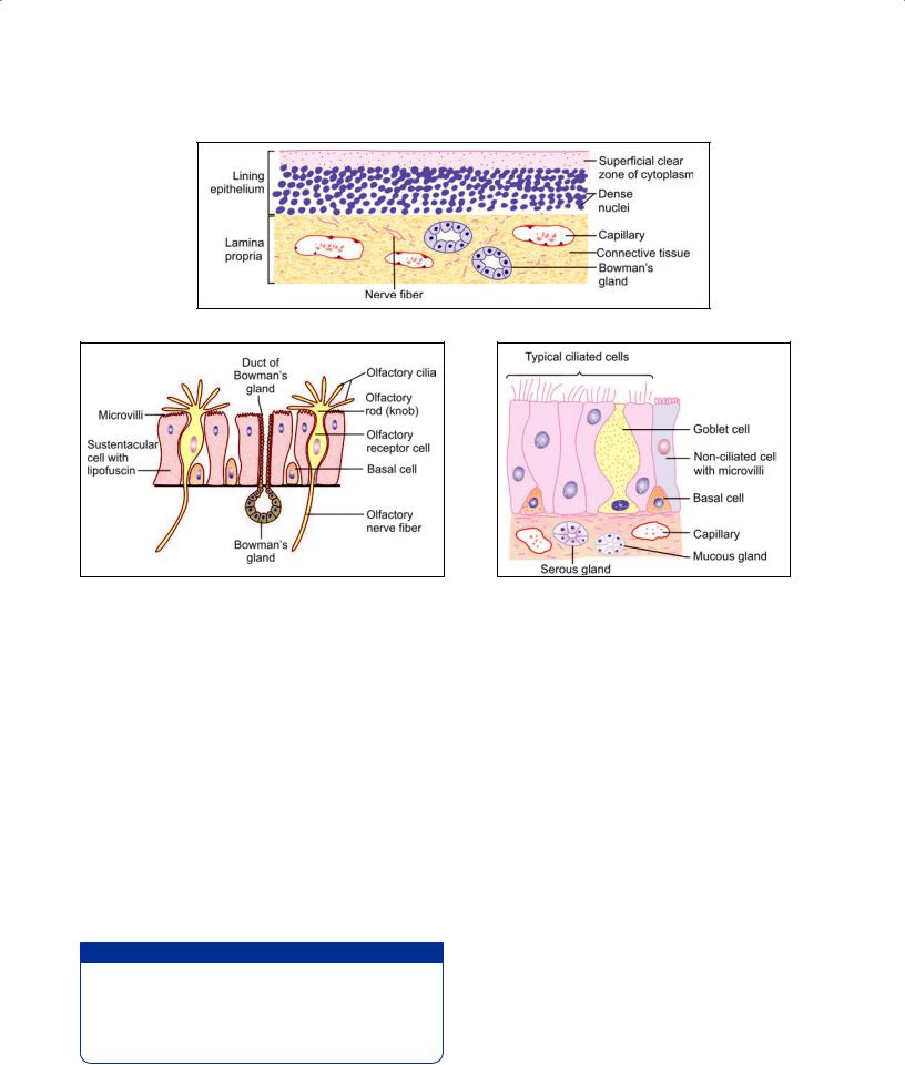
The olfactory epithelium is pseudostratified. It is much thicker than the epithelium lining the respiratory mucosa (about 100 µm). Within the epithelium there is a superficial zone of clear cytoplasm below which there are several rows of nuclei (Fig. 13.1). Using special methods three types of cells can be recognized in the epithelium (Fig. 13.2).
The olfactory cells are modified neurons. Each cell has a central part containing a rounded nucleus. Two pro cesses, distal and proximal, arise from this central part. The distal process (representing the dendrite) passes toward the surface of the olfactory epithelium. It ends in a thickening (called the rod or knob) from which a number of nonmotile olfactory cilia arise and project into a layer of fluid covering the epithelium. (Some of them pass laterally in between the microvilli of adja cent sustentacular cells). The proximal process of each olfactory cell represents the axon. It passes into the
150 Textbook of Human Histology
Fig. 13.1: Olfactory mucosa seen in section stained by routine methods (Schematic representation)
Fig. 13.2: Cells to be seen in olfactory epithelium |
Fig. 13.3: Structure of respiratory part of |
(Schematic representation) |
nasal mucosa (Schematic representation) |
subjacent connective tissue where it forms one fiber of the olfactory nerve. The nuclei of olfactory cells lie at various levels in the basal twothirds of the epithelium.
The sustentacular cells support the olfactory cells. Their nuclei are oval, and lie near the free surface of the epithelium. The free surface of each cell bears numerous microvilli (embedded in overlying mucous). The cytoplasm contains yellow pigment (lipofuscin) that gives olfactory mucosa its yellow color. In addition to their supporting function sustentacular cells may be phagocytic, and the pigment in them may represent remnants of phagocytosed olfactory cells.
The basal cells lie deep in the epithelium and do not reach the luminal surface. They divide to form new olfactory cells to replace those that die. Some basal cells have a supporting function.
Added Information
In vertebrates, olfactory cells are unique in being the only neurons that have cell bodies located in an epithelium.
Olfactory cells are believed to have a short life. Dead olfactory cells are replaced by new cells produced by division of basal cells. This is the only example of regeneration of neurons in mammals.
Lamina Propria
The lamina propria, lying deep to the olfactory epithelium consistsofconnectivetissuewithinwhichbloodcapillaries, lymphatic capillaries and olfactory nerve bundles are present. It also contains serous glands (of Bowman) the secretions of which constantly “wash” the surface of the olfactory epithelium. This fluid may help in transferring smell carrying substances from air to receptors on olfactory cells. The fluid may also offer protection against bacteria.
Respiratory Mucosa
The rest of the wall of each half of the nasal cavity is covered by respiratory mucosa lined by pseudostratified ciliated columnar epithelium.
This mucosa is lined by a pseudostratified ciliated colu mnar epithelium resting on a basal lamina. In the epithe lium, the following cells are present (Fig. 13.3):
Ciliated cells are the columnar cells with cilia on their free surfaces and are the most abundant cell type.
Goblet cells (flaskshaped cells) scattered in the epithelium produce mucous.
Nonciliated columnar cells with microvilli on the free surface probably secrete a serous fluid that keeps the mucosa moist.

Basal cells lying near the basal lamina probably give rise to ciliated cells to replace those lost.
At places the respiratory mucosa may be lined by a
simple ciliated columnar epithelium, or even a cuboidal epithelium.
Deep to the basal lamina supporting the epithelium lining, the mucosa contains a layer of fibrous tissue, through which the mucosa is firmly connected to underlying periosteum or perichondrium. The fibrous tissue may con tain numerous lymphocytes. It also contains mucous and serous glands that open on to the mucosal surface. Some serous cells contain basophilic granules, and probably secrete amylase. Others with eosinophilic granules pro duce lysozyme.
The deeper parts of the mucosa contain a rich capillary network that constitutes a cavernous tissue. Blood flow ing through the network warms inspired air. Variations in blood flow can cause swelling or shrinkage of the mucosa.
Respiratory mucosa also lines the paranasal air sinuses. Here it is closely bound to underlying periosteum forming a mucoperiosteum.
Lamina Propria
The lamina propria of nasal mucosa contains lymphocytes, plasma cells, macrophages, a few neutrophils and eosino phils. Eosinophils increase greatly in number in persons suffering from allergic rhinitis.
Clinical Correlation
Acute Rhinitis (Common Cold): Acute rhinitis or common cold is the common inflammatory disorder of the nasal cavities that may extend into the nasal sinuses. It begins with rhinorrhea, nasal obstruction and sneezing. Initially, the nasal discharge is watery, but later it becomes thick and purulent.
Nasal Polyps: Nasal polyps are common and are pedunculated grape-like masses of tissue. They are the end-result of prolonged chronic inflammation causing polypoid thickening of the mucosa.Theymaybeallergicorinflammatory.Theyarefrequently bilateral and the middle turbinate is the common site.
THE PHARYNX
The pharynx consists of nasal, oral, and laryngeal parts. The nasal part is purely respiratory in function, but the oral and laryngeal parts are more intimately concerned with the alimentary system. The wall of the pharynx is fibromuscular.
Epithelium
Inthenasopharynxtheepithelialliningisciliatedcolumnar, or pseudostratified ciliated columnar. Over the inferior
Chapter 13 The Respiratory System 151
surface of the soft palate, and over the oropharynx and laryngopharynx the epithelium is stratified squamous (as these parts come in contact with food during swallowing).
Lymphoid Tissue
Subepithelial aggregations of lymphoid tissue are present specially on the posterior wall of the nasopharynx, and around the orifices of the auditory tubes, forming the nasopharyngeal and tubal tonsils. The palatine tonsils are present in relation to the oropharynx.
Submucosa
Numerous mucous glands are present in the submucosa, including that of the soft palate.
Clinical Correlation
Ludwig’s Angina: This is a severe, acute streptococcalcellulitis involvingtheneck,tongue,andbackofthethroat.Thecondition was more common in the pre-antibiotic era as a complication of compound fracture of the mandible and periapical infection of the molars. The condition often proves fatal due to glottic edema, asphyxia, and severe toxaemia.
Diphtheria: Diphtheria is an acute communicable disease caused by Corynebacterium diphtheriae. It usually occurs in children and results in the formation of a yellowish-grey pseudomembraneinthemucosaofnasopharynx,oropharynx,tonsils,larynx, and trachea.
Tonsillitis: Tonsillitis caused by staphylococci or streptococci may be acute or chronic. Acute tonsillitis is characterized by enlargement, redness and inflammation. Acute tonsillitis may progress to acute follicular tonsillitis in which crypts are filled with debris and pus giving it follicular appearance. Chronic tonsillitis is caused by repeated attacks of acute tonsillitis in which case the tonsils are small and fibrosed. Acute tonsillitis may pass on to tissues adjacent to tonsils to form peritonsillar abscess or quinsy.
THE LARYNX
Larynx is a specialized organ responsible for production of voice. It houses the vocal cords. The wall of the larynx has a complex structure made up of a number of cartilages, membranes, and muscles.
Mucous Membrane
The epithelium lining the mucous membrane of the larynx is predominantly pseudostratified ciliated colum nar. However, over some parts that come in contact with swallowed food the epithelium is stratified squamous. These parts include the epiglottis (anterior surface and upper part of the posterior surface), and the upper parts of the aryepiglottic folds. The vocal folds do not come in
152Textbook of Human Histology
contact with swallowed food, but their lining epithelium is exposed to considerable stress during vibration of the folds. These folds are also covered with stratified squamous epithelium.
Numerous goblet cells and subepithelial mucous glands provide a mucous covering to the epithelium. Mucous glands are specially numerous over the epiglottis; in the lower part of the aryepiglottic folds (where they are called arytenoid glands); and in the saccule. The glands in the saccule provide lubrication to the vocal folds. Serous glands and lymphoid tissue are also present.
EM studies have shown that epithelial cells lining the vocal folds bear microvilli and ridgelike foldings of the surface plasma membrane (called microplicae). It is believed that these help to retain fluid on the surface of the cells keeping them moist.
Added Information
The connective tissue subjacent to the epithelial lining of vocal folds is devoid of lymph vessels. This factor slows down lymphatic spread of cancer arising in the epithelium of the vocal folds.
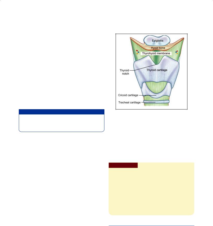
Cartilages of the Larynx
The larynx has a cartilaginous framework which is made of nine cartilages (3 paired and 3 unpaired) that are connected to each other by membranes and ligaments (Fig. 13.4). The cartilages are either hyaline or elastic in nature. These are:
Hyaline cartilages
Thyroid (unpaired)Cricoid (unpaired)Arytenoid (paired)
Elastic cartilages
Epiglottis (unpaired)Cuneiform (paired)Corniculate (paired)
With advancing age, calcification may occur in hyaline cartilage, but not in elastic cartilage.
The Epiglottis
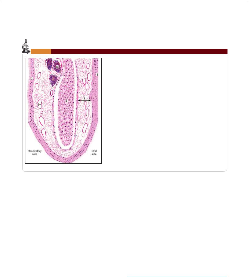
The epiglottis is considered separately because sections through it are usually included in sets of class slides. The epiglottis has a central core of elastic cartilage. Overlying the cartilage there is mucous membrane. The greater part of the mucous membrane is lined by stratified squamous epithelium (nonkeratinizing). The mucous membrane over the lower part of the posterior surface of the epiglottis
Fig. 13.4: Anterior view of the larynx (Schematic representation)
is lined by pseudostratified ciliated columnar epithelium (Plate 13.1). This part of the epiglottis does not come in contact with swallowed food as it is overlapped by the aryepiglottic folds. Some taste buds are present in the epithelium of the epiglottis. (A few taste buds may be seen in the epithelium elsewhere in the larynx).
Numerous glands, predominantly mucous, are present in the mucosa deep to the epithelium. Some of them lie in depressions present on the epiglottic cartilage.
Clinical Correlation
Acute Laryngitis: Thismayoccurasapartoftheupperorlower respiratory tract infection. Atmospheric pollutants like cigarette smoke, exhaust fumes, industrial and domestic smoke, etc, predispose the larynx to acute bacterial and viral infections. Streptococci and H. influenzae cause acute epiglottitis which may be life-threatening.
Chronic Laryngitis: Chronic laryngitis may occur from repeated attacks of acute inflammation, excessive smoking, chronic alcoholism or vocal abuse. The surface is granular due to swollen mucous glands. There may be extensive squamous metaplasia due to heavy smoking, chronic bronchitis, and atmospheric pollution.
THE TRACHEA AND PRINCIPAL BRONCHI
Trachea
The trachea is a fibroelastic cartilaginous tube. It extends from the lower border of cricoid cartilage (C6) to its level of bifurcation (T4) into right and left bronchi. The trachea consists of four layers (Plate 13.2).
Chapter 13 The Respiratory System 153
Plate 13.1: Epiglottis
The surface of epiglottis is covered on oral side by stratified squamous epithelium and on respiratory side by pseudostratified ciliated columnar epithelium
The core of the epiglottis is made up of a plate of elastic cartilage covered byconnectivetissueinwhichtherearenumerousbloodvesselsandglands.
Note: This figure shows the appearance of elastic cartilage when stained by hematoxylin and eosin.
Key
1.Stratified squamous epithelium
2.Pseudostratified ciliated columnar epithelium
3.Connective tissue
4.Elastic cartilage
G. Gland
Bv. Blood vessel
Epiglottis. As seen in drawing
Mucosa
The lumen of the trachea is lined by mucous membrane that consists of a lining epithelium and an underlying layer of connective tissue. The lining epithelium is pseudostrati fied ciliated columnar. It contains numerous goblet cells, and basal cells that lie next to the basement membrane. Numerous lymphocytes are seen in deeper parts of the epithelium.
Submucosa
The subepithelial connective tissue contains numerous elastic fibers. It contains serous glands that keep the epithe lium moist; and mucous glands that provide a covering of mucous in which dust particles get caught. The mucous is continuously moved toward the larynx by ciliary action. Numerous aggregations of lymphoid tissue are present in the subepithelial connective tissue. Eosinophil leucocytes are also present.
Cartilage and Smooth Muscle Layer
Occasionally, adjoining cartilages may partly fuse with each other or may have Yshaped ends. The intervals between the cartilages are filled by fibrous tissue that becomes continuous with the perichondrium covering the cartilages. The gaps between the cartilage ends, present on the poste rior aspect, are filled in by smooth muscle and fibrous tissue. The connective tissue in the wall of the trachea con tains many elastic fibers.
Adventitia
It is made of fibroelastic connective tissue containing blood vessels and nerves.
Principal Bronchi
The trachea divides at the level of T4 into right and left principal bronchi (primary or main bronchi). They have a structure similar to that of the trachea.
THE LUNGS
The skeletal basis of the trachea is made up of 16 to 20 trac heal cartilages. Each of these is a Cshaped mass of hyaline cartilage. The open end of the “C” is directed posteriorly.
The lungs are the principal respiratory organs that are situated one on either side of mediastinum in the thoracic cavity. They are covered by visceral pleura (Plate 13.3).
154 Textbook of Human Histology
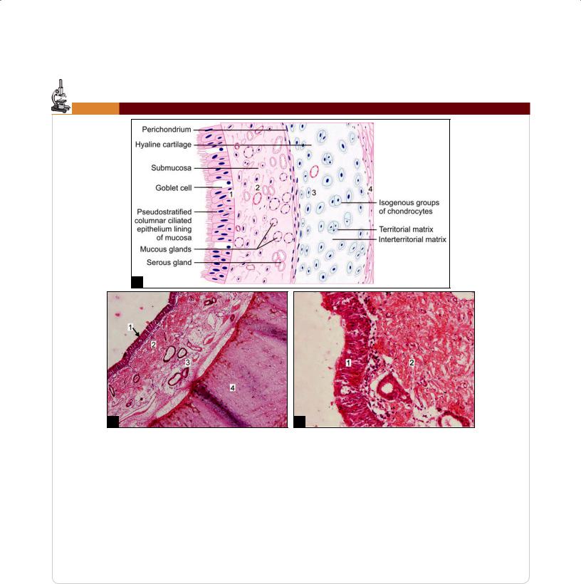
Plate 13.2: Trachea
A
B |
C |
Trachea. A. As seen in drawing; B. Photomicrograph (low magnification);
C. Photomicrograph (high magnification)
From within outwards the wall of trachea consists of:
Mucosa formed by pseudostratified ciliated columnar epithelium with goblet cells and the underlying lamina propria
Submucosa made up of loose connective tissue containing mucous glands and serous glands, blood vessels, and ducts
A “C” shaped plate of hyaline cartilage. Perichondrium has outer fibrous and inner chondrogenic layers. Chondrocytes increase in size from periphery to center. They may appear as isogenous groups surrounded by darkly stained territorial matrix
Adventitia consisting of collagen fibers (Not shown here).
Key
1.Pseudostratified ciliated columnar epithelium with goblet cells}Mucosa
2.Lamina propria
3.Submucosa
4.Hyaline cartilage
The structure of the lungs has to be understood keeping in mind their function of oxygenation of blood. The follow ing features are essential for this purpose.
A surface at which air (containing oxygen) can be brought into close contact with circulating blood. The barrier between air and blood has to be very thin to allow oxygen (and carbon dioxide) to pass through it.
The surface has to be extensive enough to meet the oxygen requirements of the body.
A system of tubes to convey air to and away from the surface at which exchanges take place.
A rich network of blood capillaries present in intimate relationship to the surface at which exchanges take place.
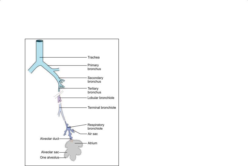
Fig. 13.5: Some terms used to describe the terminal ramifications of
the bronchial tree (Schematic representation)
Intrapulmonary Passages
On entering the lung the principal bronchus divides into secondary, or lobar bronchi (one for each lobe). Each lobar bronchus divides into tertiary, or segmental bronchi (one for each segment of the lobe). The segmental bronchi divide into smaller and smaller bronchi, which ultimately end in bronchioles.
The lung substance is divided into numerous lobules each of which receives a lobular bronchiole. The lobular bronchiole gives off a number of terminal bronchioles (Fig. 13.5). As indicated by their name the terminal bron chioles represent the most distal parts of the conducting passage.
Each terminal bronchiole ends by dividing into respi ratory bronchioles. These are so called because they are partly respiratory in function as some air sacs (see below) arise from them.
Each respiratory bronchiole ends by dividing into a few alveolar ducts. Each alveolar duct ends in a passage, the atrium, which leads into a number of rounded alveolar sacs. Each alveolar sac is studded with a number of air sacs or alveoli.
Chapter 13 The Respiratory System 155
The alveoli are blind sacs having very thin walls through which oxygen passes from air into blood, and carbon dioxide passes from blood into air.
The structure of the larger intrapulmonary bronchi is similar to that of the trachea. As these bronchi divide into smaller ones the following changes in structure are observed.
The cartilages in the walls of the bronchi become irregular in shape, and are progressively smaller. Cartilage is absent in the walls of bronchioles: this is the criterion that distinguishes a bronchiole from a bronchus.
The amount of muscle in the bronchial wall increases as the bronchi become smaller. The presence of muscle in the walls of bronchi is of considerable clinical signi ficance. Spasm of this muscle constricts the bronchi and can cause difficulty in breathing.
Subepithelial lymphoid tissue increases in quantity as bronchi become smaller. Glands become fewer, and are absent in the walls of bronchioles.
The trachea and larger bronchi are lined by pseudo stratified ciliated columnar epithelium. As the bronchi become smaller the epithelium first becomes simple ciliated columnar, then nonciliated columnar, and finally cuboidal (in respiratory bronchioles). The cells contain
lysosomes and numerous mitochondria. Plate 13.3 illustrates the salient microscopic features of the lung parenchyma.
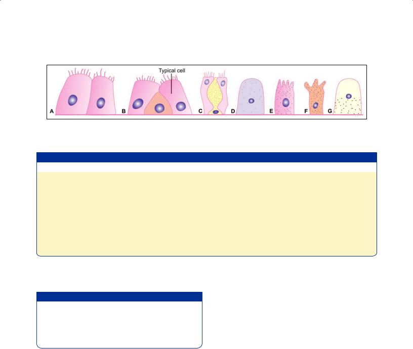
EM studies have shown that apart from typical ciliated columnar cells, various other types of cells are to be seen in the epithelium lining the air passages. Some of the cells encountered are as follows (Fig. 13.6):
Goblet cells are numerous. They provide mucous which helps to trap dust entering the passages and is moved by ciliary action toward the larynx and pharynx.
Nonciliated serous cells secrete fluid that keeps the epithelium moist.
Basal cells multiply and transform into other cell types to replace those that are lost.
Some nonciliated cells present predominantly in ter minal bronchioles (see below) produce a secretion that spreads over the alveolar cells forming a film that reduces surface tension. These include the cells of Clara.
Cells similar to diffuse endocrine cells of the gut, and containing argyrophil granules are present. They secrete hormones and active peptides including serotonin and bombesin.
Lymphocytes and other leucocytes may be present in the epithelium. They migrate into the epithelium from surrounding tissues.
156 Textbook of Human Histology
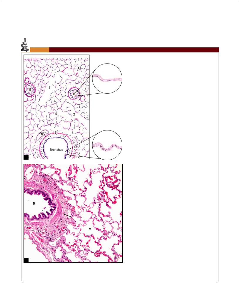
Plate 13.3: Lung
Thelungsurfaceiscoveredbypleura.Itconsistsofalining of mesothelium resting on a layer of connective tissueThe lung parenchyma is made up of numerous thin-
walled spaces or alveoli
The alveoli give a honey comb appearance and are lined by flattened squamous cells. They are filled with air
Theintrapulmonarybronchusislinedbypseudostratified ciliated columnar epithelium with few goblet cells. Its structureissimilartotrachea,i.e.ithassmoothmuscles, cartilage and glands present in its wall
The bronchiole is lined by simple columnar or cuboidal epitheliumsurroundedbybundlesofsmoothmusclecells (see arrow in photomicrograph)
Bronchiolessubdivideandwhentheirdiameterisapproximately 1 mm or less, they are called terminal bronchiole
Arteriesareseennearthebronchioles
Respiratory bronchiole, alveolar duct and atrium are also present
This slide shows a medium size bronchiole surrounded by alveoli.
A
Key
1. Mesothelium resting on connective tissue
2. Respiratory bronchiole
3. Alveolar duct
4. Atrium
5. Smooth muscle
6. Plates of cartilage
7. Glands
A. Alveoli
B. Bronchus bronchiole
B
Lung. A. As seen in drawing; B. Photomicrograph.
Courtesy: Atlas of Histopathology, 1st Edition. Ivan Damjanov.
Jaypee Brothers. 2012. p37
Chapter 13 The Respiratory System 157
Fig. 13.6: Various types of cells to be seen lining the respiratory passages.
(A) Typical ciliated columnar, (B) Basal, (C) Goblet, (D) Serous, (E) Brush, (F) Clara, (G) Argyrophil (Schematic representation)
Table 13.1: Differences between Bronchus and Bronchiole
Characteristics |
Bronchus |
Bronchiole |
|
Diameter |
Larger diameter (more than 1 mm) |
Smaller diameter (less than 1 mm) |
|
Lining epithelium |
Pseudostratified ciliated columnar epithelium with |
• |
Large size bronchioles: simple columnar cells with |
|
goblet cells |
|
few cilia and few goblet cells |
|
|
• |
Small size bronchioles: simple columnar or simple |
|
|
|
cuboidal cells with no cilia or no goblet cells |
Smooth muscle layer |
Present between mucosa and cartilage layer |
Smooth muscles and elastic fibers form a well-defined |
|
|
|
layer beneath mucosa |
|
Cartilage |
Present in irregular patches |
Absent |
|
|
|
|
|
Glands in |
Both serous and mucous acini present between |
Absent |
|
submucosa |
cartilage and muscle layer |
|
|
The differences between bronchus and bronchioles are given in Table 13.1.
Added Information
Some other functions attributed to cells of Clara include:Protection against harmful substances that are inhaled.Protection against development of emphysema by
opposing the action of substances (proteases) that tend to destroy walls of lung alveoli.
Stem cell function.
Alveoli
There are about 200 million alveoli in a normal lung. The total area of the alveolar surface of each lung is extensive. It has been estimated to be about 75 square meters. The total capillary surface area available for gaseous exchanges is about 125 square meters.
Structure of Alveolar Wall
Each alveolus has a very thin wall. The wall is lined by an epithelium consisting mainly of flattened squamous cells. The epithelium rests on a basement membrane. Deep to the basement membrane there is a layer of delicate con nective tissue through which pulmonary capillaries run. These capillaries have the usual endothelial lining that rests on a basement membrane.
The barrier between air and blood is made up of the epithelial cells and their basement membrane; by endo thelial cells and their basement membrane; and by inter vening connective tissue. At many places the two basement membranes fuse greatly reducing the thickness of the barrier.
The endothelial cells lining the alveolar capillaries are remarkable for their extreme thinness. With the EM they are seen to have numerous projections extending into the capillary lumen. These projections greatly increase the surface of the cell membrane that is exposed to blood and is, therefore, available for exchange of gases. At many places the basement membrane of the endothelium fuses with that of the alveolar epithelium greatly reducing the thickness of the barrier between blood and air in alveoli.
Pneumocytes
EM studies have shown that the cells forming the lining epithelium of alveoli (pneumocytes) are of various types (Fig. 13.7).
The most numerous cells are the squamous cells already referred to. They are called type I alveolar epithelial cells. Except in the region of the nucleus, these cells are reduced to a very thin layer (0.05 to 0.2 µm). The edges of adjoining cells overlap and are united by tight