

Human_Histology
.pdf108 Textbook of Human Histology
A B
Figs. 10.5A and B: ! / 0 1 %
Added Information
Axon hillock and initial segment:
( %% %% ( % %% % #) # %
! #%( + %,- $ # % )-,, .,, %
a
Myelinated and Unmyelinated Axons
Axon may be myelinated or unmyelinated. Axons having a myelin sheath are called myelinated axons.
Myelin Sheath
Myelin sheath when present is seen outside the axolemma.
The cells providing this sheath for axons lying outside in the peripheral nervous system are called Schwann cells. Axons lying within the central nervous system are provided a similar covering by a kind of neuroglial cell called an oligodendrocyte.
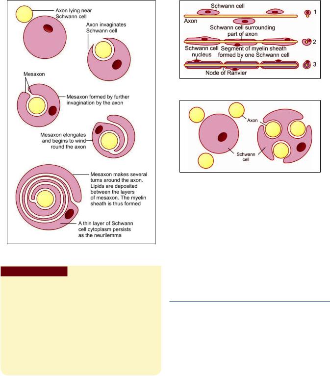
The nature of this sheath is best understood by considering the mode of its formation. An axon lying near a Schwann cell invaginates into the cytoplasm of the Schwann cell. In this process the axon comes to be suspended by a fold of the cell membrane of the Schwann
cell: this fold is called the mesaxon (Figs. 10.5A and B). In some situations the mesaxon becomes greatly elongated and comes to be spirally wound around the axon, which is thus surrounded by several layers of cell membrane. Lipids are deposited between adjacent layers of the membrane. These layers of the mesaxon, along with the lipids, form the myelin sheath. Outside the myelin sheath a thin layer of Schwann cell cytoplasm persists to form an additional sheath that is called the neurilemma (also called the neurilemmal sheath or Schwann cell sheath).
The presence of a myelin sheath increases the velocity of conduction (for a nerve fiber of the same diameter). It also reduces the energy expended in the process of conduction.
An axon is related to a large number of Schwann cells over its length (Fig. 10.6). Each Schwann cell provides the myelin sheath for a short segment of the axon. At the junction of any two such segments there is a short gap in the myelin sheath. These gaps are called the nodes of Ranvier.
The nodes of Ranvier have great physiological importance. When an impulse travels down a nerve fiber it does not proceed uniformly along the length of the axis cylinder, but jumps from one node to the next. This is called saltatory conduction (In unmyelinated neurons the impulse travels along the axolemma. Such conduction is much slower than saltatory conduction and consumes more energy).
The segment of myelin sheath between two nodes of Ranvier is called internode.
Composition of Myelin Sheath
Myelin contains protein, lipids, and water. The main lipids present include cholesterol, phospholipids, and glycosphingolipids. Other lipids are present in smaller amounts.
Chapter 10 Nervous System 109
Fig. 10.6:%
Pathological Correlation
Myelination can be seriously impaired, and there can be abnormal collections of lipids, in disorders of lipid metabolism. Various in them can be the basis of some neuropathies.
The composition and structure of myelin sheaths formed by oligodendrocytes show differences from those formed by Schwann cells. The two are different in protein content and can be distinguished by immunocytochemical methods. As damage to neurons within the CNS is not followed by regeneration, oligodendrocytes have no role to play in this respect. Also note that, in multiple sclerosis, myelin formed by oligodendrocytes undergoes degeneration, but that derived from Schwann cells is spared.
Functions of the Myelin Sheath
The presence of a myelin sheath increases the velocity of conduction (for a nerve fiber of the same diameter)
Fig. 10.7:2 %
Fig. 10.8:3
It reduces the energy expended in the process of conduction
It is responsible for the color of the white matter of the brain and spinal cord.
Unmyelinated Axons
There are some axons that are devoid of myelin sheaths. These unmyelinated axons invaginate into the cytoplasm of Schwann cells, but the mesaxon does not spiral around them (Figs. 10.7 and 10.8). Another difference is that several such axons may invaginate into the cytoplasm of a single Schwann cell (Table 10.2).
TYPES OF NEURONS (TABLE 10.3)
On the basis of number of processes
Unipolar neurons: These neurons have single process (which is highly convoluted). After a very short course, this process divides into two. One of the divisions represents the axon; the other is functionally a dendrite, but its structure is indistinguishable from that of an axon.
E.g. Neurons in dorsal root ganglion (Fig. 10.9).
Bipolar neurons: These neurons have only one axon and one dendrite.
E.g. Neurons in vestibular and spiral ganglia.
Multipolar neurons: It is most common type of neurons; the neuron gives off several processes, i.e.

110 Textbook of Human Histology
Myelinated nerves |
Unmyelinated nerves |
! % |
4 |
|
|
|
|
|
|
* |
% |
|
|
|
5 |
% |
% |
|
|
! "
Morphology |
Location and example |
|
% |
" % % & % % |
|
6 |
4 7 |
0 & % % |
6 |
0 |
! % |
6 |
! |
|
% 8 |
" ( * & & " |
|
6 |
9 % ) |
|
6 |
9 % )) |
5 |
Fig. 10.9: 4 & &
these neurons have one axon and many dendrites. E.g. Motor neurons (Fig. 10.9)
On the basis of function
Sensory neuron: They carry impulses from receptor organ to the central nervous system (CNS).
Motor neuron: They transmit impulses from the CNS to the muscles and glands
On the basis of length of axons
E.g. pyramidal cells of motor cortex in cerebrum
Golgi type II: These neurons have short axons which end near the cell body.
E.g. Cerebral and cerebellar cortex.
PERIPHERAL NERVES
Peripheral nerves are collections of nerve fibers (axons). These may be myelinated or unmyelinated.
Some nerve fibers carry impulses from the spinal cord or brain to peripheral structures like muscle or gland, these are called efferent or motor fibers. Efferent fibers are axons of neurons (the cell bodies of which are) located in the grey matter of the spinal cord or of the brainstem.
Other nerve fibers carry impulses from peripheral organs to the brain or spinal cord: these are called afferent fibers. Afferent nerve fibers are processes of neurons that are located (as a rule) in sensory ganglia. In the case of spinal nerves these ganglia are located on the dorsal nerve roots. In the case of cranial nerves they are located on ganglia situated on the nerve concerned. The neurons in these ganglia are usually of the unipolar type.
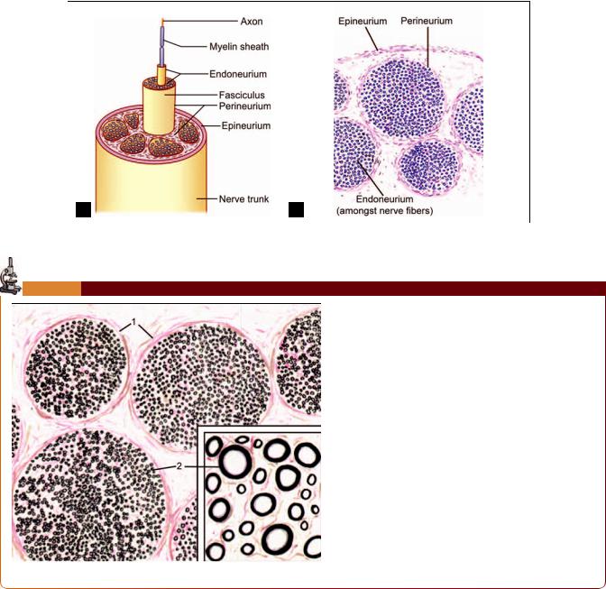
Basic Structure of Peripheral Nerves
Golgi type I: These neurons have long axons, and connect remote regions.
In the peripheral nerves each nerve fiber with its Schwann cell and basal lamina is surrounded by a layer of
Chapter 10 Nervous System 111
A B
Figs. 10.10A and B: % 1 % / 0
PLATE 10.1: Single Nerve Fibre
|
|
|
peripheral nerve. |
|
|
|
|
|
! ! " |
|
|
|
# |
|
|
|
" $ |
|
% |
|
neurium. |
Key
1. Perineurium& '
Peripheral nerve, special stain (as seen in drawing)
connective tissue called the endoneurium (Figs. 10.10A and B and Plate 10.1). The endoneurium contains collagen, fibroblasts, Schwann cells, endothelial cells, and macrophages.
Many nerve fibers together form bundles or fasciculi. Endoneurium holds adjoining nerve fibers together and facilitates their aggregation to form fasciculi.
Each fasciculus is surrounded by a thicker layer of connective tissue called the perineurium. The perineurium
is made up of layers of flattened cells separated by layers of collagen fibers. The perineurium probably controls diffusion of substances in and out of axons. A very thin nerve may consist of a single fasciculus, but usually a nerve is made up of several fasciculi.
The fasciculi are held together by a fairly dense layer of connective tissue that surrounds the entire nerve and is called the epineurium.

112 Textbook of Human Histology
Flowchart 10.2:%
Clinical Correlation
ofthis fat in bedridden patients can lead to pressure on nerve
Blood vessels to a nerve travel through the connective tissue that surrounds it. Severe reduction in blood supply can lead to ischemic neuritis and pain.
NEUROGLIA
In addition to neurons, the nervous system contains several types of supporting cells called neuroglia (Flowchart 10.2).
Types of Neuroglia
Astrocytes, oligodendrocytes, and microglia found in the parenchyma of the brain and spinal cord.
Ependymal cells lining the ventricular system.
Schwann cells lemmocytes or peripheral glia forming myelin sheaths around axons of peripheral nerves. It is important to note that both neurilemma and myelin sheaths are components of Schwann cells.
Capsular cells (also called satellite cells or capsular gliocytes) that surround neurons in peripheral ganglia.
Various types of supporting cells found in relation to
motor and sensory terminals of nerve fibers.
Some workers use the term neuroglia for all these categories, while others restrict the term only to supporting cells present within the brain and spinal cord. The latter convention is used in the description that follows.
Neuroglia of Brain and Spinal Cord
Neuroglial cells present in the parenchyma of brain and spinal cord are mainly of four types:
Astrocytes, that may be subdivided into fibrous and protoplasmic astrocytes.
Fig. 10.11: %
Oligodendrocytes
Ependymal cells
Microglia.
All neuroglial cells are much smaller in size than neurons. However, they are far more numerous. It is interesting to note that the number of glial cells in the brain and spinal cord is 10–50 times as much as that of neurons. Neurons and neuroglia are separated by a very narrow extracellular space.
In ordinary histological preparations, only the nuclei of neuroglial cells are seen. Their processes can be demonstrated by special techniques.
Astrocytes
These are small star-shaped cells that give off a number of processes (Fig. 10.11). The processes are often flattened into leaf-like laminae that may partly surround neurons and separate them from other neurons. The processes frequently end in expansions in relation to blood vessels or

in relation to the surface of the brain. Small swellings called gliosomes are present on the processes of astrocytes. These swellings are rich in mitochondria.
Astrocytes are of two types: fibrous and protoplasmic.
Fibrous astrocytes are seen mainly in white matter. Their processes are thin and are asymmetrical.
Protoplasmic astrocytes are seen mainly in grey matter. Their processes are thicker than those of fibrous astrocytes and are symmetrical.
Intermediate forms between fibrous and protoplasmic
astrocytes are also present. Protoplasmic extensions of astrocytes surround nodes of Ranvier.
The processes of astrocytes are united to those of other astrocytes through gap junctions. Astrocytes communicate with one another through calcium channels. Such communication plays a role in regulation of synaptic activity and in the metabolism of neurotransmitters and neuromodulators.
Function
They provide mechanical support to neurons.
In view of their nonconducting nature, they serve as insulators and prevent neuronal impulses from spreading in unwanted directions.
They are believed to help in neuronal function by playing an important role in maintaining a suitable metabolic environment for the neurons. They can absorb neurotransmitters from synapses, thus, terminating their action
They help in the formation of blood–brain barrier.
Substances secreted by end feet of astrocytes probably assist in maintaining a membrane, the glia limitans externa, which covers the exposed surfaces of the brain. They also help to maintain the basal laminae of blood vessels that they come in contact with.
Astroglial cells are also responsible for repair of damaged areas of nervous tissue. They proliferate in such regions (gliosis). The microglia act as macrophages to engulf and destroy unwanted material.
Oligodendrocytes
These cells are rounded or pear-shaped bodies with relatively few processes (oligo—scanty) (Fig. 10.12).
These cells provide myelin sheaths to nerve fibers that lie within the brain and spinal cord. Their relationship to nerve fibers is basically similar to that of Schwann cells to peripheral nerve fibers. However, in contrast to a Schwann cell that ensheaths only one axon, an oligodendrocyte may enclose several axons.
Chapter 10 Nervous System 113
Fig. 10.12:2 % % %
%
Oligodendrocytes are classified into several types depending on the number of neurons they provide sheaths to. As a rule, oligodendrocytes present in relation to large diameter axons provide sheaths to fewer axons than those related to axons of small diameter. The plasma membrane of oligodendrocytes comes into contact with axolemma at nodes of Ranvier.
Function
Oligodendrocytes provide myelin sheaths to nerve fibers within the CNS for fast conduction of nerve impulses.
Ependymal Cells
Ependymal cells line the ventricles of the brain and central canal of the spinal cord. Ependymal cells are mainly of three types:
Ependymocytes
Choroid epithelial cells
Tanycytes.
The ependymocytes constitute the majority of the ependymal cells. The specialized ependymal cells in choroid plexuses (choroidal epithelial cells) secrete cerebrospinal fluid. The ependymal cells lining the floor of the fourth ventricle have long basal processes and are termed “tanycytes”.
Function
Ependymal cells are concerned in exchange of material between the brain and the cerebrospinal fluid at the braincerebrospinal fluid barrier. The blood in the capillaries of the choroid plexus is filtered through choroid epithelial cells at the blood–cerebrospinal fluid barrier to secrete cerebrospinal fluid.

114Textbook of Human Histology
Microglia
These are the smallest neuroglial cells (Fig. 10.11). The cell body is flattened. The processes are short. These cells are frequently seen in relation to capillaries. As already stated, they differ from other neuroglial elements in being mesodermal in origin. They are probably derived from monocytes that invade the brain during fetal life. They are more numerous in grey matter than in white matter.
Function
They act as phagocytes and become active after damage to nervous tissue by trauma or disease. The microglia act as macrophages to engulf and destroy unwanted material.
Pathological Correlation
Tumors of nervous tissue
Precursors of neural cells can give rise to medulloblastomas. Once mature neurons are formed they lose the power of mitosis and do not give origin to tumors
Certain tumors called germinomas appear near the midline, mostly near the third ventricle. They arise from germ cells that also give rise to teratomas
Most tumors of the brain arise from neuroglial cells. Astrocytomas are most common. Oligodendromas are also frequentTumors can also arise from ependyma and from Schwann
cells.
THE SYNAPSE
Synapses are sites of junction between neurons.
Synapses can be broadly classified into:
Chemical synapses
Electrical synapses.
Synapses involving the release of neurotransmitters are referred to as chemical synapses.
At some sites one cell may excite another without the release of a transmitter. At such sites adjacent cells have direct channels of communication through which ions can pass from one cell to another altering their electrical status. Such synapses are called electrical synapses.
At the site of an electrical synapse plasma membranes (of the two elements taking part) are closely applied, the gap between them being about 4 nm. Proteins called connexins project into this gap from the membrane on either side of the synapse. The proteins are so arranged that small open channels are created between the two synaptic elements.
Electrical synapses are common in lower vertebrates and invertebrates. They have been demonstrated at some
sites in the brains of mammals (for example, in the inferior olive and cerebellum).
Junctions between receptors and neurons, or between neurons and effectors, share some of the features of typical synapses and may also be regarded as synapses. Junctions between cardiac myocytes and between smooth muscle cells, are regarded as electrical synapses.
Classification of a Chemical Synapse Based on Neuronal Elements Taking Part
Synapses may be of various types depending upon the parts of the neurons that come in contact.
Axodendritic synapse: It is the most common type of synapse. In this type, an axon terminal establishes contact with the dendrite of a receiving neuron to form a synapse (Fig. 10.13A).
Axosomatic synapse: The axon terminal synapses with the cell body (Fig. 10.13B).
Axoaxonal synapse: The axon terminal synapses with the axon of the receiving neuron. An axoaxonal synapse may be located either on the initial segment (of the receiving axon) or just proximal to an axon terminal (Fig. 10.13C).
Dendroaxonic synapse: In some parts of the brain (for example, the thalamus), we see some synapses in which the presynaptic element is a dendrite, instead of an axon, which synapses with the axon of the receiving neuron.
Dendrodendritic synapse: Synapse between two dendrites.
Somatosomatic synapse: The soma of a neuron may synapse with the soma of another neuron.
Somatodendritic synapse: Synapse between a soma and a dendrite.
Structure of a Chemical Synapse
A synapse transmits an impulse only in one direction. The two elements taking part in a synapse can, therefore, be spoken of as presynaptic and postsynaptic (Figs. 10.14A and B).
In an axodendritic synapse, the terminal enlargement of the axon may be referred to as the presynaptic bouton or synaptic bag. The region of the dendrite receiving the axon terminal is the postsynaptic process. The two are separated by a space called the synaptic cleft. Delicate fibers or granular material may be seen within the cleft. On either side of the cleft, there is a region of dense cytoplasm. On the presynaptic side, this dense cytoplasm is broken up into several bits. On the postsynaptic side, the dense cytoplasm is continuous and is associated with a meshwork of filaments called the synaptic web.

Chapter 10 Nervous System 115
A |
|
B |
|
C |
Figs. 10.13A to C:: / 0 / 5
A B
Figs. 10.14A and B: /
0 !
The thickened areas of membrane on the presynaptic and postsynaptic sides constitute the active zone of a synapse. Neurotransmission takes place through this region.
GANGLIA
Aggregations of cell bodies of neurons, present outside the brain and spinal cord are known as ganglia. Ganglia are of two main types: sensory and autonomic.
Sensory ganglia (Plate 10.2) are present on the dorsal nerve roots of spinal nerves, where they are called dorsal nerve root ganglia or spinal ganglia. They are also present on the 5th, 7th, 8th, 9th, and 10th cranial nerves. We have seen that the neurons in these ganglia are of the unipolar type (except in the case of ganglia associated with the vestibulocochlear nerve in which they are bipolar). The peripheral process of each neuron forms an afferent (or sensory) fiber of a peripheral nerve. The central process

116 Textbook of Human Histology
PLATE 10.2: Sensory Ganglia
#
Each neuron has a vesicular nucleus with a prominent nucleolus.
A
Key
( )
& "
3.Satellite cells " " " "
B
Sensory ganglia. A. As seen in drawing; B. Photomicrograph
enters the spinal cord or brainstem. (For further details of the connections of these neurons see the author’s Textbook of Human Neuroanatomy).
Autonomic ganglia (Plate 10.3) are concerned with the nerve supply of smooth muscle or of glands. The pathway for this supply always consists of two neurons: preganglionic and postganglionic. The cell bodies of preganglionic neurons are always located within the spinal cord or brainstem. Their axons leave the spinal cord or brainstem and terminate by synapsing with postganglionic neurons, the cell bodies of which are located in autonomic ganglia. Autonomic ganglia are, therefore, aggregations of the cell bodies of postganglionic neurons. These neurons are multipolar. Their axons leave the ganglia as postganglionic fibers to reach and supply smooth muscle or gland. Autonomic ganglia are subdivisible into two major types: sympatheticandparasympathetic.Sympatheticgangliaare located on the right and left sympathetic trunks. Parasympathetic ganglia usually lie close to the viscera supplied through them (For further details of the connections of sympathetic and parasympathetic ganglia see the author’s Textbook of Human Neuroanatomy).
Structure of Sensory Ganglia
In hematoxylin and eosin stained sections the neurons of sensory ganglia are seen to be large and arranged in groups chiefly at the periphery of the ganglion (Plate 10.2). In sections stained by silver impregnation the neurons can be seen to be unipolar. The groups of cells are separated by groups of myelinated nerve fibers.
The cell body of each neuron is surrounded by a layer of flattened capsular cells or satellite cells. Outside the satellite cells there is a layer of delicate connective tissue (The satellite cells are continuous with the Schwann cells covering the processes arising from the neuron. The connective tissue covering each neuron is continuous with the endoneurium).
The entire ganglion is pervaded by fine connective tissue. The ganglion is covered on the outside by a connective tissue capsule.
Structure of Autonomic Ganglia
The neurons of autonomic ganglia are smaller than those in sensory ganglia (Plate 10.3). With silver impregnation they are seen to be multipolar. The neurons are not

Chapter 10 Nervous System 117
PLATE 10.3: Autonomic Ganglia
|
* |
|
! + |
|
|
|
Satellite cells are present, but are less prominent than in |
|
sensory ganglia. |
A
Key
( -& "
3. Satellite cells
B
Autonomic ganglia. A. As seen in drawing;
B. Photomicrograph
arranged in definite groups as in sensory ganglia, but are scattered throughout the ganglion. The nerve fibers are non-myelinated and thinner. They are, therefore, much less conspicuous than in sensory ganglia.
Satellite cells are present around neurons of autonomic ganglia, but they are not so well defined. The ganglion is permeated by connective tissue that also provides a capsule for it (just as in sensory ganglia).
The Nissl substance of the neurons is much better defined in autonomic ganglia than in sensory ganglia. In sympathetic ganglia the neuronal cytoplasm synthesizes catecholamines; and in parasympathetic ganglia it synthesizes acetylcholine. These neurotransmitters travel down the axons to be released at nerve terminals.
SPINAL CORD; CEREBELLAR CORTEX;
CEREBRAL CORTEX
GREY AND WHITE MATTER
Sections through the spinal cord or through any part of the brain show certain regions that appear whitish, and others that have a darker greyish color. These constitute the white and grey matter respectively. Microscopic examination shows that the cell bodies of neurons are located only in grey matter that also contains dendrites and axons starting from or ending on the cell bodies. Most of the fibers within the grey matter are unmyelinated. On the other hand the white matter consists predominantly of myelinated fibers. It is the reflection of light by myelin that