Human_Histology
.pdf
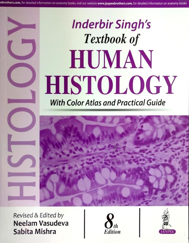
Inderbir Singh’s
Textbook of
HUMAN HISTOLOGY

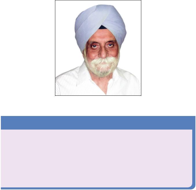
Late Professor Inderbir Singh
(1930–2014)
Tribute to a Legend
Professor Inderbir Singh, a legendary anatomist, is renowned for being a pillar in the education of genera tions of medical graduates across the globe. He was one of the greatest teachers of his times. He was a passionate writer who poured his soul into his work. His eagle’s eye for details and meticulous way of writing made his books immensely popular amongst students. He managed to become enmeshed in millions of hearts in his lifetime. He was conferred the title of Professor Emeritus by Maharishi Dayanand Univer sity, Rohtak, Haryana, India.
On 12th May 2014, he has been awarded posthumously with Emeritus Teacher Award by National Board of Examination for making invaluable contribution in teaching of Anatomy. This award is given to honor legends who have made tremendous contribution in the field of medical education and their work had vast impact on the education of medical graduates. He was a visionary for his times and the legacies he left behind are his various textbooks on gross anatomy, histology, neuroanatomy, and embryology. Although his mortal frame is not present amongst us, his genius will live on forever.

Inderbir Singh’s
Textbook of
HUMAN HISTOLOGY
With Color Atlas and Practical Guide
Eighth Edition
Revised and Edited by
NEELAM VASUDEVA MBBS MD
Director Professor and Head
Department of Anatomy
Maulana Azad Medical College
New Delhi, India
SABITA MISHRA MBBS DNB PhD (AIIMS)
Professor
Department of Anatomy
Maulana Azad Medical College
New Delhi, India
The Health Sciences Publisher
New Delhi | London | Philadelphia | Panama

Jaypee Brothers Medical Publishers (P) Ltd
Headquarters
Jaypee Brothers Medical Publishers (P) Ltd. 4838/24, Ansari Road, Daryaganj
New Delhi 110 002, India
Phone: +91-11-43574357 Fax: +91-11-43574314
E-mail: jaypee@jaypeebrothers.com
Overseas Offices |
|
J.P. Medical Ltd. |
Jaypee-Highlights Medical Publishers Inc. |
83, Victoria Street, London |
City of Knowledge, Bld. 235, 2nd Floor, Clayton |
SW1H 0HW (UK) |
Panama City, Panama |
Phone: +44-20 3170 8910 |
Phone: +1 507-301-0496 |
Fax: +44(0)20 3008 6180 |
Fax: +1 507-301-0499 |
E-mail: info@jpmedpub.com |
E-mail: cservice@jphmedical.com |
Jaypee Medical Inc. |
Jaypee Brothers Medical Publishers (P) Ltd. |
325 Chestnut Street |
17/1-B, Babar Road, Block-B, Shaymali |
Suite 412 |
Mohammadpur, Dhaka-1207 |
Philadelphia, PA 19106, USA |
Bangladesh |
Phone: +1 267-519-9789 |
Mobile: +08801912003485 |
E-mail: support@jpmedus.com |
E-mail: jaypeedhaka@gmail.com |
Jaypee Brothers Medical Publishers (P) Ltd. |
|
Bhotahity, Kathmandu, Nepal |
|
Phone: +977-9741283608 |
|
E-mail: kathmandu@jaypeebrothers.com |
|
Website: www.jaypeebrothers.com |
|
Website: www.jaypeedigital.com |
|
© 2016, Jaypee Brothers Medical Publishers |
|
The views and opinions expressed in this book are solely those of the original contributor(s)/author(s) and do not necessarily represent those of editor(s) of the book.
All rights reserved. No part of this publication may be reproduced, stored or transmitted in any form or by any means, electronic, mechanical, photocopying, recording or otherwise, without the prior permission in writing of the publishers.
All brand names and product names used in this book are trade names, service marks, trademarks or registered trademarks of their respective owners. The publisher is not associated with any product or vendor mentioned in this book.
Medical knowledge and practice change constantly. This book is designed to provide accurate, authoritative information about the subject matter in question. However, readers are advised to check the most current information available on procedures included and check information from the manufacturer of each product to be administered, to verify the recommended dose, formula, method and duration of administration, adverse effects and contraindications. It is the responsibility of the practitioner to take all appropriate safety precautions. Neither the publisher nor the author(s)/editor(s) assume any liability for any injury and/or damage to persons or property arising from or related to use of material in this book.
This book is sold on the understanding that the publisher is not engaged in providing professional medical services. If such advice or services are required, the services of a competent medical professional should be sought.
Every effort has been made where necessary to contact holders of copyright to obtain permission to reproduce copyright material. If any have been inadvertently overlooked, the publisher will be pleased to make the necessary arrangements at the first opportunity.
Inquiries for bulk sales may be solicited at: jaypee@jaypeebrothers.com
Inderbir Singh’s Textbook of Human Histology |
|
|
|
|
|
|||
First Edition |
: |
1987 |
Fifth Edition |
: |
2006 |
Seventh Edition |
: |
2014 |
Second Edition |
: 1992 |
Reprint |
: 2008 |
Eighth Edition |
: |
2016 |
||
Third Edition |
: |
1992 |
Reprint |
: |
2009 |
|
|
|
Fourth Edition |
: |
2002 |
Sixth Edition |
: |
2011 |
|
|
|
Reprint |
: 2005 |
|
|
|
|
|
|
|
ISBN 978-93-85999-32-1
Printed at

Preface
Textbook of Human Histology by Professor Inderbir Singh has remained an authoritative and standard textbook for the past many decades and it is our proud privilege to revise this book and bring out the Eighth Edition. The strength and popularity of this textbook has been its simple language and comprehensiveness that has essentially remained unchanged since its inception. Professor Singh’s eye for details and his meticulous writing style has always been popular amongst the generations of medical students. Although all the chapters have been revisited and thoroughly revised, we have taken special care to retain the basic essence of the book.
To make this standard textbook fulfill the needs of today's generation of students, some new features have been introduced in this edition. A new chapter on Light Microscopy and Tissue Preparation has been added to acquaint the students with the basics of histology. Every student of histology is expected to identify the slides and differentiate amongst them in a perfect manner. To make the students familiar with the various slides, Histological Plates have been added in each chapter that include a photomicrograph, line drawing, and salient features that are visible while examining under the microscope.
Each chapter has been rearranged to provide sequential learning to the students. All the diagrams have been redrawn and many new illustrations have been added for easy comprehension of the basic concepts. Clinical and Pathological Correlations have been added at relevant places for creating an interest of the students in the understanding of pathologies associated with various tissues.
For providing an overview of histology to the student and for quick recall, an atlas has been provided at the beginning of the book. The atlas includes more than 80 slides of histological importance along with their important features.
As envisioned by Professor Inderbir Singh, this textbook is of utmost utility not only for the undergraduate students but also for the students pursuing postgraduation in Anatomy. Keeping this in mind, advanced information on various topics has been included as Added Information to cater to the needs of postgraduate students.
The revision of this book was a team effort. We are thankful to our colleagues for their constant encouragement throughout our venture. We extend our heartfelt thanks to our staff in the Histology laboratory for preparing the slides for photography. We are thankful to Dr Swati Tiwari Bakshi for her important contribution in drawing some of the figures.
We are grateful to Professor Ivan Damjanov, an esteemed teacher and expert in the field of pathology well known across the globe, for allowing us to use some of the slides from his collection. We gratefully acknowledge Professor Harsh Mohan, a well known surgical pathologist of India, for providing pathological correlations in the book. We are thankful to Dr Sunayna Misra [M.D. (Path.), PGI Chandigarh] for her valuable suggestions and inputs especially in the pathological correlations.
We extend our heartfelt thanks to Shri Jitendar P Vij (Group Chairman) and Mr Ankit Vij (Group President) for providing us the opportunity to revise Text of Human Histology and for their persistent support in publication of this book.
Dr Madhu Choudhary (Senior Content Strategist), the driving force of this endeavor, deserves a special thanks for her tireless efforts. She has persevered throughout this venture with a smile on her face. We are thankful to Ms Chetna Malhotra Vohra (Associate Director – Content Strategy), Mr. Vipin Kaushik (Team Leader – Typesetters), Pawan Kishor Tiwari (Team Leader – Proof Readers) and Rajeev Joshi (Team Leader – Designers) of Jaypee Brothers Medical Publishers (P) Ltd. for providing insights and creative ideas that helped in polishing this book to best meet the needs of students and faculty alike.
We present the 8th Edition of this most popular textbook to the medical fraternity as our tribute to a legendary anatomist, Late Professor Inderbir Singh for being a pillar in the education of generations of doctors throughout the world.
Neelam Vasudeva MBBS MD
Sabita Mishra MBBS DNB PhD (AIIMS)

Contents
Color Atlas |
A1–A40 |
Chapter 1: Light Microscopy and Tissue Preparation
Components of a light microscope |
1 |
Principles of a conventional bright field microscope |
2 |
Practical tips in using a bright field microscope |
3 |
Types of microscopes |
3 |
Tissue processing |
4 |
Steps involved in tissue preparation |
4 |
Steps in tissue processing |
4 |
|
|
Chapter 2: Cell Structure |
|
|
|
The cell membrane |
5 |
Contacts between adjoining cells |
8 |
Cell organelles |
12 |
The cytoskeleton |
16 |
The nucleus |
18 |
Chromosomes |
20 |
|
|
Chapter 3: Epithelia |
|
|
|
Characteristic features of epithelial tissue |
25 |
Functions |
25 |
Classification of epithelia |
25 |
Simple epithelium |
26 |
Pseudostratified epithelium |
29 |
Stratified epithelium |
30 |
Basement membrane |
34 |
Projections from the cell surface |
34 |
|
|
Chapter 4: Glands |
|
|
|
Classification of glands |
37 |
Classification of exocrine glands |
37 |
Structural organization |
39 |
Development of glands |
40 |
Chapter 5: General Connective Tissue
|
Fibers of connective tissue |
41 |
|
Cells of connective tissue |
44 |
viii Textbook of Human Histology |
|
|
|
Intercellular ground substance of connective tissue |
48 |
|
Different forms of connective tissue |
48 |
|
Summary of the functions of connective tissue |
53 |
|
|
|
|
Chapter 6: Cartilage |
|
|
|
|
|
General features of cartilage |
55 |
|
Components of cartilage |
55 |
|
Types of cartilage |
56 |
|
|
|
|
Chapter 7: Bone |
|
|
|
|
|
General features |
60 |
|
The periosteum |
60 |
|
Elements comprising bone tissue |
61 |
|
Types of bone |
63 |
|
Formation of bone |
67 |
|
How bones grow |
70 |
|
|
|
|
Chapter 8: Muscular Tissue |
|
|
|
|
|
Types of muscular tissue |
75 |
|
Skeletal muscle |
75 |
|
Cardiac muscle |
84 |
|
Smooth muscle |
86 |
|
Myoepithelial cells |
88 |
|
|
|
|
Chapter 9: Lymphatics and Lymphoid Tissue |
|
|
|
|
|
Lymphatic vessel |
90 |
|
Lymphoid tissue |
90 |
|
Lymph |
90 |
|
Lymphocytes |
91 |
|
Mononuclear phagocyte system |
93 |
|
Lymphatic vessels |
94 |
|
Lymph capillaries |
94 |
|
Larger lymph vessels |
95 |
|
Lymph nodes |
95 |
|
The spleen |
97 |
|
The thymus |
100 |
|
Mucosa-associated lymphoid tissue |
103 |
|
Tonsils |
103 |
|
|
|
|
Chapter 10: Nervous System |
|
|
|
|
|
Tissues constituting the nervous system |
105 |
|
Structure of a neuron |
105 |
|
Types of neurons |
109 |
|
Peripheral nerves |
110 |
|
Neuroglia |
112 |
|
Contents ix |
|
The synapse |
114 |
|
Ganglia |
115 |
|
Spinal cord; cerebellar cortex; cerebral cortex |
116 |
|
Grey and white matter |
116 |
|
The spinal cord |
117 |
|
The cerebellar cortex |
119 |
|
The cerebral cortex |
122 |
|
|
|
|
Chapter 11: Skin and its Appendages |
|
|
|
|
|
Skin |
126 |
|
Types of skin |
126 |
|
Structure of skin |
126 |
|
Blood supply of the skin |
132 |
|
Nerve supply of the skin |
132 |
|
Functions of the skin |
132 |
|
Appendages of the skin |
133 |
|
Hair |
133 |
|
Sebaceous glands |
135 |
|
Sweat glands |
136 |
|
Nails |
137 |
|
|
|
|
Chapter 12: The Cardiovascular System |
|
|
|
|
|
Endothelium |
139 |
|
Arteries |
139 |
|
Arterioles |
141 |
|
Capillaries |
142 |
|
Sinusoids |
144 |
|
Veins |
145 |
|
Venules |
146 |
|
Blood vessels, lymphatics and nerves supplying blood vessels |
146 |
|
Mechanisms controlling blood flow through the capillary bed |
146 |
|
The heart |
148 |
|
|
|
|
Chapter 13: The Respiratory System |
|
|
|
|
|
Common features of air passages |
149 |
|
The nasal cavities |
149 |
|
The pharynx |
151 |
|
The larynx |
151 |
|
The trachea and principal bronchi |
152 |
|
The lungs |
153 |
|
|
|
|
Chapter 14: Digestive System: Oral Cavity and Related Structures |
|
|
|
|
|
Oral cavity |
160 |
|
The lips |
160 |
|
The teeth |
160 |
|
The tongue |
165 |
|
Salivary glands |
168 |
|
