

Module 1. Care of the patients, its role in the treatment process and organization in the hospital.
Content module 1. Structure and the main tasks of the patients care in general system of the therapeutic profile patients treatment.
Topic 6. Estimation of the patient’s condition and the main parameters of his vital activity. Study of the main parameters of hemodynamics and respiration
STUDY OF ARTERIAL PULSE
Pulse [L, pulsare, to beat] is the regular, recurrent expansion and contraction of an artery produced by waves of pressure caused by the ejection of blood from the left ventricle of the heart as it contracts.
T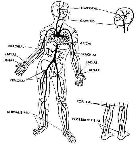 here
are several sites on the body where a pulse is normally taken. All
arteries have a pulse, but it is easier to palpate the pulse at
certain locations. It is easier to feel the pulse when the artery is
near the surface of the skin and when there is firm tissue (such as a
bone) beneath the artery. The three most common sites are the radial
(wrist), carotid (throat), and brachial (inside of elbow). These and
other sites are discussed below and illustrated in figure.
The site or sites that you choose to use may vary depending upon the
condition of the patient. You may take the pulse at the popliteal
(behind the knee) site, the dorsalis pedis (top of the foot) site,
and/or the posterior tibial (back of the ankle) site.
here
are several sites on the body where a pulse is normally taken. All
arteries have a pulse, but it is easier to palpate the pulse at
certain locations. It is easier to feel the pulse when the artery is
near the surface of the skin and when there is firm tissue (such as a
bone) beneath the artery. The three most common sites are the radial
(wrist), carotid (throat), and brachial (inside of elbow). These and
other sites are discussed below and illustrated in figure.
The site or sites that you choose to use may vary depending upon the
condition of the patient. You may take the pulse at the popliteal
(behind the knee) site, the dorsalis pedis (top of the foot) site,
and/or the posterior tibial (back of the ankle) site.
Figure. Sites for taking a pulse.
a. Radial. The radial pulse (the pulse taken using the radial artery) is taken at a point where the radial artery crosses the bones of the wrist. If the patient's hand is turned so that the palm is up, the radial pulse is taken on the thumb side of top side of the wrist.
b. Carotid. The carotid pulse is taken on either side of the trachea. The best location is the grooves located to the right and to the left of the larynx (Adam's apple).
c. Brachial. The brachial pulse is taken in the depression located about one-half inch above the crease on the inside (not the bony side) of the elbow. This site is used when taking the patient's blood pressure.
NOTE: All pulse sites except apical exist on both sides of the body. For example, one radial site exists on the right wrist and one exists on the left wrist.
d. Temporal. The temporal pulse is taken in the temple area on either side of the head. The temple area is located in front of the upper part of the ear. The pulse is felt just above a large, raised bony area called the zygomatic arch.
e. Ulnar. Like the radial pulse, the ulnar pulse is taken at the wrist. The radial pulse is taken over the artery on the thumb side of the wrist while the ulnar pulse is taken on the other side of the wrist. Both pulses are taken on the palm side of the wrist. The radial artery is normally preferred over the ulnar artery for taking the pulse because the radial artery is somewhat larger.
f. Femoral. The femoral pulse is taken in the groin area by pressing the right or left femoral artery against the ischium (the lower part of the pelvic bones located in the front part of the body).
g. Popliteal. The popliteal pulse is taken in the middle of the area located on the inside of the knee (the area opposite the kneecap).
h. Posterior Tibial. The posterior tibial pulse is taken at the top of the ankle or just above the ankle on the back, inside part of the ankle.
i. Dorsalis Pedis. The dorsalis pedis pulse is taken on the top portion of the foot just below the ankle. The pulse is taken in the middle of this area (not to the inside or outside).
j. Apical. Unlike the other sites, the apical pulse is not taken over an artery. Instead, it is taken over the heart itself. The apical pulse (actually, the heartbeat) can be felt over the apex of the heart (the pointed lower end of the heart.) This site is located to the (patient's) left of the breastbone and two to three inches above the bottom of the breastbone. The apical pulse is easily heard when a stethoscope is used.
Palpation of the pulse. Technique. Pulse is commonly studied on the radial artery between the styloid process of the radial bone and the tendon of the internal radial muscle.
T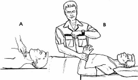 he
carotid pulse is best examined with the sternocleidomastoid muscle
relaxed and with the head rotated slightly toward the examiner.
Palpate the carotid pulse with the patient lying on a bed or couch.
Never compress both carotids simultaneously. Use the left thumb for
the right carotid and vise versa. Place the tip of the thumb between
the larynx and the anterior border of the sternocleidomastoid muscle.
Press the thumb gently backwards to feel the pulse. In palpating the
brachial arterial pulse, the examiner can support the subject’s
relaxed elbow with the arm while compressing the brachial pulse with
the thumb with the fingers cupped round the back of the elbow. Feel
just medial to the tendon of the biceps muscle to detect the pulse.
he
carotid pulse is best examined with the sternocleidomastoid muscle
relaxed and with the head rotated slightly toward the examiner.
Palpate the carotid pulse with the patient lying on a bed or couch.
Never compress both carotids simultaneously. Use the left thumb for
the right carotid and vise versa. Place the tip of the thumb between
the larynx and the anterior border of the sternocleidomastoid muscle.
Press the thumb gently backwards to feel the pulse. In palpating the
brachial arterial pulse, the examiner can support the subject’s
relaxed elbow with the arm while compressing the brachial pulse with
the thumb with the fingers cupped round the back of the elbow. Feel
just medial to the tendon of the biceps muscle to detect the pulse.
Fig. Taking a patient's pulse. A - carotid pulse. B - radial pulse.
The usual technique is to compress the artery with the fingers until the maximum pulse is sensed. Varying degrees of pressure should then be applied while concentrating on the separate phases of the pulse wave.
This method is used for assessing the following characteristics of the pulse: symmetry, rhythm, rate, correlation of the pulse and heart rate, tension, filling (volume), size (Tab.).