

Borchers Andrea Ann (ed.) Handbook of Signs & Symptoms 2015
.pdfObesity. Hepatomegaly can result from fatty infiltration of the liver. Weight loss reduces the liver’s size.
Pancreatic cancer. In pancreatic cancer, hepatomegaly accompanies such classic signs and symptoms as anorexia, weight loss, abdominal or back pain, and jaundice. Other findings include nausea, vomiting, a fever, fatigue, weakness, pruritus, and skin lesions (usually on the legs).
Pericarditis. In chronic constrictive pericarditis, an increase in systemic venous pressure produces marked congestive hepatomegaly. Distended jugular veins (more prominent on inspiration) are a common finding. The usual signs of cardiac disease typically are absent; other features include peripheral edema, ascites, fatigue, and decreased muscle mass.
Special Considerations
Prepare the patient for liver enzyme, alkaline phosphatase, bilirubin, albumin, and globulin studies to evaluate liver function and for X-rays, a liver scan, celiac arteriography, a computed tomography scan, and ultrasonography to confirm hepatomegaly.
Bed rest, relief from stress, and adequate nutrition are important for the patient with hepatomegaly to help protect liver cells from further damage and to allow the liver to regenerate functioning cells. Dietary protein may need to be monitored and possibly restricted. Ammonia, a major cause of hepatic encephalopathy, is a by-product of protein metabolism. Hepatotoxic drugs or drugs metabolized by the liver should be given in very small doses, if at all. These treatment measures should be explained to the patient.
Patient Counseling
Explain the treatment plan for the underlying disorder. Stress the need to avoid alcohol and exposure to people with infections. Discuss the importance of pacing activities and rest.
Pediatric Pointers
Assess hepatomegaly in children the same way you do in adults. Childhood hepatomegaly may stem from Reye’s syndrome; biliary atresia; rare disorders, such as Wilson’s, Gaucher’s, and NiemannPick diseases; or poorly controlled type 1 diabetes mellitus.
REFERENCES
Musso, G., Gambino, R., & Cassader, M. (2011). Need for a three-focused approach to nonalcoholic fatty liver disease. Hepatology, 53(5), 1773.
Stepanova, M., & Younossi, Z. M. (2012). Independent association between nonalcoholic fatty liver disease and cardiovascular disease in the US population. Clinical Gastroenterology and Hepatology, 10(6), 646–650.
Hoarseness
Hoarseness — a rough or harsh sound to the voice — can result from infections, inflammatory lesions, or exudates of the larynx; laryngeal edema; and compression or disruption of the vocal cords or recurrent laryngeal nerve. This common sign can also result from a thoracic aortic aneurysm, vocal cord paralysis, and systemic disorders such as rheumatoid arthritis. It’s characteristically worsened

by excessive alcohol intake, smoking, inhaling noxious fumes, excessive talking, and shouting. Hoarseness can be acute or chronic. For example, chronic hoarseness and laryngitis result when
irritating polyps or nodules develop on the vocal cords. Gastroesophageal reflux into the larynx should also be considered as a possible cause of chronic hoarseness. Hoarseness may also result from progressive atrophy of the laryngeal muscles and mucosa due to aging, which leads to diminished control of the vocal cords.
History and Physical Examination
Obtain a patient history. First, consider his age and sex; laryngeal cancer is most common in men between ages 50 and 70. Be sure to ask about the onset of hoarseness. Has the patient been overusing his voice? Has he experienced shortness of breath, a sore throat, a dry mouth, a cough, or difficulty swallowing dry food? In addition, ask if he has been in or near a fire within the past 48 hours. Be aware that an inhalation injury can cause sudden airway obstruction.
Next, explore associated symptoms. Does the patient have a history of cancer, rheumatoid arthritis, or aortic aneurysm? Does he regularly drink alcohol or smoke?
Inspect the oral cavity and pharynx for redness or exudate, possibly indicating an upper respiratory infection. Palpate the neck for masses and the cervical lymph nodes and thyroid for enlargement. Palpate the trachea — is it midline? Ask the patient to stick out his tongue; if he can’t, he may have paralysis from cranial nerve involvement. Examine the eyes for corneal ulcers and enlarged lacrimal ducts (signs of Sjögren’s syndrome). Dilated jugular and chest veins may indicate compression by an aortic aneurysm.
Take the patient’s vital signs, noting especially a fever and bradycardia. Inspect for asymmetrical chest expansion or signs of respiratory distress — nasal flaring, stridor, and intercostal retractions. Then auscultate for crackles, rhonchi, wheezing, and tubular sounds, and percuss for dullness.
Medical Causes
Gastroesophageal reflux. With gastroesophageal reflux, retrograde flow of gastric juices into the esophagus may then spill into the hypopharynx. This, in turn, irritates the larynx, resulting in hoarseness as well as a sore throat, a cough, throat clearing, and a sensation of a lump in the throat. The arytenoids and the vocal cords may appear red and swollen.
Hypothyroidism. With hypothyroidism, hoarseness may be an early sign. Others include fatigue, cold intolerance, weight gain despite anorexia, and menorrhagia.
Laryngeal cancer. Hoarseness is an early sign of vocal cord cancer but may not occur until later in cancer of other laryngeal areas. The patient usually has a long history of smoking. Other common findings include a mild, dry cough; minor throat discomfort; otalgia; and, sometimes, hemoptysis.
Laryngeal leukoplakia. Leukoplakia is a common cause of hoarseness, especially in smokers. Histologic examination from direct laryngoscopy usually reveals mild, moderate, or severe dysphagia.
Laryngitis. Persistent hoarseness may be the only sign of chronic laryngitis. With acute laryngitis, hoarseness or a complete loss of voice develops suddenly. Related findings include pain (especially during swallowing or speaking), a cough, a fever, profuse diaphoresis, a sore throat, and rhinorrhea.

Rheumatoid arthritis. Hoarseness may signal laryngeal involvement. Other findings include pain, dysphagia, a sensation of fullness or tension in the throat, dyspnea on exertion, and stridor. Thoracic aortic aneurysm. Thoracic aortic aneurysm typically produces no symptoms but may cause hoarseness. Its most common symptom is penetrating pain that’s especially severe when the patient is supine. Other clinical features include a brassy cough; dyspnea; wheezing; a substernal aching in the shoulders, lower back, or abdomen; a tracheal tug; facial and neck edema; jugular vein distention; dysphagia; prominent chest veins; stridor; and, possibly, paresthesia or neuralgia.
Tracheal trauma. Torn tracheal mucosa may cause hoarseness, hemoptysis, dysphagia, neck pain, airway occlusion, and respiratory distress.
Vocal cord paralysis. Unilateral vocal cord paralysis causes hoarseness and vocal weakness. Paralysis may accompany signs of trauma, such as pain and swelling of the head and neck.
Vocal cord polyps or nodules. Raspy hoarseness, the chief complaint, accompanies a chronic cough and a crackling voice.
Other Causes
Inhalation injury. Inhalation injury from a fire or explosion produces hoarseness and coughing, singed nasal hairs, orofacial burns, and soot-stained sputum. Subsequent signs and symptoms include crackles, rhonchi, and wheezing, which rapidly deteriorate to respiratory distress.
Treatments. Occasionally, surgical trauma to the recurrent laryngeal nerve results in temporary or permanent unilateral vocal cord paralysis, leading to hoarseness. Prolonged intubation may cause temporary hoarseness.
Special Considerations
Carefully observe the patient for stridor, which may indicate bilateral vocal cord paralysis. When hoarseness lasts for longer than 2 weeks, indirect or fiber-optic laryngoscopy is indicated to observe the larynx at rest and during phonation.
Patient Counseling
Explain the importance of resting the voice, and teach the patient alternative ways to communicate. Stress the need to avoid alcohol, smoking, and secondhand smoke.
Pediatric Pointers
In children, hoarseness may result from congenital anomalies, such as laryngocele and dysphonia plicae ventricularis. In prepubescent boys, it can stem from juvenile papillomatosis of the upper respiratory tract.
In infants and young children, hoarseness commonly stems from acute laryngotracheobronchitis (croup). Acute laryngitis in children younger than age 5 may cause respiratory distress because the larynx is small and, if irritated or infected, subject to spasm. This may cause partial or total obstruction of the larynx. Temporary hoarseness usually results from laryngeal irritation due to aspiration of liquids, foreign bodies, or stomach contents. Hoarseness may also stem from diphtheria, although immunization has made this disease rare.
Help the child with hoarseness rest his voice. Comfort an infant to minimize crying, play quiet games with him, and humidify his environment.
REFERENCES
Holtzman, M. J. (2012). Asthma as a chronic disease of the innate and adaptive immune systems responding to viruses and allergens.
Journal of Clinical Investigation, 122, 2741–2748.
Sato, S., & Kiyono, H. (2012). The mucosal immune system of the respiratory tract. Current Opinion in Virology, 2, 225–232.
Homans’ Sign
Homans’ sign is positive when deep calf pain results from strong and abrupt dorsiflexion of the ankle. This pain results from venous thrombosis or inflammation of the calf muscles. However, because a positive Homans’ sign appears in only 35% of patients with these conditions, it's an unreliable indicator. (See Eliciting Homans’ Sign .) Even when accurate, a positive Homans’ sign doesn’t indicate the extent of the venous disorder.
This elicited sign may be confused with continuous calf pain, which can result from strains, contusions, cellulitis, or arterial occlusion, or with pain in the posterior ankle or Achilles tendon (for example, in a woman with Achilles tendons shortened from wearing high heels).
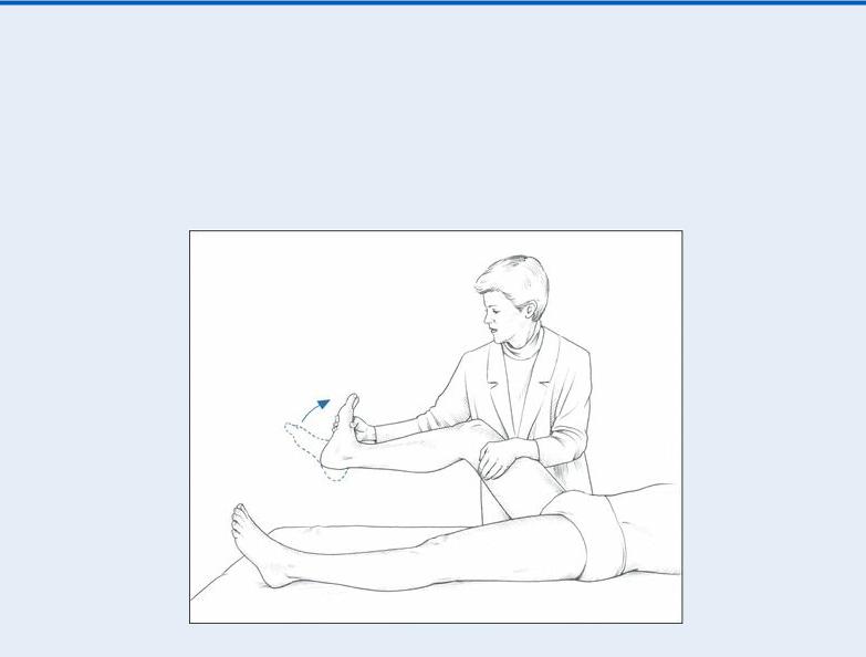
 EXAMINATION TIP Eliciting Homans’ Sign
To elicit Homans’ sign, first support the patient’s thigh with one hand and his foot with the other. Bend his leg slightly at the knee, and then firmly and abruptly dorsiflex the ankle. Resulting deep calf pain indicates a positive Homans’ sign. (The patient may also resist ankle dorsiflexion or flex the knee involuntarily if Homans’ sign is positive.)

History and Physical Examination
When you detect a positive Homans’ sign, focus the patient history on signs and symptoms that can accompany deep vein thrombosis (DVT) or thrombophlebitis. These include throbbing, aching, heavy, or tight sensations in the calf and leg pain during or after exercise or routine activity. Also, ask about shortness of breath or chest pain, which may indicate pulmonary embolism. Make sure to ask about predisposing events, such as a leg injury, recent surgery, childbirth, use of hormonal contraceptives, associated diseases (cancer, nephrosis, hypercoagulable states), and prolonged inactivity or bed rest.
Next, inspect and palpate the patient’s calf for warmth, tenderness, redness, swelling, and the presence of a palpable vein. If you strongly suspect DVT, elicit Homans’ sign very carefully to avoid dislodging the clot, which could cause pulmonary embolism, a life-threatening condition.
In addition, measure the circumferences of both the patient’s calves. The calf with the positive Homans’ sign may be larger because of edema and swelling.
Medical Causes
Deep vein thrombophlebitis. A positive Homans’ sign and calf tenderness may be the only clinical features of deep vein thrombophlebitis. However, the patient may also have severe pain, heaviness, warmth, and swelling of the affected leg; visible, engorged superficial veins or palpable, cordlike veins; and a fever, chills, and malaise.
DVT. DVT causes a positive Homans’ sign along with tenderness over the deep calf veins, slight edema of the calves and thighs, a low-grade fever, and tachycardia. If DVT affects the femoral and iliac veins, you’ll notice marked local swelling and tenderness. If DVT causes venous obstruction, you’ll notice cyanosis and possibly cool skin in the affected leg.
Popliteal cyst (ruptured). Rupture of this synovial cyst may produce a positive Homans’ sign as well as a sudden onset of calf tenderness, swelling, and redness.
Cellulitis (superficial). Superficial cellulitis typically affects the legs but can also affect the arms, producing pain, redness, tenderness, and edema. Some patients also experience a fever, chills, tachycardia, a headache, and hypotension.
Special Considerations
Be sure to place the patient on bed rest, with the affected leg elevated above heart level. Apply warm, moist compresses to the affected area, and administer mild oral analgesics. In addition, prepare the patient for further diagnostic tests, such as Doppler studies and venograms.
When the patient is ambulatory, advise him to wear elastic support stockings after his discomfort decreases (usually in 5 to 10 days) and to continue wearing them for at least 3 months. In addition, instruct the patient to keep the affected leg elevated while sitting and to avoid crossing his legs at the knees to prevent impairing circulation to the popliteal area. (Crossing at the ankles is acceptable.)
Patient Counseling
Explain which signs of prolonged clotting time the patient should report if an anticoagulant is ordered. Explain the benefits and use of elastic support stockings. Emphasize necessary dietary restrictions (green leafy vegetables), alcohol avoidance, and the importance of checking with the
practitioner before taking any new drugs. Stress the importance of keeping follow-up appointments.
Pediatric Pointers
Homans’ sign is seldom assessed in children, who rarely have DVT or thrombophlebitis.
REFERENCES
Kuipers, S., Cannegieter, S. C., Doggen, C. M., & Rosendaal, F. R. (2009). Effect of elevated levels of coagulation factors on the risk of venous thrombosis in long-distance travelers. Blood, 113, 2064–2069.
Silverman, D., & Gendreau, M. (2009). Medical issues associated with commercial flights. Lancet, 373, 2067–2077.
Hyperpnea
Hyperpnea indicates increased respiratory effort for a sustained period — a normal rate (at least 12 breaths/minute) with increased depth (a tidal volume greater than 7.5 mL/kg), an increased rate (more than 20 breaths/minute) with normal depth, or increased rate and depth. This sign differs from sighing (intermittent deep inspirations) and may or may not be associated with tachypnea (increased respiratory frequency).
The typical patient with hyperpnea breathes at a normal or increased rate and inhales deeply, displaying marked chest expansion. He may complain of shortness of breath if a respiratory disorder is causing hypoxemia, or he may not be aware of his breathing if a metabolic, psychiatric, or neurologic disorder is causing involuntary hyperpnea. Other causes of hyperpnea include profuse diarrhea or dehydration, loss of pancreatic juice or bile from GI drainage, and ureterosigmoidostomy. All these conditions and procedures cause a loss of bicarbonate ions, resulting in metabolic acidosis. Of course, hyperpnea may also accompany strenuous exercise, and voluntary hyperpnea can promote relaxation in the patient experiencing stress or pain — for example, a woman in labor.
Hyperventilation, a consequence of hyperpnea, is characterized by alkalosis (arterial pH above 7.45 and partial pressure of arterial carbon dioxide below 35 mm Hg). In central neurogenic hyperventilation, brain stem dysfunction (such as results from a severe cranial injury) increases the rate and depth of respirations. In acute intermittent hyperventilation, the respiratory pattern may be a response to hypoxemia, anxiety, fear, pain, or excitement. Hyperpnea may also be a compensatory mechanism to metabolic acidosis. Under these conditions, it’s known as Kussmaul’s respirations.
(See Kussmaul’s Respirations: A Compensatory Mechanism.)
History and Physical Examination
If you observe hyperpnea in a patient whose other signs and symptoms signal a life-threatening emergency, you must intervene quickly and effectively. (See Managing Hyperpnea, page 408.) However, if the patient’s condition isn’t grave, first determine his level of consciousness (LOC). If he’s alert (and if his hyperpnea isn’t interfering with speaking), ask about recent illnesses or infections, ingestion of aspirin, and ingestion or inhalation of other drugs or chemicals. Find out if the patient has diabetes mellitus, renal disease, or pulmonary condition. Is he excessively thirsty or hungry? Has he recently had severe diarrhea or an upper respiratory tract infection?
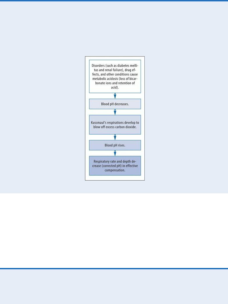
Kussmaul’s Respirations: A Compensatory Mechanism
Kussmaul’s respirations — fast, deep breathing without pauses — characteristically sound labored, with deep breaths that resemble sighs. This breathing pattern develops when respiratory centers in the medulla detect decreased blood pH, thereby triggering compensatory fast and deep breathing to remove excess carbon dioxide and restore pH balance.
Next, observe the patient for clues to his abnormal breathing pattern. Can he speak, or does he speak only in brief, choppy phrases? Is his breathing abnormally rapid? Examine the patient for cyanosis (especially of the mouth, lips, mucous membranes, and earlobes), restlessness, and anxiety
— all signs of decreased tissue oxygenation, as occurs in shock. In addition, observe the patient for intercostal and abdominal retractions, use of accessory muscles, and diaphoresis, all of which may indicate deep breathing related to an insufficient oxygen supply. Next, inspect for draining wounds or signs of infection, and ask about nausea and vomiting. Take the patient’s vital signs, including oxygen saturation, noting a fever, and examine his skin and mucous membranes for turgor, possibly indicating dehydration. Auscultate the patient’s heart and lungs.
 EXAMINATION TIP Managing Hyperpnea
Carefully examine the patient with hyperpnea for related signs of life-threatening conditions,

such as increased intracranial pressure (ICP), metabolic acidosis, diabetic ketoacidosis, and uremia. Be prepared for rapid intervention.
INCREASED ICP
If you observe hyperpnea in a patient who has signs of head trauma (soft tissue injury, edema, or ecchymoses on the face or head) from a recent accident and has lost consciousness, act quickly to prevent further brain stem injury and irreversible deterioration. Then take the patient’s vital signs, noting bradycardia, increased systolic blood pressure, and a widening pulse pressure — signs of increased ICP.
Examine the patient’s pupillary reaction. Elevate the head of the bed 30 degrees (unless you suspect spinal cord injury), and insert an artificial airway. Connect the patient to a cardiac monitor, and continuously observe his respiratory pattern. (Irregular respirations signal deterioration.) Start an I.V. line at a slow infusion rate, and prepare to administer an osmotic diuretic, such as mannitol, to decrease cerebral edema. Catheterize the patient to measure urine output, administer supplemental oxygen, and keep emergency resuscitation equipment close by. Obtain an arterial blood gas analysis to help guide treatments.
METABOLIC ACIDOSIS
If the patient with hyperpnea doesn’t have a head injury, his increased respiratory rate probably indicates metabolic acidosis. If the patient’s level of consciousness is decreased, check his chart for history data to help you determine the cause of his metabolic acidosis, and intervene appropriately. Suspect shock if the patient has cold, clammy skin. Palpate for a rapid, thready pulse and take his blood pressure, noting hypotension. Elevate the patient’s legs 30 degrees, apply pressure dressings to any obvious hemorrhage, start several large-bore I.V. lines, and prepare to administer fluids, vasopressors, and blood transfusions.
A patient with hyperpnea who has a history of alcohol abuse, is vomiting profusely, has diarrhea or profuse abdominal drainage, has ingested an overdose of aspirin, or is cachectic and has a history of starvation may also have metabolic acidosis. Inspect his skin for dryness and poor turgor, indicating dehydration. Take his vital signs, looking for a low-grade fever and hypotension. Start an I.V. line for fluid replacement. Draw blood for electrolyte studies, and prepare to administer sodium bicarbonate.
DIABETIC KETOACIDOSIS
If the patient has a history of diabetes mellitus, is vomiting, and has a fruity breath odor (acetone breath), suspect diabetic ketoacidosis. Catheterize him to monitor increased urine output. Infuse an I.V. saline solution. Perform a fingerstick to estimate blood glucose levels with a reagent strip. Obtain a urine specimen to test for glucose and acetone, and draw blood for glucose and ketone tests. Also, administer fluids, insulin, potassium, and sodium bicarbonate I.V.
UREMIA
If the patient has a history of renal disease, an ammonia breath odor (uremic fetor), and a fine, white powder on his skin (uremic frost), suspect uremia. Start an I.V. line at a slow rate, and prepare to administer sodium bicarbonate. Monitor his electrocardiogram for arrhythmias due to hyperkalemia. Monitor his serum electrolyte, blood urea nitrogen, and creatinine levels as

well until hemodialysis or peritoneal dialysis begins.
Medical Causes
Head injury. Hyperpnea that results from a severe head injury is called central neurogenic hyperventilation. Whether its onset is acute or gradual, this type of hyperpnea indicates damage to the lower midbrain or upper pons. Accompanying signs reflect the site and extent of injury and can include loss of consciousness; soft tissue injury or bony deformity of the face, head, or neck; facial edema; clear or bloody drainage from the mouth, nose, or ears; raccoon eyes; Battle’s sign; an absent doll’s eye sign; and motor and sensory disturbances.
Signs of increased intracranial pressure include decreased response to painful stimulation, loss of pupillary reaction, bradycardia, increased systolic pressure, and a widening pulse pressure. Hyperventilation syndrome. Acute anxiety triggers episodic hyperpnea, resulting in respiratory alkalosis. Other findings may include agitation, vertigo, syncope, pallor, circumoral and peripheral paresthesia, muscle twitching, carpopedal spasm, weakness, and arrhythmias. Hypoxemia. Many pulmonary disorders that cause hypoxemia — for example, pneumonia, pulmonary edema, chronic obstructive pulmonary disease, and pneumothorax — may cause hyperpnea and episodes of hyperventilation with chest pain, dizziness, and paresthesia. Other effects include dyspnea, a cough, crackles, rhonchi, wheezing, and decreased breath sounds. Ketoacidosis. Alcoholic ketoacidosis (occurring most commonly in females with a history of alcohol abuse) typically follows cessation of drinking after a marked increase in alcohol consumption has caused severe vomiting. Kussmaul’s respirations begin abruptly and are accompanied by vomiting for several days, a fruity breath odor, slight dehydration, abdominal pain and distention, and absent bowel sounds. The patient is alert and has a normal blood glucose level, unlike the patient with diabetic ketoacidosis.
Diabetic ketoacidosis is potentially life threatening and typically produces Kussmaul’s respirations. The patient usually experiences polydipsia, polyphagia, and polyuria before the onset of acidosis; he may or may not have a history of diabetes mellitus. Other clinical features include a fruity breath odor; orthostatic hypotension; a rapid, thready pulse; generalized weakness; a decreased LOC (lethargy to coma); nausea; vomiting; anorexia; and abdominal pain. Starvation ketoacidosis is also potentially life threatening and can cause Kussmaul’s respirations. Its onset is gradual; typical findings include signs of cachexia and dehydration, a decreased LOC, bradycardia, and a history of severely limited food intake.
Renal failure. Acute or chronic renal failure can cause life-threatening acidosis with Kussmaul’s respirations. Signs and symptoms of severe renal failure include oliguria or anuria, uremic fetor, and yellow, dry, scaly skin. Other cutaneous signs include severe pruritus, uremic frost, purpura, and ecchymoses. The patient may complain of nausea and vomiting, weakness, burning pain in the legs and feet, and diarrhea or constipation.
As acidosis progresses, corresponding clinical features include frothy sputum, pleuritic chest pain, and signs of heart failure and pleural or pericardial effusion. Neurologic signs include an altered LOC (lethargy to coma), twitching, and seizures. Hyperkalemia and hypertension, if present, require rapid intervention to prevent cardiovascular collapse.
Sepsis. A severe infection may cause lactic acidosis, resulting in Kussmaul’s respirations. Other findings include tachycardia, a fever or a low temperature, chills, a headache, lethargy, profuse

diaphoresis, anorexia, a cough, wound drainage, burning on urination, confusion or a change in mental status, and other signs of local infection.
Shock. Potentially life-threatening metabolic acidosis produces Kussmaul’s respirations, hypotension, tachycardia, narrowed pulse pressure, a weak pulse, dyspnea, oliguria, anxiety, restlessness, stupor that can progress to coma, and cool, clammy skin. Other clinical features may include external or internal bleeding (in hypovolemic shock); chest pain or arrhythmias and signs of heart failure (in cardiogenic shock); a high fever, chills and, rarely, hypothermia (in septic shock); or stridor due to laryngeal edema (in anaphylactic shock). The onset is usually acute in hypovolemic, cardiogenic, or anaphylactic shock, but it may be gradual in septic shock.
Other Causes
Drugs. Toxic levels of salicylates, ammonium chloride, acetazolamide, and other carbonic anhydrase inhibitors can cause Kussmaul’s respirations. So can ingestion of methanol and ethylene glycol, found in antifreeze solutions.
Special Considerations
Monitor vital signs, including oxygen saturation, in every patient with hyperpnea, and observe for increasing respiratory distress, an irregular respiratory pattern, or hypoxia — all of which signal deterioration. Prepare for immediate intervention to prevent cardiovascular collapse: Start an I.V. line for administration of fluids, blood transfusions, and vasopressor drugs for hemodynamic stabilization, as ordered, and prepare to give ventilatory support. Prepare the patient for arterial blood gas analysis and blood chemistry studies.
Patient Counseling
Teach the patient how to monitor his blood glucose level, and stress the importance of compliance with diabetes therapy, if applicable. Explain which foods and fluids he should avoid. Teach him to avoid respiratory infections. Emphasize the importance of avoiding alcohol, and provide information about support groups or other resources on alcohol cessation if the patient has a history of alcohol abuse.
Pediatric Pointers
Hyperpnea in children indicates the same metabolic or neurologic causes as in adults and requires the same prompt intervention. The most common cause of metabolic acidosis in children is diarrhea, which can cause a life-threatening crisis. In infants, Kussmaul’s respirations may accompany acidosis due to inborn errors of metabolism.
REFERENCES
Hayward, J. (2011). Common genetic conditions in primary care. Pulse, 26, 824–825.
McCahon, D., Holder, R., Metcalfe, A., Clifford, S., Gill, P., Cole, T., … Wilson, S. (2009). General practitioners’ attitudes to assessment of genetic risk of common disorders in routine primary care. Clinical Genetics, 76, 544–551.