

Учебники / Lasers in Otorhinolaryngology Huttenbrink
.pdf90 Lasers for Benign Diseases of the Larynx, Hypopharynx, and Trachea
T Abstract
The use of endoscopic laser surgery for benign lesions of the larynx has led to the modification of numerous operative procedures, which are briefly reviewed in this article. The CO2 laser meets the criteria for use for most benign laryngeal lesions, particularly in phonosurgery, stenosis surgery, swallowing rehabilitation, and for the resection of most benign laryngeal tumors. Optimum instruments and equipment are even more important in surgical procedures for benign laryngeal lesions than in the surgery of malignant tumors. While the use of surgical lasers in phonosurgery is still somewhat controversial, laser surgery has gained an established place in the treatment of glottic laryngeal stenoses, selected tracheal stenoses, and Zenker diverticula. Today, CO2 lasers are an indispensable tool for endolaryngeal surgery.
T Operative Technique: Considerations
Historical Background
Modern endoscopic laryngeal surgery traces its origins to the second half of the nineteenth century. Turck and Czermak of Vienna first used angled mirrors for clinical examination of the larynx in 1857, and two years later Stoerk used these mirrors for endolaryngeal cauterization of the larynx. As a result of these efforts, endolaryngeal surgery became more widely practiced and techniques were advanced by McKenzie, Fränkel, Kirstein, Killian, and Jackson. Thus, the development of endolaryngeal surgery was contemporaneous with, but essentially separate from, that of open laryngeal surgery.
In the U.S., the separation between endolaryngeal surgery and open laryngeal surgery is still evident since while head and neck surgeons in the U.S. are skilled in open laryngeal surgery, they may take a skeptical view toward endoscopic laryngeal surgery, which is the domain of “laryngologists.” This “overdivision” into subspecialties helps to explain the long delay in the acceptance of endolaryngeal laser surgery in the U.S.
The decisive breakthrough for endolaryngeal surgery as a universal approach for laryngeal operations occurred when Oskar Kleinsasser of Cologne, Germany, introduced microlaryngoscopy in the early 1960s. This method provides access to most of the mucosal lesions of the larynx and a number of submucous lesions. At the same time, it requires a specialized set of microinstruments that are controlled with both hands, requiring a high degree of manual skills. Moreover, the rich vascular supply of the tissues in the larynx makes it difficult to carry out more extensive procedures, tending to cause troublesome bleeding from the cut tissues, obscuring the operative field and making precision microsurgery difficult or impossible. An early solution to this problem involved using monopolar electrocautery probes in endolaryngeal surgery to improve hemostasis in operations involving the relatively extensive division of submucous tissues (e. g., cordectomy, arytenoidectomy).
In the light of the above situation, the introduction of laser systems into laryngeal surgery marks an important milestone in the development of this operative approach. By coupling a CO2 laser to the operating microscope and controlling the invisible treatment beam with a micromanipulator, guided by a visible-wavelength coaxial aiming beam, a relatively stationary, noncontact technique was acheived for dividing endolaryngeal tissues while simultaneously coagulating smaller blood vessels. This approach overcame several basic problems in endolaryngeal surgery: the laser beam does not obstruct the surgeon’s view of the confined operating field, it provides adequate tissue hemostasis for greater visibility, and it facilitates the surgical dissection.
The obvious advantages of endolaryngeal laser surgery were not generally appreciated at first. A major objection was that laser incisions always caused thermal tissue damage (burns) and therefore did not meet the requirement of clean, atraumatic tissue division with margins that could be histologically evaluated. Over time, technical refinements eliminated most of these objections, and laser use not only made it easier to carry out known surgical procedures but also laid the foundation for developing new therapeutic concepts, especially in the field of oncologic surgery (see Chapter 6). Remarkably, these concepts were not developed in the U.S., where lasers were first used in laryngology [1]. It was left to clinical pioneers in Europe such as Burian and Höfler [2], Rudert and Werner [3, 4], Thumfart [5], and especially Steiner and Ambrosch [6, 7] in the Ger- man-speaking world, Motta in Italy [8], Grossenbacher in Switzerland [9], and others to research, describe, apply, and publicize the clinical potential of endolaryngeal laser surgery.
In addition, endoscopic laser surgery has led to a modification of numerous operative procedures for benign lesions of the larynx. These are briefly reviewed in this chapter.
Fundamentals of Endolaryngeal Surgery
and Equipment
The necessary ingredients for successful endolaryngeal surgery include:
•a refined operating technique,
•appropriate anesthesia and ventilation,
•a complete instrument set for endolaryngeal surgery.
Other important factors are proper positioning of the patient, preoperative assessment of dental status, adequate mouth protection during the procedure to prevent dental injuries, and a trained, proficient surgeon. Suitable options for anesthesia–ventilation are general endotracheal anesthesia (using a laser-safe ventilation tube!), jet ventilation, or mask ventilation with intermittent apnea [10, 11]. The necessary equipment includes an assortment of laryngoscopes and stands, an operating microscope, microinstruments, the CO2 laser, monopolar cautery probes, rigid telescopes, and suction. Some procedures also require implants and their corresponding application instruments

|
Operative Technique: Considerations 91 |
|
|
|
|
(collagen, fat, cartilage, dispersed silicone) and photo- |
fined algorithm, the robotic mechanism scans the laser |
|
graphic or video equipment [12]. |
beam over the targeted area in a cruciform pattern at a pre- |
|
|
set speed and constant power output. This ensures that |
|
The essential problems in any minimally invasive proce- |
equal power densities and exposure times are delivered to |
|
dure, regardless of whether it is in the abdominal cavity, |
all subareas of the selected scan area. The rapid movement |
|
knee joint, or larynx, are obtaining an adequate view of the |
of the laser beam leads to rapid heat dispersion in the tis- |
|
operative field and the ability to use the necessary instru- |
sue. Meanwhile the surface of the selected mucosal area is |
|
ments at the surgical site. In laryngeal surgery, operating |
vaporized completely and uniformly while the underlying |
|
laryngoscopes are employed for this purpose. Since no |
anatomic structures are largely preserved. This mode of la- |
|
laryngoscope can satisfy all requirements, the laryngeal |
ser use is suitable for the selective, superficial removal of |
|
surgeon should have an assortment of different models on |
mucosal lesions in cases where histologic examination is |
|
hand. The most important are listed below: |
not required and the main aim is to achieve uniform tissue |
|
• Kleinsasser laryngoscopes (sizes A–C, DN, J, JL) |
ablation with the least possible collateral injury. An exam- |
|
• Bivalved laryngoscopes in various sizes (Rudert, Steiner, |
ple is the removal of papillomas or patchy areas of leuko- |
|
Weerda) |
plakia, which can be histologically confirmed prior to ac- |
|
• Lindholm laryngoscope—when this is inserted into the |
tual laser ablation. The result of this procedure is a super- |
|
vallecula epiglottica, it affords an excellent view of su- |
ficial mucosal wound with no thermal alteration of the un- |
|
praglottic structures (ideally, of the whole larynx) |
derlying tissue. This type of wound undergoes rapid |
|
• Diverticuloscope (Holinger-Benjamin) for endoscopic |
secondary epithelialization and can heal to an excellent |
|
cricopharyngeal myotomy |
functional result. Scanners based on a similar working |
|
• Pediatric laryngoscopes (various models) |
principle are also available for producing straight or curved |
|
|
incisions, as in a cricopharyngeal myotomy for a Zenker di- |
|
Optimum instrumentation is just as important in the treat- |
verticulum (Accublade, Lumenis). |
|
ment of benign laryngeal lesions as it is in the surgery of |
For the selection of suitable laser parameters (pulse shape |
|
malignant tumors. It may be even more so, inasmuch as |
||
tumor surgery is basically a destructive process while the |
and duration, power output, etc.), the reader is referred to |
|
surgery of benign laryngeal lesions is often a tailoring pro- |
selected publications [see references 19–22] and to Chap- |
|
cedure. The goal in this type of surgery is not just to remove |
ters 1, 2, and 6 of this volume. |
|
abnormalities but rather to modify and adapt (tailor) ana- |
In the treatment of laryngeal hemangiomas, especially |
|
tomic changes to allow for optimum functional rehabilita- |
||
tion of the voice, swallowing, and respiration. This can be |
when large, the neodymium:yttrium aluminum garnet |
|
achieved, as in phonosurgery, only by means of precise, |
(Nd:YAG) laser has the advantage of a greater penetration |
|
noncharring laser tissue cutting without collateral thermal |
depth in tissue, producing deeper coagulation of the hem- |
|
tissue damage, requiring the use of optimum instruments |
angioma [23, 24]. However, circumscribed hemangiomas |
|
and equipment. |
can be successfully excised locally with the CO2 laser. Ar- |
|
|
gon lasers can also be used to treat vascular neoplasms ow- |
|
|
ing to the absorption of the light by red blood pigment. |
|
Laser Systems Used in the Endoscopic Surgery of Benign Laryngeal and Tracheal Lesions
The CO2 laser is the undisputed workhorse of laser surgery in laryngology. Owing to its frequent use in tumor surgery, nowadays it is available in the ENT departments of most larger hospitals. The CO2 laser meets the requirements for use for most benign laryngeal lesions, particularly in phonosurgery, stenosis surgery, swallowing rehabilitation, and in the resection of most benign laryngeal tumors [3, 13– 17].
Besides the laser unit itself, accessories are available for the optimum delivery of laser energy to the operative site. These include devices for optimum focusing of the surgical beam (e. g., AccuSpot, Lumenis) [18], which focus the beam to an extremely small spot for precision cutting. Scanner systems (e. g., Surgitouch, Lumenis) [16] are also available for making a precise linear incision or for coagulating predefined mucosal areas while preserving the underlying tissue (e. g., in the treatment of laryngeal papillomatosis). A scanner system consists of a robotic guidance mechanism that tracks the CO2 laser beam over a rectangular, round, or elliptical mucosal area selected by the operator on the micromanipulator of the laser head. Following a prede-
A special laser treatment modality is photodynamic therapy (tissue lasing following the selective uptake of a photosensitizing agent) [25–28]. The efficacy of photodynamic therapy has been documented for a number of benign, preneoplastic, and neoplastic mucosal lesions. On the other hand, the costs of the procedure and concerns about unpredictable mucosal scarring still limit its application in the treatment of benign lesions of the larynx and trachea.
Alternatives to Surgical Laser Use
Surgical lasers, particularly the CO2 laser, can be used in various ways for the treatment of benign laryngeal and tracheal lesions:
•Tissue ablation (precise cutting with minimal coagulation for the resection of abnormal tissue)
•Coagulation (of blood vessels or very vascular neoplasms)
•Vaporization (of tissues, as for papilloma removal)
•Induction of photochemical processes (in photodynamic therapy)
Alternative techniques are available for all of these applications and will be briefly described below.

92 Lasers for Benign Diseases of the Larynx, Hypopharynx, and Trachea
Tissue Ablation
Tissue ablation can be accomplished with sharp cutting instruments (knives, scissors)—the essential tools for selective tissue ablation in every surgical discipline. Special small knives and microscissors are available for traditional tissue ablation in the larynx and trachea [29], but these instruments require expert manual skills and rigorous practice. They are designed to have a long shaft that must be passed down the operating laryngoscope, making them less effective than the scalpels and scissors used in open operations. At the same time, the division of tissues with cutting instruments always involves capillaries and small arterial or venous vessels, leading to diffuse bleeding at the surgical site. Although the blood loss in the larynx is not quantitatively significant in itself, the bleeding nevertheless obscures the operative field and makes it difficult to assess the progress of the operation. This type of bleeding is particularly troublesome in the situation of maximumprecision microsurgery. Not infrequently, it prevents the surgical precision that is desired from a functional standpoint and prolongs the operating time. The bleeding, and the associated obstacles created during the surgery, can result in functional failures and persistent, undesired anatomic changes (scars, synechiae).
On the other hand, “cold” instruments offer the significant advantage of precise cutting with no thermal damage to surrounding tissues. This makes it easier for the pathologist to evaluate the margins of the excised tissues and eliminates the deleterious effects of collateral thermal damage on wound healing [30, 31]. Moreover, the use of cutting microinstruments in laryngeal surgery does not require the elaborate technical precautions necessitated by laser use.
In summary, there is no question that the ability of cold instruments to remove small lesions confined to the mucosa, especially in phonosurgery, is the equal of surgical lasers and is even superior to lasers in some situations. The CO2 laser should be used for the phonosurgical ablation of mucosal lesions only by a highly skilled surgeon and only in settings where optimum technical facilities are available (Accuspot micromanipulator, precisely adjusted laser system, proficiency in laser use) [32–35].
Besides conventional instruments, powered instruments (microdebriders, shavers) for use in laryngeal and tracheal surgery have been described recently [36–38]. These instruments have been widely used in paranasal sinus surgery. Basically, they consist of a motorized blade rotating in a sheath with suction at the tip. Tissue is excised superficially by the rotating blade and simultaneously aspirated out of the sheath. Those advocating this type of instrument for endolaryngeal surgery (especially papilloma removal) have noted that shaver systems require less time for tissue ablation and do not generate a smoke plume. This argument is worth mentioning because human papillomavirus (HPV) DNA has been identified in the plume produced during the CO2 laser vaporization of respiratory tract papillomas [39, 40]. Its clinical significance is uncertain however.
The difficult healing of “tissue burns” caused by endolaryngeal laser surgery has been cited as another argument for
the use of microdebriders [41]. One fact should be emphasized at this point: surgical lasers cause burns in the larynx and trachea only if the surgeon is using an outdated device or is not well versed in modern laser surgery. In other words, burns following laser surgery are the fault of the operator, not of the method. Modern, fully equipped CO2 laser systems enable the surgeon to divide tissue with absolute precision with a focused beam, without causing clinically significant thermal damage to normal surrounding tissue. Thus, the concern voiced by many authors that lasers cause “burns” is based on observations either from the early days of laser surgery or of improper laser use. When all facts are considered, the argument of thermal tissue damage and its implications for wound healing can no longer be taken seriously.
Moreover, it is unclear whether motorized instruments used on the very fine structures of the vocal cord mucosa pose an equal or even greater risk of inadvertent tissue damage than present-day laser technology. Let us consider an analogy with middle ear surgery: CO2 lasers have gained an established place in stapes surgery, and today no ear surgeon would think of using shaver systems on the auditory ossicles. At the same time, microdebriders are probably an effective supplement to the surgical armamentarium for the rapid removal of larger neoplasms that do not require further histologic evaluation.
Coagulation
Besides surgical lasers (CO2, Nd:YAG), electrocautery probes are commonly used to coagulate blood vessels and tissues having a rich blood supply. Before the advent of surgical lasers, electrocautery probes were widely used in endolaryngeal surgery [29, 42, 43]. They were used for cutting (e. g., arytenoidectomy) and tissue ablation (suction–coag- ulation during papilloma removal) and for the selective cauterization of small blood vessels. Today, classic electrosurgery is more of an adjunct to endolaryngeal laser surgery than a competing modality. It is no longer considered an acceptable tool for most procedures because it causes far more extensive thermal damage and cauterization of specimen margins than the CO2 laser and cannot be manipulated as precisely as the laser beam.
When used as an adjunct to the CO2 laser, electrosurgery is indispensable in all major endolaryngeal procedures because the CO2 laser can only coagulate blood vessels up to a certain size. It is unable to coagulate large bleeders, such as those encountered in an arytenoidectomy or endolaryngeal partial laryngectomy, even when applied in the continuous mode with a defocused beam. In these cases the electrocautery probe is an essential adjunct to the laser for obtaining surgical hemostasis.
Electrosurgery as a stand-alone method has undergone technical refinements in recent years, and these improvements have eliminated some of the inherent disadvantages of electrosurgical procedures. For example, thermal tissue damage can be substantially reduced by irrigating the surgical site with water [44]. Good clinical results have been achieved by applying a continuous stream of noble gas to
|
Surgical Endoscopy 93 |
|
|
|
|
the tissue to be coagulated (argon beamer, argon plasma |
T Surgical Endoscopy |
|
coagulation) [45–47]. This technique permits optimum |
||
electrocoagulation of the tissue while preventing carboni- |
Diagnostic laryngoscopy is indicated for all laryngeal dis- |
|
zation. It has become an accepted alternative to laser sur- |
||
gery, particularly in operations on the nose and paranasal |
eases in which the larynx cannot be examined in the con- |
|
sinuses. So far there has been very little clinical experience |
scious patient (children) or in which mirror laryngoscopy, |
|
with argon plasma coagulation in laryngeal surgery. But it |
telescopic laryngoscopy, or flexible endoscopy has re- |
|
is entirely conceivable that the method could be effective |
vealed findings in the larynx, hypopharynx, or trachea that |
|
in treating vascular lesions located away from the vocal |
require further investigation. Suspension laryngoscopy |
|
cords. |
provides ideal access for many diagnostic and therapeutic |
|
|
procedures in the trachea, in which the laryngoscope can |
|
Vaporization |
serve as a “sheath” for the introduction of tracheoscopes |
|
and operating instruments (Figs. 5.1, 5.2). Besides inspec- |
||
Aside from the argon plasma coagulation systems de- |
tion of the mucosal surface with the operating microscope |
|
and introduction of rigid endoscopes, this technique allows |
||
scribed above, basically there is no alternative to the CO2 |
selective excisional biopsies, even enabling the surgeon to |
|
laser for tissue vaporization. |
take large samples if required. In additional, visual inspec- |
|
|
tion can be supplemented by tactile examination (e. g., to |
|
Photodynamic Therapy |
assess the mobility of an ankylosed arytenoid cartilage). |
|
|
|
|
Laser systems have ideal technical properties for inducing |
|
|
the desired photochemical reactions in tissue. Photody- |
|
|
namic therapy would not be possible without lasers. The |
|
|
question is which laser system is the most suitable. This |
|
|
question, just as that of choosing the best photosensitizing |
|
|
agent, is a topic of current research [48] and is beyond the |
|
|
scope of this chapter. |
|
|
Anesthesia, Perioperative Care, and
Adjunctive Medical Therapy
Laser surgery can be done under general endotracheal anesthesia and using jet ventilation [10, 11]. Endotracheal intubation is generally preferred in operations where there is likely to be heavy bleeding (tumor resections, arytenoidectomies, laryngeal papillomatosis). Special laser-safe tubes can be used to protect against tube combustion and airway fires, but they are expensive. Jet ventilation is preferred in operations for airway stenosis and in many phonosurgical procedures, as it provides a better view of the glottis and subglottis.
Even extensive endolaryngeal procedures can almost always be done without a prior tracheotomy when surgical lasers are used [49–51]. The present author has achieved good results with the routine i.v. administration of 250 mg methylprednisolone before endolaryngeal procedures. Antibiotic prophylaxis (e. g., 3 g ampicillin–sulbactam or 600 mg clindamycin i. v.) is also given for more extensive procedures, especially those involving the exposure of laryngeal cartilages. Patients generally require postoperative monitoring in an intensive care unit (ICU) following laser surgery for airway stenosis. At this time the patient should be awake and extubated. For all other procedures, ICU monitoring is generally unnecessary from a surgical standpoint.
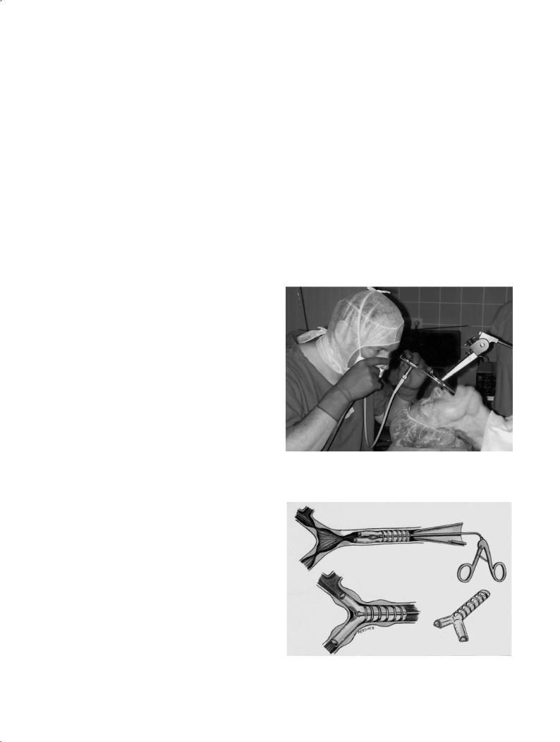
Fig. 5.1 Suspension laryngoscopy for tracheal access. A rigid telescope is passed into the trachea through the laryngoscope. Tracheal secretions are simultaneously aspirated.
Fig. 5.2 Endoscopic tracheal surgery through the laryngoscope using jet ventilation. A rigid tracheoscope is introduced through the laryngoscope, allowing dilatation and stenting of a neoplastic tracheal stenosis.

94 Lasers for Benign Diseases of the Larynx, Hypopharynx, and Trachea
Currently, the CO2 laser can be used for excisional biopsies, completely resecting smaller mucosal lesions of indeterminate nature in a procedure that is both diagnostic and therapeutic.
T Phonosurgery
Phonosurgery is the term applied to all surgical measures aimed at improving the voice (in a broad sense, it also includes voice rehabilitation after laryngectomy).
Classification
Based on a proposal by the European Laryngological Society (ELS), phonosurgical procedures can be grouped into four classes [52]:
•Vocal fold surgery: surgical procedures carried out directly on the vocal cords (generally endolaryngeal) with the goal of improving the vibrational properties of the vocal cords or glottic closure.
•Laryngeal framework surgery: procedures done on the cartilaginous skeleton of the larynx or the laryngeal musculature to correct the position, shape, or tension of the vocal cords.
•Neuromuscular surgery: procedures done on the nerves of the larynx with the goal of restoring or improving the mobility and/or tension of the vocal cords.
•Reconstructive surgery addressing partial or total laryngeal defects: procedures done to restore or improve the voice after surgically induced loss of function.
This chapter deals with the predominant use of lasers in phonosurgery: vocal cord surgery. Other works should be consulted for details of laryngeal framework procedures and neuromuscular surgery.
Preoperative Diagnosis
In otologic surgery, one or more preoperative tests are done to provide a functional description of the affected ear. The preoperative audiogram describes the existing functional impairment and provides a baseline for evaluating the outcome of the tympanoplasty. The same basic principles should be applied in phonosurgery. These cases also require a preoperative functional description, although there is no universal measuring tool comparable to the pure-tone audiogram in otology. The author feels that the minimal workup before a planned phonosurgical procedure should include an examination of the larynx by telescopic laryngoscopy and stroboscopy, a perceptual voice analysis (based on the HBR scale), and generally a determination of vocal pitch and loudness range and maximum phonation time. Videoendoscopic documentation and a digital voice recording provide an ideal baseline for evaluating the treatment outcome, although these resources will not be available at every center.
Laser Surgery of the Vocal Cords
Laser surgery on the vocal cords may be done for a number of conditions such as epithelial changes (vocal nodules, leukoplakia, hyperkeratosis, acanthosis, dysplasia, etc.), exudative changes in the Reinke space (vocal cord polyps, Reinke edema), granulomas (contact granuloma, intubation granuloma), scarring, and subepithelial lesions (cysts) [16, 53–57]. The use of the CO2 laser in particular is highly controversial for these indications [31, 33, 57, 58]. While the advocates of laser surgery state that lasers using noncontact technique are ideal for making precise, atraumatic, bloodless mucosal incisions, the proponents of “cold instruments” claim that laser incisions always cause poorly healing burns followed by unpredictable scarring, often with adverse functional consequences. It is likely that both views are correct,
With a modern CO2 laser operated by a well-trained surgeon using optimum equipment and state-of-the-art peripheral devices (AccuSpot), the laser can indeed cut tissues atraumatically and without charring [15, 16, 18, 20, 30]. Under these ideal conditions, CO2 lasers are excellent instruments for use in phonosurgery, although operating time can be considerably prolonged in some cases. The use of less optimal lasers by inadequately trained surgeons can cause significant thermal damage to the mucosa far beyond that caused by microsurgical incisions with conventional instruments. Thus, vocal cord surgery with or without a CO2 laser (i. e., using hot or cold instruments) is less a matter of deciding in favor of one technique or the other than a matter of the availability of equipment and the surgeon’s training and proficiency. Moreover, the use of a laser system does not compel the surgeon to use the laser exclusively. The laser surgeon is free to use the instrument (hot or cold) that is best for any given situation.
Several common surgical lesions of the vocal cords are discussed briefly in the sections below.
Idiopathic Granulomas (Contact Granulomas)
The etiology of contact granulomas is not yet fully understood. The disease affects males almost exclusively. The typical configuration of the male larynx, where there is a relatively small angle between the thyroid laminae, is believed to contribute to the more frequent occurrence of contact granulomas in males. The causal mechanism may be repeated microtrauma to the mucosa on the medial side of the arytenoid cartilage during phonation. The microtrauma, whose repetitive nature keeps the mucosal wound from healing once it has developed, apparently incites the formation of granulation tissue. Gastroesophageal reflux into the pharynx has also been cited as a possible etiologic factor [59–61].
Telescopic laryngoscopy generally reveals a typical granuloma located on the medial side of an arytenoid cartilage (Fig. 5.3). In any given case, it is often unclear whether the dysphonia is related to the granuloma or whether it is a sign of abnormal functional processes in the larynx, which

Phonosurgery 95
Fig. 5.3 Large contact granuloma on the left vocal cord. a Microlaryngoscopic view. b Appearance after laser ablation.
in turn may contribute to the granuloma formation. Granulomas should be surgically removed to establish the diagnosis. This can be done in ideal fashion with the CO2 laser (using jet ventilation), because the laser allows precise ablation with good hemostasis without damaging the cartilaginous tissue. Medical treatment with proton pump inhibitors is often recommended in the literature to prevent a recurrence, but the author has not had encouraging results with these agents. While this conclusion is not based on a systematic analysis, it is the author’s impression that this therapy does not reduce the number of recurrences compared with patients who have not received proton pump inhibitors. Also, the phenotype and psychological profile of patients with contact granuloma do not appear to support reflux disease as the cause of the lesions. The disease is particularly common in patients with a very correct, conscientious personality type who are not overweight, generally do not smoke, and do not drink excessively. In contrast, vocal cord granulomas occur very rarely in patients with a known history of severe reflux disease. Thus, once the lesions have been removed by laser abla-
tion, the patient often undergoes counseling until the condition resolves spontaneously, often after a period of years.
Intubation Granulomas
Intubation granulomas resemble contact granulomas in their gross and microscopic features, but they may be bilateral and also occur in women and children. Removal follows the principles outlined above with the lesions much less likely to recur than in contact granulomas.
Vocal Nodules
Functional voice disorders and vocal overuse often lead to circumscribed epithelial thickening of the free edges of the vocal cords, usually located at the junction of the anterior and middle thirds and situated at opposite sites on both cords. All vocal nodules do not require surgical removal. They should be removed only if they obstruct complete glottic closure. Whether the nodules are resected (very sparingly, always preserving the underlying vocal ligament) with microinstrumentation or a meticulous laser technique (single pulse, low power density) depends less on theoretical considerations than on the personal experience of the surgeon [34, 35].
Reinke Edema
Fluid accumulation in Reinke’s space beneath the surface of the vocal cords increases the volume and mass of the vibrating portions of the cords, lowering the pitch of the voice in affected individuals [62, 63]. Many patients accept this low-pitched voice as a personality trait. Surgery should be withheld in these cases unless the edema is hampering respiration or there are concomitant mucosal changes suspicious for malignancy (leukoplakia, etc.). But if the edema is causing a subjective deterioration of voice quality, decreased voice performance, or a fluctuating voice quality, it should be surgically corrected.
In all cases the vocal effects of surgical ablation should be discussed with the patient preoperatively, giving particular attention to the anticipated (irreversible) increase in vocal pitch, the transient worsening of dysphonia, and the fact that surgical procedures for Reinke edema generally do not completely restore the voice [64]. The patient should be informed that the surgical treatment and the subsequent period of voice rest are only part of the overall treatment plan, which should include functional voice therapy and long-term vocal hygiene which involves avoiding vocal misuse and smoking.
The operation begins by making a mucosal incision lateral to the free edge of the vocal cord, near the floor of Morgagni’s pouch, extending the full length of the cord. The incision can be made with a microsurgical instrument or a surgical laser. The CO2 laser can also provide point coagulation of thickened blood vessels in the mucosa. Next the gelatinous edema is suctioned from Reinke’s space without causing further damage to the mucosa. The mucosal flaps are
96 Lasers for Benign Diseases of the Larynx, Hypopharynx, and Trachea
then reapproximated, resecting (with a laser or cold instrument) the mucosa made redundant by the decreased volume. Ideally, the mucosal flaps should be apposed edge to edge. The free edges of the vocal cords should be protected from injury and should be covered with intact epithelium at the end of the operation. In this way both sides can be treated in one sitting. If there is very extensive edema with scarring, it can be difficult or impossible to carry out the technique as described. If there is epithelial tearing or the vocal ligament is exposed at the free edge on one side, it is better to defer operating on the second side to avoid the risk of synechia formation. These cases require a two-stage procedure in which the second side is treated after the first side has healed.
Vocal Cord Polyps
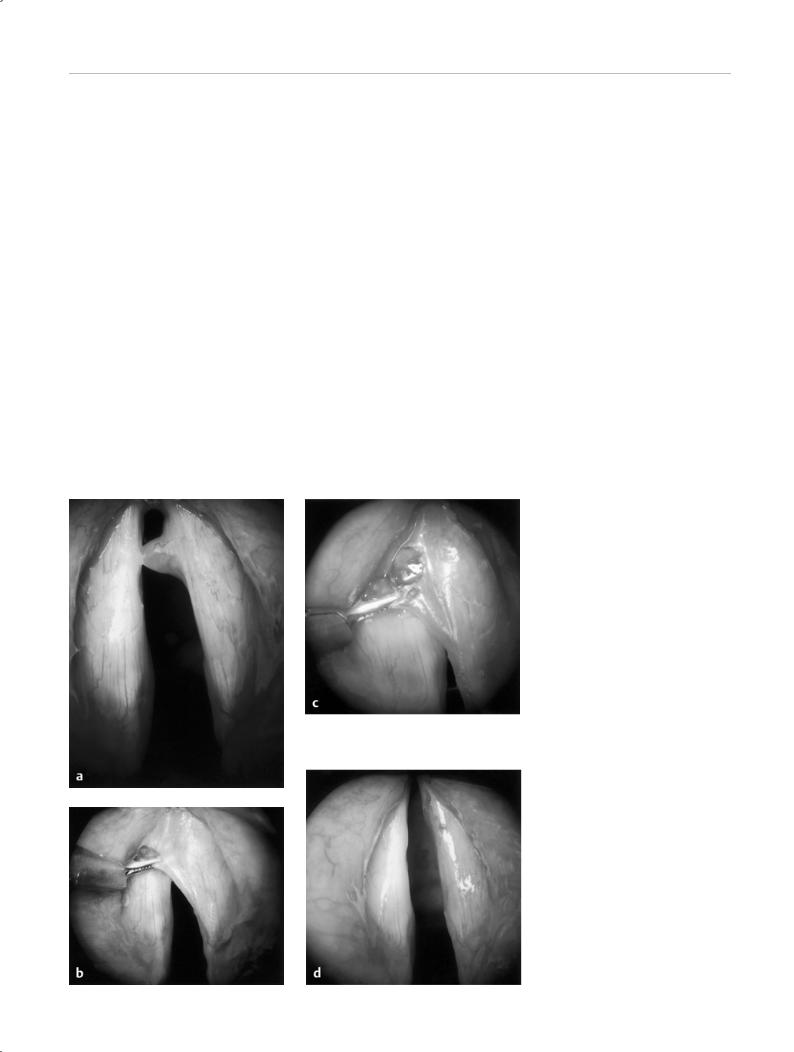
Vocal cord polyps, as Reinke edema, are exudative lesions of Reinke’s space but are more circumscribed than the edema. They can be removed with microscissors or with a CO2 laser beam in the single-pulse mode (Fig. 5.4 a–d) [65].
Vocal Cord Cysts
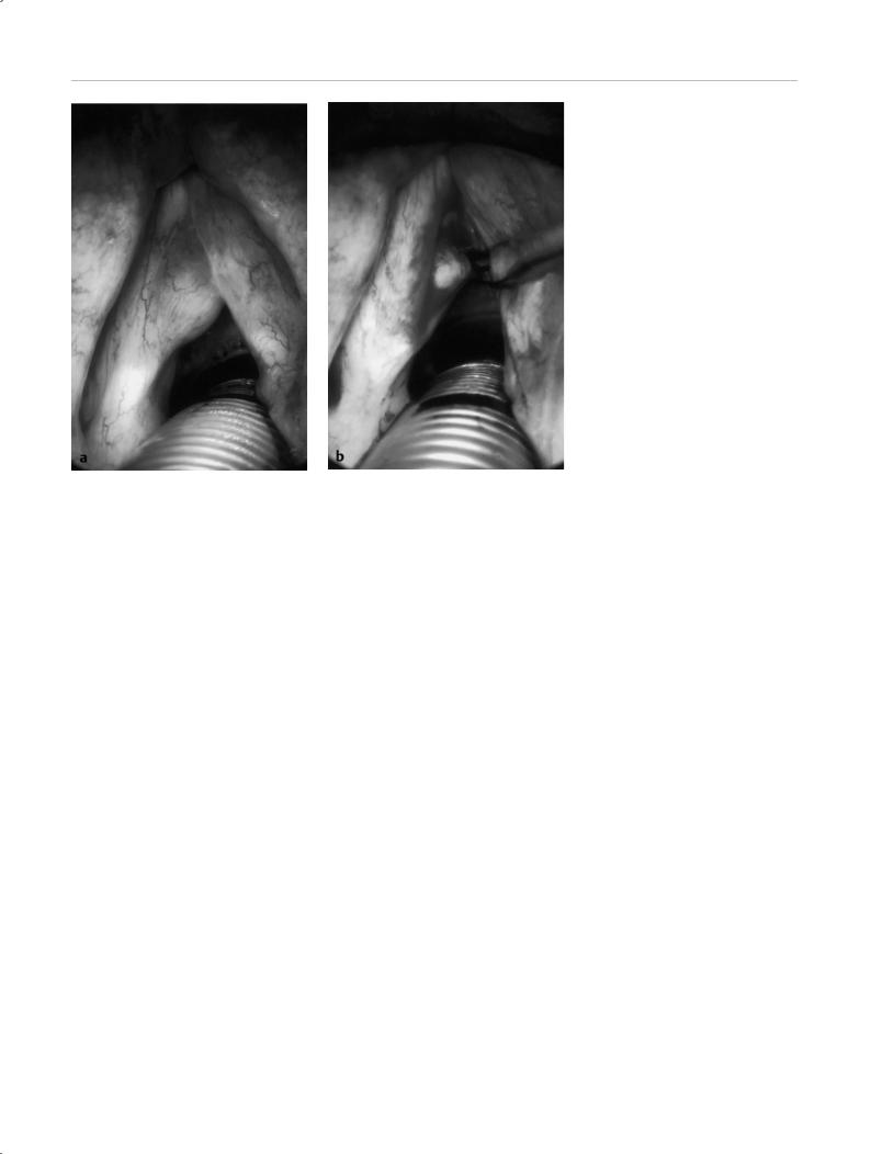
Typical subepithelial vocal cord cysts should be exposed by careful incision of the mucosa, isolated by blunt dissection, and removed in one piece without opening the cyst wall to reduce the risk of recurrence (similar to middle ear cholesteatomas) (Fig. 5.5 a, b). The author prefers to use the Kleinsasser microinstrument set (Karl Storz) or Bouchayer set (Micro-France, Xomed) for this purpose, and not the laser.
Surgical Voice Rehabilitation
Surgical lasers are occasionally used to create a speech fistula in patients who have undergone a total laryngectomy [66]. The author does not feel that this procedure offers any special advantages over established methods of prosthetic voice restoration, and these options will not be described here.
T Surgery to Improve Swallowing
Difficulty in swallowing (dysphagia) is a relatively common presenting complaint in the practice of otolaryngolo-
Fig. 5.4 Polyp on the right vocal cord. a Microlaryngoscopic view. b The mucosa is grasped with a Bouchayer forceps. c The
mucosa is incised with the CO2 laser in sin- gle-pulse mode, using the AccuSpot system for bloodless cutting without charring. d Appearance at the end of the operation.
Surgery to Improve Swallowing 97
gy. It is reasonable to expect that, with the growing percentage of older individuals in the overall population, the numbers will increase in the future.
Classification
Dysphagia may present with any one of three main symptoms:
•Obstruction (impairment of bolus transport)
•Aspiration (inhalation of food particles or saliva)
•Globus sensation (vague feeling of fullness in the throat, often perceived subjectively as “difficult swallowing” although solid foods and fluids are still swallowed normally)
Surgical treatment is an option only for obstruction or as- piration-related dysphagia. No sound rationale exists for the operative treatment of globus sensation.
Obstruction
Swallowing of food may be obstructed by mass lesions in the oral cavity, oropharynx, hypopharynx, or esophagus or by disturbances of neuromuscular regulation, especially absent or delayed opening of the upper esophageal sphincter (cricopharyngeus muscle) during swallowing. Pharyngeal masses leading to dysphagia may be due to hyperplasia of the lymphatic Waldeyer ring (hyperplasia of the tonsils or lymphatic tissue at the base of the tongue) or benign and malignant tumors of the oral cavity, pharynx, and esophagus. The treatment of benign tumors is covered later and the treatment of malignant tumors is discussed in Chapter 6.
Obstruction of the foodway by tonsillar hyperplasia can be treated medically (for acute inflammation) or surgically
Fig. 5.5 Large subepithelial cyst of the left vocal cord. a Microlaryngoscopic view. b The cyst is exposed by microsurgical (not laser) incision of the mucosa and removed in toto.
(tonsillectomy) (see Chapter 4). In children, laser tonsillotomy may be appropriate if the goal is to reduce the volume of the tonsils while preserving tonsillar remnants for immunologic reasons [67].
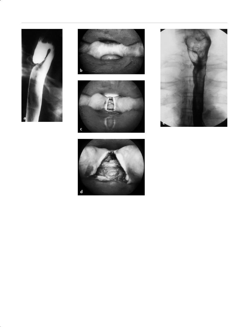
The treatment of Zenker diverticulum has become an established domain of laser surgery during recent years. These diverticula develop as a result of deficient or delayed relaxation of the cricopharyngeus muscle [68] during swallowing. Until a few years ago, open cricopharyngeus myotomy combined with diverticulectomy through a left transcervical approach was the standard surgical treatment for Zenker diverticula [69–71]. Although endoscopic myotomy (division of the posterior part of the annular cricopharyngeus muscle) was described as early as 1917, the endoscopic approach was abandoned later due to excessive complications. Van Overbeek, in particular, showed that endoscopic crico-pharyngeal myotomy could be undertaken with a high degree of precision using the CO2 laser (Fig. 5.6 a–e) [71]. According to the literature, the complications and results of endoscopic myotomies appear to be comparable to or better than those achieved with open surgery [72–76]. In any case, endoscopic myotomy obviates the need for an external neck incision and practically eliminates the risk of recurrent nerve paralysis. Generally the myotomy can be done without difficulty when a special diverticuloscope (Dohlman type) is used. There is disagreement as to whether patients should be fed through a temporary nasogastric tube (placed preoperatively and carefully protected during laser use) or by temporary parenteral nutrition. Opinions also vary as to the timing of the initial feeding after endoscopic myotomy. An interval of 2–7 days is most commonly recommended. In all cases, oral contrast examination with a water-soluble medium (Gastrografin) should be done before the resumption of oral intake to exclude paravasation into the upper mediastinum.
98 Lasers for Benign Diseases of the Larynx, Hypopharynx, and Trachea
Although endoscopic myotomy is technically straightforward, it should be emphasized that this procedure is by no means without risks. Contrary to van Overbeek’s belief that the anterior wall of the diverticular sac is always fused to the posterior wall of the esophagus by inflammatory adhesions, minimizing the risk of opening the mediastinum during endoscopic myotomy, in the author’s experience in the open surgery of Zenker diverticula such adhesions are rarely if ever present. In any case, they do not offer adequate protection against opening the mediastinum. It should be assumed, rather, that any complete cricopharyngeal myotomy will open the upper mediastinum. This underscores the urgent need for preoperative antibiotic prophylaxis. Local mucosal antisepsis in the region of the hypopharynx and diverticular sac may also help lower the risk of infection.
Mild wound pain, leukocytosis, and temperature elevation are common after the operation, especially during the first night [72]. These findings alone do not indicate significant mediastinitis. But if the temperature remains elevated beyond the first day or does not respond promptly to antipyretic agents, and if the patient has res-
Fig. 5.6 A typical Zenker diverticulum. a Preoperative contrast radiograph of the esophagus. b Diverticuloscope demonstrates the common wall between the diverticular sac and esophagus. c Laser incision of the common wall in the midline. d Complete cricopharyngeal myotomy. e No swallowing difficulties
3 months after endoscopic laser myotomy.
piration-dependent back pain, local warmth in the neck region, or other manifestations of a septic systemic process, appropriate imaging studies should be done at once to assess the need for surgical intervention (opening the upper mediastinum through a left cervical incision, oversewing an esophageal perforation, local irrigation) (Fig. 5.7). Thus, endoscopic surgery for Zenker diverticula is relatively easy to do but is not risk free. The surgeon should take note of the potential risks and should be prepared to manage them.
Aspiration
Aspiration may result from loss of substance, especially in the larynx (e. g., after a supraglottic partial laryngectomy), or it may have a neurogenic cause (e. g., lesions of the superior laryngeal nerve or vagus nerve). These forms of dysphagia are not amenable to correction by laser surgery [70].

Laser Treatment of Airway Stenosis 99
Fig. 5.7 Contrast extravasation from the pharynx after endoscopic myotomy, with incipient mediastinitis—treated surgically by mediastinal drainage through a lateral neck incision.
Cricopharyngeus Motility Disorders without Diverticula
Delayed relaxation of the cricopharyngeus muscle during swallowing can occasionally lead to dysphagia, which often is accompanied by secondary aspiration. A diverticulum is not (yet) demonstrable in all cases. The disorder is more common in older individuals. Video cinematography is generally necessary to make a diagnosis. Unlike a conventional oral contrast examination, the temporal resolution of this technique is high enough to demonstrate the underlying functional disorder. As for Zenker diverticula, the recommended treatment is endoscopic myotomy [77]. The author has no personal experience with the endoscopic treatment of this disorder and would prefer an openneck myotomy (to avoid the risk of opening the pharynx and possibly causing mediastinitis).
Surgical Alternatives to Laser Use (Stapler, Botulinum Toxin, Open Myotomy)
A number of alternatives to laser surgery are available for the treatment of Zenker diverticulum and cricopharyngeal dysfunction. Endoscopic myotomy with a stapler has been recommended from a surgical and laryngologic standpoint in recent years [69, 78, 79]. The device is an automatic stapler of the type commonly used in open abdominal surgery. A special stapler has beed developed for the treatment of Zenker diverticula and can be introduced and operated through a diverticuloscope. It divides the common wall between the diverticular sac and esophagus, simultaneously closing the wound surfaces on both sides with staples. This technique is supposed to prevent leakage into the mediastinum. Because of the way the stapler is designed, a part of the wound always remains open at the lowest point of the incision [80]. The author has no personal basis for judging whether this method actually offers advantages over endolaryngeal laser surgery. The results in the literature are comparable to those of endolaryngeal laser surgery, and there is no published evidence showing definite advantages.
Open myotomy is always available as an alternative to endoscopic myotomy in cases where it is important to avoid opening the pharynx. The surgery is done through a left transcervical approach, like the classic operation for treating a Zenker diverticulum. Following meticulous division of the cricopharyngeus muscle, the pharynx is closed. There is virtually no risk of infection under these conditions (with proper attention to surgical asepsis). The procedure can thus provide an alternative to endoscopic myotomy and the classic open operation for Zenker diverticula in all patients considered to be at increased risk for mediastinitis (patients with multiple morbidities, older patients). To the author’s knowledge, however, no comparative studies on this technique have been published.
An elegant, minimally invasive treatment option is to temporarily paralyze the cricopharyngeus muscle by the localized injection of botulinum toxin [81]. The advantage of this method is that it is practically free of complications when carried out correctly. The disadvantage is that its effect lasts only a few months, at which time the treatment may have to be repeated (generally necessitating brief general anesthesia). Thus botulinum toxin injection is not a permanent treatment option. It does, however, provide an ideal test method in equivocal cases to determine whether the proposed surgical division of the cricopharyngeus muscle will improve the patient’s symptoms. If the symptoms improve following transient paralysis of the muscle with botulinum toxin and then worsen again after the effect has subsided, the patient can be scheduled for a definitive cricopharyngeal myotomy.
T Laser Treatment of Airway Stenosis
The larynx forms the narrowest part of the central respiratory tract [82]. As a result, anatomic changes can easily lead to clinically significant airway narrowing [83]. The relative narrowness of the respiratory tract at this level is based on the physiologic function of the larynx as a safety valve between the upper respiratory and alimentary tracts. Because of its dual functions in preventing aspiration and producing speech, the larynx must be able to close the airway temporarily when needed. Disturbances of this physiologic process can lead to permanent narrowing of the airway lumen. Inflammatory swelling, scarring, and tumor masses can also narrow or obstruct the airway.
Classification
The symptoms of central airway stenosis depend on the location and type of the stenosis and the degree of airway narrowing. The main etiologic factors are summarized in Table 5.1. Several etiologic categories of airway stenosis are described below.
Malformations
The most common laryngeal malformation causing respiratory obstruction is laryngomalacia. Based on a congenital