

Intracellular Signaling Complexes Enhance the Speed, Efficiency, and Specificity of the Response
Even a single type of extracellular signal acting through a single type of G-protein-linked or enzyme-linked receptor usually activates multiple parallel signaling pathways and can thereby influence multiple aspects of cell behaviorsuch as shape, movement, metabolism, and gene expression. Indeed, these two main classes of cell-surface receptors often activate some of the same signaling pathways, and there is usually no obvious reason why a particular extracellular signal utilizes one class of receptors rather than the other.
The complexity of these signal-response systems, with multiple interacting relay chains of signaling proteins, is daunting. It is not clear how an individual cell manages to display specific responses to so many different extracellular signals, many of which bind to the same class of receptor and activate many of the same signaling pathways. One strategy that the cell uses to achieve specificity involves scaffold proteins (see Figure 15-16), which organize groups of interacting signaling proteins into signaling complexes (Figure 15-19A). Because the scaffold guides the interactions between the successive components in such a complex, the signal can be relayed with precision, speed, and efficiency; moreover, unwanted cross-talk between signaling pathways is avoided. In order to amplify a signal, however, and spread it to other parts of the cell, at least some of the components in most signaling pathways are likely to be freely diffusible.
In other cases, signaling complexes form only transiently, as when signaling proteins assemble around a receptor after an extracellular signal molecule has activated it. In some of these cases, the cytoplasmic tail of the activated receptor is phosphorylated during the activation process, and the phosphorylated amino acids then serve as docking sites for the assembly of other signaling proteins (Figure 15-19B). In yet other cases, receptor activation leads to the production of modified phospholipid molecules in the adjacent plasma membrane, and these lipids then recruit specific intracellular signaling proteins to this region of membrane. All such signaling complexes form only transiently and rapidly disassemble after the extracellular ligand dissociates from the receptor.
Interactions Between Intracellular Signaling Proteins Are Mediated by Modular Binding Domains
The assembly of both stable and transient signaling complexes depends on a variety of highly conserved, small binding domains that are found in many intracellular signaling proteins. Each of these compact protein modules binds to a particular structural motif in the protein (or lipid) with which the signaling protein interacts. Because of these modular domains, signaling proteins bind to one another in multiple combinations, like Lego bricks, with the proteins often forming a three-dimensional network of interactions that determines the route followed by the signaling pathway. By joining existing domains together in novel combinations, the use of such modular binding domains has presumably facilitated the rapid evolution of new signaling pathways.
Src homology 2 (SH2) domains and phosphotyrosine-binding (PTB) domains, for example, bind to phosphorylated tyrosines in a particular peptide sequence on activated receptors or intracellular signaling proteins. Src homology 3 (SH3) domains bind to a short proline-rich amino acid sequence. Pleckstrin homology (PH) domains (first described in the Pleckstrin protein in blood platelets) bind to the charged headgroups of specific phosphorylated inositol phospholipids that are produced in the plasma membrane in response to an extracellular signal; they thereby enable the protein they are part of to dock on the membrane and interact with other recruited signaling proteins. Some signaling proteins function only as adaptors to link two other proteins together in a signaling pathway, and they consist solely of two or more binding domains (Figure 15-20).
Scaffold proteins often contain multiple PDZ domains (originally found in a region of a synapse called the postsynaptic density), each of which binds to a specific motif on a receptor or signaling protein. The InaD scaffold protein in Drosophila photoreceptor cells is a striking example. It contains five PDZ domains, one of which binds a light-activated ion channel, while the others each bind to a different signaling protein involved in the response of the cell to light. If any of these PDZ domains are missing, the corresponding signaling protein fails to assemble in the complex, and the fly's vision is defective.
Some cell-surface receptors and intracellular signaling proteins are thought to cluster together transiently in specific microdomains in the lipid bilayer of the plasma membrane that are enriched in cholesterol and glycolipids. Some of the proteins are directed to these lipid rafts by covalently attached lipid molecules. Like scaffold proteins, these lipid scaffolds may promote speed and efficiency in the signaling process by serving as sites where signaling molecules can assemble and interact (see Figure 10-13).
Cells Can Respond Abruptly to a Gradually Increasing Concentration of an Extracellular Signal
Some cellular responses to extracellular signal molecules are smoothly graded in simple proportion to the concentration of the molecule. The primary responses to steroid hormones (see Figure 15-14) often follow this pattern, presumably because the nuclear hormone receptor protein binds a single molecule of hormone and each specific DNA recognition sequence in a steroid-hormone-responsive gene acts independently. As the concentration of hormone increases, the concentration of activated receptor-hormone complexes increases proportionally, as does the number of complexes bound to specific recognition sequences in the responsive genes; the response of the cell is therefore a gradual and linear one.
Many responses to extracellular signal molecules, however, begin more abruptly as the concentration of the molecule increases. Some may even occur in a nearly all-or-none manner, being undetectable below a threshold concentration of the molecule and then reaching a maximum as soon as this concentration is exceeded. What might be the molecular basis for such steep or even switchlike responses to graded signals?
One mechanism for sharpening the response is to require that more than one intracellular effector molecule or complex bind to some target macromolecule to induce a response. In some steroid-hormone-induced responses, for example, it seems that more than one activated receptor-hormone complex must bind simultaneously to specific regulatory sequences in the DNA to activate a particular gene. As a result, as the hormone concentration rises, gene activation begins more abruptly than it would if only one bound complex were sufficient for activation (Figure 15-21). A similar cooperative mechanism often operates in the signaling cascades activated by cell-surface receptors. As we discuss later, four molecules of the small intracellular mediator cyclic AMP, for example, must bind to each molecule of cyclic-AMP-dependent protein kinase to activate the kinase. Such responses become sharper as the number of cooperating molecules increases, and if the number is large enough, responses approaching the all-or-none type can be achieved (Figures 15-22 and 15-23).
Responses are also sharpened when an intracellular signaling molecule activates one enzyme and, at the same time, inhibits another enzyme that catalyzes the opposite reaction. A well-studied example of this common type of regulation is the stimulation of glycogen breakdown in skeletal muscle cells induced by the hormone adrenaline (epinephrine). Adrenaline's binding to a G-protein-linked cell-surface receptor leads to an increase in intracellular cyclic AMP concentration, which both activates an enzyme that promotes glycogen breakdown and inhibits an enzyme that promotes glycogen synthesis.
All of these mechanisms can produce responses that are very steep but, nevertheless, always smoothly graded according to the concentration of the extracellular signal molecule. Another mechanism, however, can produce true all-or-none responses, such that raising the signal above a critical threshold level trips a sudden switch in the responding cell. All-or-none threshold responses of this type generally depend on positive feedback; by this mechanism, nerve and muscle cells generate all-or-none action potentials in response to neurotransmitters (discussed in Chapter 11). The activation of ion-channel-linked acetylcholine receptors at a neuromuscular junction, for example, results in a net influx of Na+ that locally depolarizes the muscle plasma membrane. This causes voltage-gated Na+ channels to open in the same membrane region, producing a further influx of Na+, which further depolarizes the membrane and thereby opens more Na+ channels. If the initial depolarization exceeds a certain threshold value, this positive feedback has an explosive "runaway" effect, producing an action potential that propagates to involve the entire muscle membrane.
An accelerating positive feedback mechanism can also operate through signaling proteins that are enzymes rather than ion channels. Suppose, for example, that a particular intracellular signaling ligand activates an enzyme located downstream in a signaling pathway and that two or more molecules of the product of the enzymatic reaction bind back to the same enzyme to activate it further (Figure 15-24). The consequence is a very low rate of synthesis of the product in the absence of the ligand. The rate increases slowly with the concentration of ligand until, at some threshold level of ligand, enough of the product has been synthesized to activate the enzyme in a self-accelerating, runaway fashion. The concentration of the product then suddenly increases to a much higher level. Through these and a number of other mechanisms not discussed here, the cell will often translate a gradual change in the concentration of a signaling ligand into a switchlike change, creating an all-or-none response by the cell.
A Cell Can Remember The Effect of Some Signals
The effect of an extracellular signal on a target cell can, in some cases, persist well after the signal has disappeared. The enzymatic accelerating positive feedback system just described represents one type of mechanism that displays this kind of persistence. If such a system has been switched on by raising the concentration of intracellular activating ligand above threshold, it will generally remain switched on even when the extracellular signal disappears; instead of faithfully reflecting the current level of signal, the response system displays a memory. We shall encounter a specific example of this later, when we discuss a protein kinase that is activated by Ca2+ to phosphorylate itself and other proteins; the autophosphorylation keeps the kinase active long after Ca2+ levels return to normal, providing a memory trace of the initial signal.
Transient extracellular signals often induce much longer-term changes in cells during the development of a multicellular organism. Some of these changes can persist for the lifetime of the organism. They usually depend on self-activating memory mechanisms that operate further downstream in a signaling pathway, at the level of gene transcription. The signals that trigger muscle cell determination, for example, turn on a series of muscle-specific gene regulatory proteins that stimulate the transcription of their own genes, as well as genes producing many other muscle cell proteins. In this way, the decision to become a muscle cell is made permanent (see Figure 7-72B).
Cells Can Adjust Their Sensitivity to a Signal
In responding to many types of stimuli, cells and organisms are able to detect the same percentage of change in a signal over a very wide range of stimulus intensities. This requires that the target cells undergo a reversible process of adaptation, or desensitization, whereby a prolonged exposure to a stimulus decreases the cells' response to that level of exposure. In chemical signaling, adaptation enables cells to respond to changes in the concentration of a signaling ligand (rather than to the absolute concentration of the ligand) over a very wide range of ligand concentrations. The general principle is one of a negative feedback that operates with a delay. A strong response modifies the machinery for making that response, such that the machinery resets itself to an off position. Owing to the delay, however, a sudden change in the stimulus is able to make itself felt strongly for a short period before the negative feedback has time to kick in.
Desensitization to a signal molecule can occur in various ways. Ligand binding to cell-surface receptors, for example, may induce their endocytosis and temporary sequestration in endosomes. Such ligand-induced receptor endocytosis can lead to the destruction of the receptors in lysosomes, a process referred to as receptor down-regulation. In other cases, desensitization results from a rapid inactivation of the receptorsfor example, as a result of a receptor phosphorylation that follows its activation, with a delay. Desensitization can also be caused by a change in a protein involved in transducing the signal or by the production of an inhibitor that blocks the transduction process (Figure 15-25).
Having discussed some of the general principles of cell signaling, we now turn to the G-protein-linked receptors. These are by far the largest class of cell-surface receptors, and they mediate the responses to the great majority of extracellular signals. This superfamily of receptor proteins not only mediates intercellular communication; it is also central to vision, smell, and taste perception.
Summary
Each cell in a multicellular animal has been programmed during development to respond to a specific set of extracellular signals produced by other cells. These signals act in various combinations to regulate the behavior of the cell. Most of the signals mediate a form of signaling in which local mediators are secreted, but then are rapidly taken up, destroyed, or immobilized, so that they act only on neighboring cells. Other signals remain bound to the outer surface of the signaling cell and mediate contact-dependent signaling. Centralized control is exerted both by endocrine signaling, in which hormones secreted by endocrine cells are carried in the blood to target cells throughout the body, and by synaptic signaling, in which neurotransmitters secreted by nerve cell axons act locally on the postsynaptic cells that the axons contact.
Cell signaling requires not only extracellular signal molecules, but also a complementary set of receptor proteins in each cell that enable it to bind and respond to the signal molecules in a characteristic way. Some small hydrophobic signal molecules, including steroid and thyroid hormones, diffuse across the plasma membrane of the target cell and activate intracellular receptor proteins that directly regulate the transcription of specific genes. The dissolved gases nitric oxide and carbon monoxide act as local mediators by diffusing across the plasma membrane of the target cell and activating an intracellular enzymeusually guanylyl cyclase, which produces cyclic GMP in the target cell. But most extracellular signal molecules are hydrophilic and can activate receptor proteins only on the surface of the target cell; these receptors act as signal transducers, converting the extracellular binding event into intracellular signals that alter the behavior of the target cell.
There are three main families of cell-surface receptors, each of which transduces extracellular signals in a different way. Ion-channel-linked receptors are transmitter-gated ion channels that open or close briefly in response to the binding of a neurotransmitter. G-protein-linked receptors indirectly activate or inactivate plasma-membrane-bound enzymes or ion channels via trimeric GTP-binding proteins (G proteins). Enzyme-linked receptors either act directly as enzymes or are associated with enzymes; these enzymes are usually protein kinases that phosphorylate specific proteins in the target cell.
Once activated, enzyme- and G-protein-linked receptors relay a signal into the cell interior by activating chains of intracellular signaling proteins; some transduce, amplify, or spread the signal as they relay it, while others integrate signals from different signaling pathways. Many of these signaling proteins function as switches that are transiently activated by phosphorylation or GTP binding. Functional signaling complexes are often formed by means of modular binding domains in the signaling proteins; these domains allow complicated protein assemblies to function in signaling networks.
Target cells can use a variety of intracellular mechanisms to respond abruptly to a gradually increasing concentration of an extracellular signal or to convert a short-lasting signal into a long-lasting response. In addition, through adaptation, they can often reversibly adjust their sensitivity to a signal to allow the cells to respond to changes in the concentration of a particular signal molecule over a large range of concentrations.
 Figure
15-2. Budding yeast cells
responding to mating factor. (A) The cells are normally
spherical. (B) In response to mating factor secreted by neighboring
yeast cells, they put out a protrusion toward the source of the
factor in preparation for mating. (Courtesy of Michael Snyder.)
Figure
15-2. Budding yeast cells
responding to mating factor. (A) The cells are normally
spherical. (B) In response to mating factor secreted by neighboring
yeast cells, they put out a protrusion toward the source of the
factor in preparation for mating. (Courtesy of Michael Snyder.)
 Figure
15-3. The binding of
extracellular signal molecules to either cell-surface receptors or
intracellular receptors. Most signal molecules are
hydrophilic and are therefore unable to cross the plasma membrane
directly; instead, they bind to cell-surface receptors, which in turn
generate one or more signals inside the target cell. Some small
signal molecules, by contrast, diffuse across the plasma membrane and
bind to receptors inside the target cell
Figure
15-3. The binding of
extracellular signal molecules to either cell-surface receptors or
intracellular receptors. Most signal molecules are
hydrophilic and are therefore unable to cross the plasma membrane
directly; instead, they bind to cell-surface receptors, which in turn
generate one or more signals inside the target cell. Some small
signal molecules, by contrast, diffuse across the plasma membrane and
bind to receptors inside the target cell![]() either
in the cytosol or in the nucleus (as shown here). Many of these small
signal molecules are hydrophobic and nearly insoluble in aqueous
solutions; they are therefore transported in the bloodstream and
other extracellular fluids after binding to carrier proteins, from
which they dissociate before entering the target cell.
either
in the cytosol or in the nucleus (as shown here). Many of these small
signal molecules are hydrophobic and nearly insoluble in aqueous
solutions; they are therefore transported in the bloodstream and
other extracellular fluids after binding to carrier proteins, from
which they dissociate before entering the target cell.
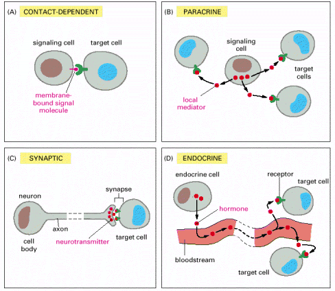 Figure
15-4. Forms of intercellular
signaling. (A) Contact-dependent signaling requires cells
to be in direct membrane-membrane contact. (B) Paracrine signaling
depends on signals that are released into the extracellular space and
act locally on neighboring cells. (C) Synaptic signaling is performed
by neurons that transmit signals electrically along their axons and
release neurotransmitters at synapses, which are often located far
away from the cell body. (D) Endocrine signaling depends on endocrine
cells, which secrete hormones into the bloodstream that are then
distributed widely throughout the body. Many of the same types of
signaling molecules are used in paracrine, synaptic, and endocrine
signaling; the crucial differences lie in the speed and selectivity
with which the signals are delivered to their targets.
Figure
15-4. Forms of intercellular
signaling. (A) Contact-dependent signaling requires cells
to be in direct membrane-membrane contact. (B) Paracrine signaling
depends on signals that are released into the extracellular space and
act locally on neighboring cells. (C) Synaptic signaling is performed
by neurons that transmit signals electrically along their axons and
release neurotransmitters at synapses, which are often located far
away from the cell body. (D) Endocrine signaling depends on endocrine
cells, which secrete hormones into the bloodstream that are then
distributed widely throughout the body. Many of the same types of
signaling molecules are used in paracrine, synaptic, and endocrine
signaling; the crucial differences lie in the speed and selectivity
with which the signals are delivered to their targets.
 Figure
15-5. The contrast between
endocrine and synaptic signaling. In complex animals,
endocrine cells and nerve cells work together to coordinate the
diverse activities of the billions of cells. Whereas different
endocrine cells must use different hormones to communicate
specifically with their target cells, different nerve cells can use
the same neurotransmitter and still communicate in a highly specific
manner. (A) Endocrine cells secrete hormones into the blood, which
signal only the specific target cells that recognize them. These
target cells have receptors for binding a specific hormone, which the
cells "pull" from the extracellular fluid. (B) In synaptic
signaling, by contrast, specificity arises from the synaptic contacts
between a nerve cell and the specific target cells it signals.
Usually, only a target cell that is in synaptic communication with a
nerve cell is exposed to the neurotransmitter released from the nerve
terminal (although some neurotransmitters act in a paracrine mode,
serving as local mediators that influence multiple target cells in
the area).
Figure
15-5. The contrast between
endocrine and synaptic signaling. In complex animals,
endocrine cells and nerve cells work together to coordinate the
diverse activities of the billions of cells. Whereas different
endocrine cells must use different hormones to communicate
specifically with their target cells, different nerve cells can use
the same neurotransmitter and still communicate in a highly specific
manner. (A) Endocrine cells secrete hormones into the blood, which
signal only the specific target cells that recognize them. These
target cells have receptors for binding a specific hormone, which the
cells "pull" from the extracellular fluid. (B) In synaptic
signaling, by contrast, specificity arises from the synaptic contacts
between a nerve cell and the specific target cells it signals.
Usually, only a target cell that is in synaptic communication with a
nerve cell is exposed to the neurotransmitter released from the nerve
terminal (although some neurotransmitters act in a paracrine mode,
serving as local mediators that influence multiple target cells in
the area).
 Figure
15-6. Autocrine signaling.
A group of identical cells produces a higher concentration of a
secreted signal than does a single cell. When this signal binds back
to a receptor on the same cell type, it encourages the cells to
respond coordinately as a group.
Figure
15-6. Autocrine signaling.
A group of identical cells produces a higher concentration of a
secreted signal than does a single cell. When this signal binds back
to a receptor on the same cell type, it encourages the cells to
respond coordinately as a group.
 Figure
15-7. Signaling via gap
junctions. Cells connected by gap junctions share small
molecules, including small intracellular signaling molecules, and can
therefore respond to extracellular signals in a coordinated way.
Figure
15-7. Signaling via gap
junctions. Cells connected by gap junctions share small
molecules, including small intracellular signaling molecules, and can
therefore respond to extracellular signals in a coordinated way.
 Figure
15-8. An animal cell's dependence
on multiple extracellular signals. Each cell type displays
a set of receptors that enables it to respond to a corresponding set
of signal molecules produced by other cells. These signal molecules
work in combinations to regulate the behavior of the cell. As shown
here, an individual cell requires multiple signals to survive (blue
arrows) and additional signals to divide (red arrow) or
differentiate (green arrows). If deprived of appropriate
survival signals, a cell will undergo a form of cell suicide known as
programmed cell death, or apoptosis.
Figure
15-8. An animal cell's dependence
on multiple extracellular signals. Each cell type displays
a set of receptors that enables it to respond to a corresponding set
of signal molecules produced by other cells. These signal molecules
work in combinations to regulate the behavior of the cell. As shown
here, an individual cell requires multiple signals to survive (blue
arrows) and additional signals to divide (red arrow) or
differentiate (green arrows). If deprived of appropriate
survival signals, a cell will undergo a form of cell suicide known as
programmed cell death, or apoptosis.
 Figure
15-9. Various responses induced
by the neurotransmitter acetylcholine. Different cell
types are specialized to respond to acetylcholine in different ways.
(A and B) For these two cell types, acetylcholine binds to similar
receptor proteins, but the intracellular signals produced are
interpreted differently in cells specialized for different functions.
(C) This muscle cell produces a distinct type of receptor protein for
acetylcholine, which generates different intracellular signals from
the receptor shown in (A) and (B), and results in a different effect.
(D) The chemical structure of acetylcholine.
Figure
15-9. Various responses induced
by the neurotransmitter acetylcholine. Different cell
types are specialized to respond to acetylcholine in different ways.
(A and B) For these two cell types, acetylcholine binds to similar
receptor proteins, but the intracellular signals produced are
interpreted differently in cells specialized for different functions.
(C) This muscle cell produces a distinct type of receptor protein for
acetylcholine, which generates different intracellular signals from
the receptor shown in (A) and (B), and results in a different effect.
(D) The chemical structure of acetylcholine.
 Figure
15-10. The importance of rapid
turnover. The graphs show the predicted relative rates of
change in the intracellular concentrations of molecules with
differing turnover times when their synthesis rates are either (A)
decreased or (B) increased suddenly by a factor of 10. In both cases,
the concentrations of those molecules that are normally being rapidly
degraded in the cell (red lines) change quickly, whereas the
concentrations of those that are normally being slowly degraded
(green lines) change proportionally more slowly. The numbers
(in blue) on the right are the half-lives assumed for each of
the different molecules.
Figure
15-10. The importance of rapid
turnover. The graphs show the predicted relative rates of
change in the intracellular concentrations of molecules with
differing turnover times when their synthesis rates are either (A)
decreased or (B) increased suddenly by a factor of 10. In both cases,
the concentrations of those molecules that are normally being rapidly
degraded in the cell (red lines) change quickly, whereas the
concentrations of those that are normally being slowly degraded
(green lines) change proportionally more slowly. The numbers
(in blue) on the right are the half-lives assumed for each of
the different molecules.
 Figure
15-11. The role of nitric oxide
(NO) in smooth muscle relaxation in a blood vessel wall.
Acetylcholine released by nerve terminals in the blood vessel wall
activates NO synthase in endothelial cells lining the blood vessel,
causing the endothelial cells to produce NO. The NO diffuses out of
the endothelial cells and into the underlying smooth muscle cells,
where it binds to and activates guanylyl cyclase to produce cyclic
GMP. The cyclic GMP triggers a response that causes the smooth muscle
cells to relax, enhancing blood flow through the blood vessel.
Figure
15-11. The role of nitric oxide
(NO) in smooth muscle relaxation in a blood vessel wall.
Acetylcholine released by nerve terminals in the blood vessel wall
activates NO synthase in endothelial cells lining the blood vessel,
causing the endothelial cells to produce NO. The NO diffuses out of
the endothelial cells and into the underlying smooth muscle cells,
where it binds to and activates guanylyl cyclase to produce cyclic
GMP. The cyclic GMP triggers a response that causes the smooth muscle
cells to relax, enhancing blood flow through the blood vessel.
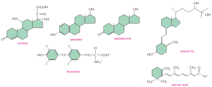 Figure
15-12. Some signaling molecules
that bind to nuclear receptors. Note that all of them are
small and hydrophobic. The active, hydroxylated form of vitamin D3
is shown. Estradiol and testosterone are steroid sex hormones.
Figure
15-12. Some signaling molecules
that bind to nuclear receptors. Note that all of them are
small and hydrophobic. The active, hydroxylated form of vitamin D3
is shown. Estradiol and testosterone are steroid sex hormones.
 Figure
15-13. The nuclear receptor
superfamily. All nuclear hormone receptors bind to DNA as
either homodimers or heterodimers, but for simplicity we show them as
monomers here. (A) The receptors all have a related structure. The
short DNA-binding domain in each receptor is shown in green.
(B) A receptor protein in its inactive state is bound to inhibitory
proteins. Domain-swap experiments suggest that many of the
ligand-binding, transcription-activating, and DNA-binding domains in
these receptors can function as interchangeable modules. (C) The
binding of ligand to the receptor causes the ligand-binding domain of
the receptor to clamp shut around the ligand, the inhibitory proteins
to dissociate, and coactivator proteins to bind to the receptor's
transcription-activating domain, thereby increasing gene
transcription. (D) The three-dimensional structure of a
ligand-binding domain with (right) and without (left)
ligand bound. Note that the blue
helix acts as a lid that snaps shut when the ligand (shown in red)
binds, trapping the ligand in place.
Figure
15-13. The nuclear receptor
superfamily. All nuclear hormone receptors bind to DNA as
either homodimers or heterodimers, but for simplicity we show them as
monomers here. (A) The receptors all have a related structure. The
short DNA-binding domain in each receptor is shown in green.
(B) A receptor protein in its inactive state is bound to inhibitory
proteins. Domain-swap experiments suggest that many of the
ligand-binding, transcription-activating, and DNA-binding domains in
these receptors can function as interchangeable modules. (C) The
binding of ligand to the receptor causes the ligand-binding domain of
the receptor to clamp shut around the ligand, the inhibitory proteins
to dissociate, and coactivator proteins to bind to the receptor's
transcription-activating domain, thereby increasing gene
transcription. (D) The three-dimensional structure of a
ligand-binding domain with (right) and without (left)
ligand bound. Note that the blue
helix acts as a lid that snaps shut when the ligand (shown in red)
binds, trapping the ligand in place.
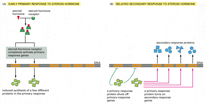 Figure
15-14. Responses induced by the
activation of a nuclear hormone receptor. (A) Early
primary response and (B) delayed secondary response. The figure shows
the responses to a steroid hormone, but the same principles apply for
all ligands that activate this family of receptor proteins. Some of
the primary-response proteins turn on secondary-response genes,
whereas others turn off the primary-response genes. The actual number
of primary- and secondary-response genes is greater than shown. As
expected, drugs that inhibit protein synthesis suppress the
transcription of secondary-response genes but not primary-response
genes, allowing these two classes of gene transcription responses to
be readily distinguished.
Figure
15-14. Responses induced by the
activation of a nuclear hormone receptor. (A) Early
primary response and (B) delayed secondary response. The figure shows
the responses to a steroid hormone, but the same principles apply for
all ligands that activate this family of receptor proteins. Some of
the primary-response proteins turn on secondary-response genes,
whereas others turn off the primary-response genes. The actual number
of primary- and secondary-response genes is greater than shown. As
expected, drugs that inhibit protein synthesis suppress the
transcription of secondary-response genes but not primary-response
genes, allowing these two classes of gene transcription responses to
be readily distinguished.
 Figure
15-15. Three classes of
cell-surface receptors. (A) Ion-channel-linked receptors,
(B) G-protein-linked receptors, and (C) enzyme-linked receptors.
Although many enzyme-linked receptors have intrinsic enzyme activity,
as shown on the left, many others rely on associated enzymes, as
shown on the right.
Figure
15-15. Three classes of
cell-surface receptors. (A) Ion-channel-linked receptors,
(B) G-protein-linked receptors, and (C) enzyme-linked receptors.
Although many enzyme-linked receptors have intrinsic enzyme activity,
as shown on the left, many others rely on associated enzymes, as
shown on the right.
 Figure
15-16. Different kinds of
intracellular signaling proteins along a signaling pathway from a
cell-surface receptor to the nucleus. In this example, a
series of signaling proteins and small intracellular mediators relay
the extracellular signal into the cell, causing a change in gene
expression. The signal is amplified, altered (transduced), and
distributed en route. Many of the steps can be modulated by
other extracellular and intracellular signals, so that the final
result of one signal depends on other factors affecting the cell (see
Figure
15-8). Ultimately, the signaling pathway
activates (or inactivates) target proteins that alter cell behavior.
In this example, the target is a gene regulatory protein.
Figure
15-16. Different kinds of
intracellular signaling proteins along a signaling pathway from a
cell-surface receptor to the nucleus. In this example, a
series of signaling proteins and small intracellular mediators relay
the extracellular signal into the cell, causing a change in gene
expression. The signal is amplified, altered (transduced), and
distributed en route. Many of the steps can be modulated by
other extracellular and intracellular signals, so that the final
result of one signal depends on other factors affecting the cell (see
Figure
15-8). Ultimately, the signaling pathway
activates (or inactivates) target proteins that alter cell behavior.
In this example, the target is a gene regulatory protein.
 Figure
15-17. Two types of intracellular
signaling proteins that act as molecular switches. In both
cases, a signaling protein is activated by the addition of a
phosphate group and inactivated by the removal of the phosphate. (A)
The phosphate is added covalently to the signaling protein by a
protein kinase. (B) A signaling protein is induced to exchange its
bound GDP for GTP. To emphasize the similarity in the two mechanisms,
ATP is shown as APPP, ADP as APP, GTP as GPPP, and GDP as GPP.
Figure
15-17. Two types of intracellular
signaling proteins that act as molecular switches. In both
cases, a signaling protein is activated by the addition of a
phosphate group and inactivated by the removal of the phosphate. (A)
The phosphate is added covalently to the signaling protein by a
protein kinase. (B) A signaling protein is induced to exchange its
bound GDP for GTP. To emphasize the similarity in the two mechanisms,
ATP is shown as APPP, ADP as APP, GTP as GPPP, and GDP as GPP.
 Figure
15-18. Signal integration.
(A) Extracellular signals A and B both activate a different series of
protein phosphorylations, each of which leads to the phosphorylation
of protein Y but at different sites on the protein. Protein Y is
activated only when both of these sites are phosphorylated, and
therefore it becomes active only when signals A and B are
simultaneously present. For this reason, integrator proteins are
sometimes called coincidence detectors. (B) Extracellular signals A
and B lead to the phosphorylation of two proteins, a and b, which
then bind to each other to create the active protein. In both of the
examples illustrated, the proteins themselves are phosphorylated. An
equivalent form of control can also occur, however, by the exchange
of GTP for GDP on GTP-binding proteins (see Figure
15-17).
Figure
15-18. Signal integration.
(A) Extracellular signals A and B both activate a different series of
protein phosphorylations, each of which leads to the phosphorylation
of protein Y but at different sites on the protein. Protein Y is
activated only when both of these sites are phosphorylated, and
therefore it becomes active only when signals A and B are
simultaneously present. For this reason, integrator proteins are
sometimes called coincidence detectors. (B) Extracellular signals A
and B lead to the phosphorylation of two proteins, a and b, which
then bind to each other to create the active protein. In both of the
examples illustrated, the proteins themselves are phosphorylated. An
equivalent form of control can also occur, however, by the exchange
of GTP for GDP on GTP-binding proteins (see Figure
15-17).
 Figure
15-19. Two types of intracellular
signaling complexes. (A) A receptor and some of the
intracellular signaling proteins it activates in sequence are
preassembled into a signaling complex by a large scaffold protein.
(B) A large signaling complex is assembled after a receptor has been
activated by the binding of an extracellular signal molecule; here
the activated receptor phosphorylates itself at multiple sites, which
then act as docking sites for intracellular signaling proteins.
Figure
15-19. Two types of intracellular
signaling complexes. (A) A receptor and some of the
intracellular signaling proteins it activates in sequence are
preassembled into a signaling complex by a large scaffold protein.
(B) A large signaling complex is assembled after a receptor has been
activated by the binding of an extracellular signal molecule; here
the activated receptor phosphorylates itself at multiple sites, which
then act as docking sites for intracellular signaling proteins.
 Figure
15-20. A hypothetical signaling
pathway using modular binding domains. Signaling protein 1
contains three different binding domains, plus a catalytic protein
kinase domain. It moves to the plasma membrane when extracellular
signals lead to the creation of various phosphorylated docking sites
on the cytosolic face of the membrane. Its SH2 domain binds to
phosphorylated tyrosines on the receptor protein, and its PH domain
binds to phosphorylated inositol phospholipids in the inner leaflet
of the lipid bilayer. Protein 1 then phosphorylates signaling protein
2 on tyrosines, which allows protein 2 to bind to the PTB domain on
protein 1 and to the SH2 domain on an adaptor protein. The adaptor
protein then links protein 2 to protein 3, causing the
phosphorylation of protein 3 by protein 2. The adaptor protein shown
consists of two binding domains
Figure
15-20. A hypothetical signaling
pathway using modular binding domains. Signaling protein 1
contains three different binding domains, plus a catalytic protein
kinase domain. It moves to the plasma membrane when extracellular
signals lead to the creation of various phosphorylated docking sites
on the cytosolic face of the membrane. Its SH2 domain binds to
phosphorylated tyrosines on the receptor protein, and its PH domain
binds to phosphorylated inositol phospholipids in the inner leaflet
of the lipid bilayer. Protein 1 then phosphorylates signaling protein
2 on tyrosines, which allows protein 2 to bind to the PTB domain on
protein 1 and to the SH2 domain on an adaptor protein. The adaptor
protein then links protein 2 to protein 3, causing the
phosphorylation of protein 3 by protein 2. The adaptor protein shown
consists of two binding domains![]() an
SH2 domain, which binds to a phosphotyrosine on protein 2, and an SH3
domain, which binds to a proline-rich motif on protein 3.
an
SH2 domain, which binds to a phosphotyrosine on protein 2, and an SH3
domain, which binds to a proline-rich motif on protein 3.
 Figure
15-21. The primary response of
chick oviduct cells to the steroid sex hormone estradiol.
When activated, estradiol receptors turn on the transcription of
several genes. Dose-response curves for two of these genes are shown,
one coding for the egg protein conalbumin and the other coding for
the egg protein ovalbumin. The linear response curve for conalbumin
indicates that each activated receptor molecule that binds to the
conalbumin gene increases the activity of the gene by the same
amount. In contrast, the lag followed by the steep increase in the
response curve for ovalbumin suggests that more than one activated
receptor (in this case, two receptors) must bind simultaneously to
the ovalbumin gene to initiate its transcription. (Adapted from E.R.
Mulvihill and R.D. Palmiter, J. Biol. Chem. 252:2060
Figure
15-21. The primary response of
chick oviduct cells to the steroid sex hormone estradiol.
When activated, estradiol receptors turn on the transcription of
several genes. Dose-response curves for two of these genes are shown,
one coding for the egg protein conalbumin and the other coding for
the egg protein ovalbumin. The linear response curve for conalbumin
indicates that each activated receptor molecule that binds to the
conalbumin gene increases the activity of the gene by the same
amount. In contrast, the lag followed by the steep increase in the
response curve for ovalbumin suggests that more than one activated
receptor (in this case, two receptors) must bind simultaneously to
the ovalbumin gene to initiate its transcription. (Adapted from E.R.
Mulvihill and R.D. Palmiter, J. Biol. Chem. 252:2060![]() 2068,
1977.)
2068,
1977.)
 Figure
15-22. Activation curves as a
function of signal-molecule concentration. The curves show
how the sharpness of the response increases with an increase in the
number of effector molecules that must bind simultaneously to
activate a target macromolecule. The curves shown are those expected
if the activation requires the simultaneous binding of 1, 2, 8, or 16
effector molecules.
Figure
15-22. Activation curves as a
function of signal-molecule concentration. The curves show
how the sharpness of the response increases with an increase in the
number of effector molecules that must bind simultaneously to
activate a target macromolecule. The curves shown are those expected
if the activation requires the simultaneous binding of 1, 2, 8, or 16
effector molecules.
 Figure
15-23. One type of signaling
mechanism expected to show a steep thresholdlike response.
Here, the simultaneous binding of eight molecules of a signaling
ligand to a set of eight protein subunits is required to form an
active protein complex. The ability of the subunits to assemble into
the active complex depends on an allosteric conformational change
that the subunits undergo when they bind their ligand. The binding of
the ligand in the formation of such a complex is generally a
cooperative process, causing a steep response as the ligand
concentration is changed, as explained in Chapter
3. At low ligand concentrations, the number of
active complexes increases roughly in proportion to the eighth power
of the ligand concentration.
Figure
15-23. One type of signaling
mechanism expected to show a steep thresholdlike response.
Here, the simultaneous binding of eight molecules of a signaling
ligand to a set of eight protein subunits is required to form an
active protein complex. The ability of the subunits to assemble into
the active complex depends on an allosteric conformational change
that the subunits undergo when they bind their ligand. The binding of
the ligand in the formation of such a complex is generally a
cooperative process, causing a steep response as the ligand
concentration is changed, as explained in Chapter
3. At low ligand concentrations, the number of
active complexes increases roughly in proportion to the eighth power
of the ligand concentration.
 Figure
15-24. An accelerating positive
feedback mechanism. In this example, the initial binding
of the signaling ligand activates the enzyme to generate a product
that binds back to the enzyme, further increasing the enzyme's
activity.
Figure
15-24. An accelerating positive
feedback mechanism. In this example, the initial binding
of the signaling ligand activates the enzyme to generate a product
that binds back to the enzyme, further increasing the enzyme's
activity.
 Figure
15-25. Five ways in which target
cells can become desensitized to a signal molecule. The
inactivation mechanisms shown here for both the receptor and the
intracellular signaling protein often involve phosphorylation of the
protein that is inactivated, although other types of modification are
also known to occur. In bacterial chemotaxis, which we discuss later,
desensitization depends on methylation of the receptor protein.
Figure
15-25. Five ways in which target
cells can become desensitized to a signal molecule. The
inactivation mechanisms shown here for both the receptor and the
intracellular signaling protein often involve phosphorylation of the
protein that is inactivated, although other types of modification are
also known to occur. In bacterial chemotaxis, which we discuss later,
desensitization depends on methylation of the receptor protein.