

Книги фарма 2 / Bertram G. Katzung-Basic & Clinical Pharmacology(9th Edition)
.pdfMitotane (Lysodren)
Oral: 500 mg tablets
1 Glucocorticoids for Aerosol Use: See Chapter 20: Drugs Used in Asthma.Glucocorticoids for Dermatologic Use: See Chapter 62: Dermatologic Pharmacology.Glucocorticoids for Gastrointestinal Use: See Chapter 63: Drugs Used in the Treatment of Gastrointestinal Diseases.
Chapter 40. The Gonadal Hormones & Inhibitors
Acronyms
ACTH: Adrenocorticotropic hormone AST: Aspartate aminotransferase
CBG: Corticosteroid-binding globulin (transcortin) DHEA: Dehydroepiandrosterone
DHEAS: Dehydroepiandrosterone sulfate ERE: Estrogen response element
FSH: Follicle-stimulating hormone GnRH: Gonadotropin-releasing hormone hCG: Human chorionic gonadotropin hMG: Human menopausal gonadotropin HRT: Hormone replacement therapy LH: Luteinizing hormone
LHRH: Luteinizing hormone-releasing hormone (synonym of GnRH) PRE: Progesterone response element
SERM: Selective estrogen receptor modulator SHBG: Sex hormone-binding globulin TBG: Thyroxine-binding globulin
Katzung PHARMACOLOGY, 9e > Section VII. Endocrine Drugs > Chapter 40. The Gonadal Hormones & Inhibitors >
The Ovary (Estrogens, Progestins, Other Ovarian Hormones, Oral Contraceptives, Inhibitors & Antagonists, & Ovulation-Inducing Agents)
The ovary has important gametogenic functions that are integrated with its hormonal activity. In the human female, the gonad is relatively quiescent during childhood, the period of rapid growth and maturation. At puberty, the ovary begins a 30to 40-year period of cyclic function called the menstrual cycle because of the regular episodes of bleeding that are its most obvious manifestation. It then fails to respond to gonadotropins secreted by the anterior pituitary gland, and the cessation of cyclic bleeding that occurs is called the menopause.
The mechanism responsible for the onset of ovarian function at the time of puberty is thought to be neural in origin, because the immature gonad can be stimulated by gonadotropins already present in the pituitary and because the pituitary is responsive to exogenous hypothalamic gonadotropinreleasing hormone. The maturation of centers in the brain may withdraw a childhood-related inhibitory effect upon hypothalamic arcuate nucleus neurons, allowing them to produce gonadotropin-releasing hormone (GnRH) in pulses with the appropriate amplitude, which stimulates the release of follicle-stimulating hormone (FSH) and luteinizing hormone (LH) (see

Chapter 37: Hypothalamic & Pituitary Hormones). At first, small amounts of the latter two hormones are released during the night, and the limited quantities of ovarian estrogen secreted in response start to cause breast development. Subsequentely, FSH and LH are secreted throughout the day and night, causing secretion of higher amounts of estrogen and leading to futher breast enlargement, alterations in fat distribution, and a growth spurt that culminates in epiphysial closure in the long bones. The beginning of ovarian function at puberty is called gonadarche.
A year or so after gonadarche, sufficient estrogen is produced to induce endometrial changes and periodic bleeding. After the first few irregular cycles, which may be anovulatory, normal cyclic function is established.
At the beginning of each cycle, a variable number of follicles (vesicular follicles), each containing an ovum, begin to enlarge in response to FSH. After 5 or 6 days, one follicle, called the dominant follicle, begins to develop more rapidly. The outer theca and inner granulosa cells of this follicle multiply and, under the influence of LH, synthesize and release estrogens at an increasing rate. The estrogens appear to inhibit FSH release and may lead to regression of the smaller, less mature follicles. The mature dominant ovarian follicle consists of an ovum surrounded by a fluid-filled antrum lined by granulosa and theca cells. The estrogen secretion reaches a peak just before midcycle, and the granulosa cells begin to secrete progesterone. These changes stimulate the brief surge in LH and FSH release that precedes and causes ovulation. When the follicle ruptures, the ovum is released into the abdominal cavity near the opening of the uterine tube.
Following the above events, the cavity of the ruptured follicle fills with blood (corpus hemorrhagicum), and the luteinized theca and granulosa cells proliferate and replace the blood to form the corpus luteum. The cells of this structure produce estrogens and progesterone for the remainder of the cycle, or longer if pregnancy occurs.
If pregnancy does not occur, the corpus luteum begins to degenerate and ceases hormone production, eventually becoming a corpus albicans. The endometrium, which proliferated during the follicular phase and developed its glandular function during the luteal phase, is shed in the process of menstruation. These events are summarized in Figure 40–1.
Figure 40–1.
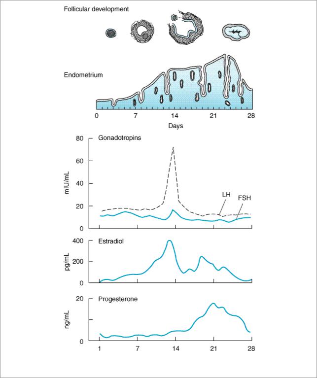
The menstrual cycle, showing plasma levels of pituitary and ovarian hormones and histologic changes.
The ovary normally ceases its gametogenic and endocrine function with time. This change is accompanied by a cessation in uterine bleeding (menopause) and occurs at a mean age of 52 years in the USA. Although the ovary ceases to secrete estrogen, significant levels of estrogen persist in many women as a result of conversion of adrenal and ovarian steroids such as androstenedione to estrone and estradiol in adipose and possibly other nonendocrine tissues.
Disturbances in Ovarian Function
Disturbances of cyclic function are common even during the peak years of reproduction. A minority

of these result from inflammatory or neoplastic processes that influence the functions of the uterus, ovaries, or pituitary. Many of the minor disturbances leading to periods of amenorrhea or anovulatory cycles are self-limited. They are often associated with emotional or physical stress and represent temporary alterations in the stress centers in the brain that control the secretion of gonadotropin-releasing hormone. Anovulatory cycles are also associated with eating disorders (bulimia, anorexia nervosa) and with severe exercise such as distance running and swimming. Among the more common organic causes of persistent ovulatory disturbances are pituitary prolactinomas and syndromes and tumors characterized by excessive ovarian or adrenal androgen production. Normal ovarian function can be modified by androgens produced by the adrenal cortex or tumors arising from it. The ovary also gives rise to androgen-producing neoplasms such as arrhenoblastomas, as well as to estrogen-producing granulosa cell tumors.
The Estrogens
Estrogenic activity is shared by a large number of chemical substances. In addition to the variety of steroidal estrogens derived from animal sources, numerous nonsteroidal estrogens have been synthesized. Many phenols are estrogenic, and estrogenic activity has been identified in such diverse forms of life as those found in ocean sediments. Estrogen-mimetic compounds (flavonoids) are found in many plants, including saw palmetto, and soybeans and other foods. Studies have shown that eating these plant products may produce slight estrogenic effects. Additionally, some compounds used in the manufacture of plastics (bisphenols, alkylphenols, phthalate phenols) have been found to be estrogenic. It has been proposed that these agents are associated with an increased breast cancer incidence in both women and men in the industrialized world.
Natural Estrogens
The major estrogens produced by women are estradiol (estradiol-17 , E2), estrone (E1), and estriol (E3) (Figure 40–2). Estradiol is the major secretory product of the ovary. Although some estrone is produced in the ovary, most estrone and estriol are formed in the liver from estradiol or in peripheral tissues from androstenedione and other androgens. As noted above, during the first part of the menstrual cycle estrogens are produced in the ovarian follicle by the theca and granulosa cells. After ovulation, the estrogens as well as progesterone are synthesized by the luteinized granulosa and theca cells of the corpus luteum, and the pathways of biosynthesis are slightly different.
Figure 40–2.
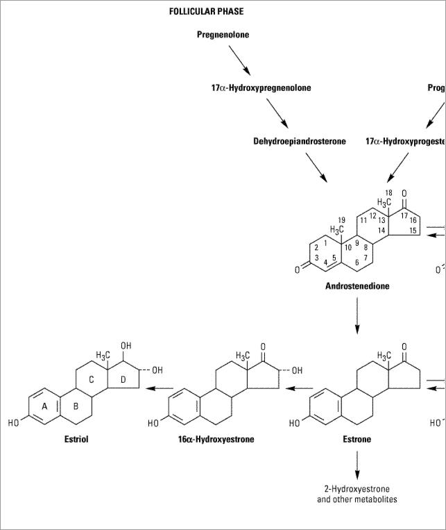
Biosynthesis and metabolism of estrogens.
During pregnancy, a large amount of estrogen is synthesized by the fetoplacental unit—consisting of the fetal adrenal zone, secreting androgen precursor, and the placenta, which aromatizes it into estrogen. The estriol synthesized by the fetoplacental unit is released into the maternal circulation and excreted into the urine. Repeated assay of maternal urinary estriol excretion has been used in the assessment of fetal well-being.
One of the most prolific natural sources of estrogenic substances is the stallion, which liberates more of these hormones than the pregnant mare or pregnant woman. The equine estrogens— equilenin and equilin—and their congeners are unsaturated in the B as well as the A ring and are
excreted in large quantities in urine, from which they can be recovered and used for medicinal purposes.
In normal women, estradiol is produced at a rate that varies during the menstrual cycle, resulting in plasma levels as low as 50 pg/mL in the early follicular phase to as high as 350–850 pg/mL at the time of the preovulatory peak (Figure 40–1).
Synthetic Estrogens
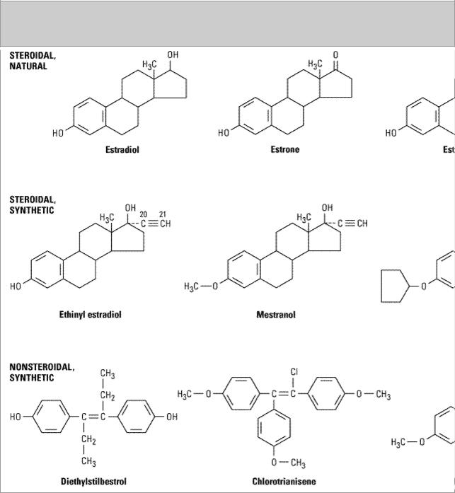
A variety of chemical alterations have been produced in the natural estrogens. The most important effect of these alterations has been to increase the oral effectiveness of the estrogens. Some structures are shown in Figure 40–3. Those with therapeutic use are listed in Table 40–1.
Figure 40–3.

Compounds with estrogenic activity.
Table 40–1. Commonly Used Estrogens.
|
Average Replacement Dosage |
|
|
Ethinyl estradiol |
0.005–0.02 mg/d |
|
|
Micronized estradiol |
1–2 mg/d |
|
|
Estradiol cypionate |
2–5 mg every 3–4 weeks |
|
|
Estradiol valerate |
2–20 mg every other week |
|
|
Estropipate |
1.25–2.5 mg/d |
|
|
Conjugated, esterified, or mixed estrogenic substances: |
|
|
|
Oral |
0.3–1.25 mg/d |
|
|
Injectable |
0.2–2 mg/d |
|
|
Transdermal |
Patch |
|
|
Diethylstilbestrol |
0.1–0.5 mg/d |
|
|
Quinestrol |
0.1–0.2 mg/week |
|
|
Chlorotrianisene |
12–25 mg/d |
|
|
Methallenestril |
3–9 mg/d |
|
|
In addition to the steroidal estrogens, a variety of nonsteroidal compounds with estrogenic activity have been synthesized and used clinically. These include dienestrol, diethylstilbestrol, benzestrol, hexestrol, methestrol, methallenestril, and chlorotrianisene (Figure 40–3).
Pharmacokinetics
When released into the circulation, estradiol binds strongly to an  2-globulin (sex hormone-binding globulin [SHBG]) and to albumin with lower affinity. Bound estrogen is relatively unavailable for diffusion into cells, so it is the free fraction that is physiologically active. Estradiol is converted by the liver and other tissues to estrone and estriol (which have low affinity for the estrogen receptor) and their 2-hydroxylated derivatives and conjugated metabolites (which are too insoluble in lipid to cross the cell membrane readily) and excreted in the bile (Figure 40–2). However, the conjugates may be hydrolyzed in the intestine to active, reabsorbable compounds. Estrogens are also excreted in small amounts in the breast milk of nursing mothers.
Because significant amounts of estrogens and their active metabolites are excreted in the bile and reabsorbed from the intestine, the resulting enterohepatic circulation ensures that orally administered estrogens will have a high ratio of hepatic to peripheral effects. As noted below, the hepatic effects are thought to be responsible for some undesirable actions such as synthesis of increased clotting factors and plasma renin substrate. The hepatic effects of estrogen can be minimized by routes that avoid first-pass liver exposure, ie, vaginal, transdermal, or by injection.
Physiologic Effects
Mechanism

Plasma estrogens in the blood and interstitial fluid are bound to sex hormone-binding globulin, from which they dissociate to enter the cell and bind to their receptor. Two genes code for two estrogen receptor isoforms, 
 and
and  , which are members of the superfamily of steroid, sterol, retinoic acid, and thyroid receptors. The estrogen receptors are found predominantly in the nucleus bound to heat shock proteins that stabilize them (Figure 39–3).
Binding of the hormone to its receptor alters its conformation and releases it from the stabilizing proteins (predominantly Hsp90). The receptor-hormone complex forms homodimers that bind to a specific sequence of nucleotides called estrogen response elements (EREs) in the promoters of various genes and regulate their transcription. The ERE is composed of two half-sites arranged as a palindrome separated by a small group of nucleotides called the spacer. The interaction of a receptor dimer with the ERE also involves a number of nuclear proteins, the coregulators, as well as components of the transcription machinery. The receptor may also bind to other transcription factors to influence the effects of these factors on their responsive genes.
The relative concentrations and types of receptors, receptor coregulators, and transcription factors confer the cell specificity of the hormone's actions. The genomic effects of estrogens are mainly due to proteins synthesized by translation of RNA transcribed from a responsive gene. Some of the effects of estrogens are indirect, mediated by the autocrine and paracrine actions of autacoids such as growth factors, lipids, glycolipids, and cytokines produced by the target cells in response to estrogen.
Rapid estrogen-induced effects such as granulosa cell Ca2+ uptake and increased uterine blood flow do not require gene activation. These appear to be mediated by nongenomic effects of the classic estrogen receptor-estrogen complex, influencing several intracellular signaling pathways.
Female Maturation
Estrogens are required for the normal sexual maturation and growth of the female. They stimulate the development of the vagina, uterus, and uterine tubes as well as the secondary sex characteristics. They stimulate stromal development and ductal growth in the breast and are responsible for the accelerated growth phase and the closing of the epiphyses of the long bones that occur at puberty. They contribute to the growth of axillary and pubic hair and alter the distribution of body fat to produce typical female body contours. Larger quantities also stimulate development of pigmentation in the skin, most prominent in the region of the nipples and areolae and in the genital region.
Endometrial Effects
In addition to its growth effects on uterine muscle, estrogen also plays an important role in the development of the endometrial lining. When estrogen production is properly coordinated with the production of progesterone during the normal human menstrual cycle, regular periodic bleeding and shedding of the endometrial lining occur. Continuous exposure to estrogens for prolonged periods leads to hyperplasia of the endometrium that is usually associated with abnormal bleeding patterns.
Metabolic and Cardiovascular Effects
Estrogens have a number of important metabolic and cardiovascular effects. They seem to be partially responsible for maintenance of the normal structure and function of the skin and blood vessels in women. Estrogens also decrease the rate of resorption of bone by promoting the apoptosis of osteoclasts and by antagonizing the osteoclastogenic and pro-osteoclastic effects of parathyroid

hormone and interleukin-6. Estrogens also stimulate adipose tissue production of leptin and are in part responsible for the higher levels of this hormone in women than in men.
In addition to stimulating the synthesis of enzymes and growth factors leading to uterine and breast growth and differentiation, estrogens alter the production and activity of many other proteins in the body. Metabolic alterations in the liver are especially important, so that there is a higher circulating level of proteins such as transcortin (CBG), thyroxine-binding globulin (TBG), sex hormonebinding globulin (SHBG), transferrin, renin substrate, and fibrinogen. This leads to increased circulating levels of thyroxine, estrogen, testosterone, iron, copper, and other substances.
Alterations in the composition of the plasma lipids caused by estrogens are characterized by an increase in the high-density lipoproteins, a slight reduction in the low-density lipoproteins, and a reduction in plasma cholesterol levels. Plasma triglyceride levels are increased. Estrogens decrease hepatic oxidation of adipose tissue lipid to ketones and increase synthesis of triglycerides.
Effects on Blood Coagulation
Estrogens enhance the coagulability of blood. Many changes in factors influencing coagulation have been reported, including increased circulating levels of factors II, VII, IX, and X and decreased antithrombin III, partially as a result of the hepatic effects mentioned above. Increased plasminogen levels and decreased platelet adhesiveness have also been found (see Hormonal Contraception).
Other Effects
Estrogens induce the synthesis of progesterone receptors. They are responsible for estrous behavior in animals and may influence behavior and libido in humans. Administration of estrogens stimulates central components of the stress system, including the production of corticotropinreleasing hormone and the activity of the sympathetic system, and promotes a sense of well-being in women who are estrogen-deficient. They also facilitate the loss of intravascular fluid into the extracellular space, producing edema. The resulting decrease in plasma volume causes a compensatory retention of sodium and water by the kidney. Estrogens also modulate sympathetic nervous system control of smooth muscle function.
Clinical Uses*
* The use of estrogens in contraception is discussed below.
Primary Hypogonadism
Estrogens have been used extensively for replacement therapy in estrogen-deficient patients. The estrogen deficiency may be due to primary failure of development of the ovaries, premature menopause, castration, or menopause.
Treatment of primary hypogonadism is usually begun at 11–13 years of age in order to stimulate the development of secondary sex characteristics and menses, to stimulate optimal growth, to prevent osteoporosis and to avoid the psychologic consequences of delayed puberty and estrogen deficiency. Treatment attempts to mimic the physiology of puberty. It is initiated with small doses of estrogen (0.3 mg conjugated estrogens or 5–10 g ethinyl estradiol) on days 1–21 each month and is slowly increased to adult doses and maintained until the age of menopause (approximately 51 years of age). A progestin is added after the first uterine bleeding. When growth is completed,
chronic therapy consists mainly of the administration of adult doses of both estrogens and progestins, as described below.
Postmenopausal Hormonal Therapy
In addition to the signs and symptoms that follow closely upon the cessation of normal ovarian function—such as loss of periods, vasomotor symptoms, sleep disturbances, and genital atrophy— there are longer-lasting changes that influence the health and well-being of postmenopausal women. These include an acceleration of bone loss, which in susceptible women may lead to vertebral, hip, and wrist fractures; and lipid changes, which may contribute to the acceleration of atherosclerotic cardiovascular disease noted in postmenopausal women. The effects of estrogens on bone have been extensively studied, and the effects of hormone withdrawal have been well-characterized. However, the role of estrogens and progestins in the cause and prevention of cardiovascular disease, which is responsible for 350,000 deaths per year, and breast cancer, which causes 35,000 deaths per year, is less well understood.
When normal ovulatory function ceases and the estrogen levels fall after menopause, oophorectomy, or premature ovarian failure, there is an accelerated rise in plasma cholesterol and LDL concentrations, while LDL receptors decline. HDL is not much affected, and levels remain higher than in men. VLDL and triglyceride levels are also relatively unaffected. Since cardiovascular disorders account for most deaths in this age group, the risk for these disorders constitutes a major consideration in deciding whether or not hormonal replacement therapy (HRT) is indicated and influences the selection of hormones to be administered. Estrogen replacement therapy has a beneficial effect on circulating lipids and lipoproteins, and this was earlier thought to be accompanied by a reduction in myocardial infarction by about 50% and of fatal strokes by as much as 40%. These findings, however, have been recently disputed by the results of a large controlled study from the Womens' Health Initative project showing no cardiovascular benefit from estrogen plus progestin replacement therapy in perimenopausal or postmenopausal patients. In fact, there may be a small increase in cardiovascular problems as well as breast cancer in women who received the replacement therapy. Interestingly, a small protective effect against colon cancer was observed. In other recent studies, a protective effect of estrogen replacement therapy against Alzheimer's disease was observed.
Progestins antagonize estrogen's effects on LDL and HDL to a variable extent. However, a large study has recently shown that the addition of a progestin to estrogen replacement therapy does not influence the cardiovascular risk.
Optimal management of the postmenopausal patient requires careful assessment of her symptoms as well as consideration of her age and the presence of (or risks for) cardiovascular disease, osteoporosis, breast cancer, and endometrial cancer. Bearing in mind the effects of the gonadal hormones on each of these disorders, the goals of therapy can then be defined and the risks of therapy assessed and discussed with the patient.
If the main indication for therapy is hot flushes and sleep disturbances, therapy with the lowest dose of estrogen required for symptomatic relief is recommended. Treatment may be required for only a limited period of time and the possible increased risk for breast cancer avoided. In women who have undergone hysterectomy, estrogens alone can be given 5 days per week or continuously, since progestins are not required to reduce the risk for endometrial hyperplasia and cancer. Hot flushes, sweating, insomnia, and atrophic vaginitis are generally relieved by estrogens; many patients experience some increased sense of well-being; and climacteric depression and other psychopathologic states are improved.